The Surgeon's Corridor to the Volar Wrist
- Origin from the medial epicondyle via the common flexor origin; insertion mainly on the base of the second metacarpal with a slip to the third and to the trapezial crest.
- Median nerve innervation, C6-C7, with motor branches entering the proximal third of the muscle belly.
- The tendon runs in its OWN fibro-osseous tunnel over the trapezium, separated from the carpal tunnel by a thick septum - it is not a carpal tunnel structure.
- Within that tunnel the tendon fills roughly 90 per cent of the available space, so any trapezial or scaphotrapezial osteophyte causes stenosing tendinopathy.
- The palmar cutaneous branch of the median nerve runs in the interval between FCR and palmaris longus and is the nerve most often injured in the FCR approach.
- “In the FCR approach the tendon is retracted ULNARWARD - this shields the palmar cutaneous branch and keeps the radial artery safely radial.
- “Deep palmar arch lies only 2 to 3 mm distal to the FCR insertion at the second metacarpal base - a hazard during distal harvest.
- “FCR tendinopathy is the classic missed cause of volar radial wrist pain and travels with scaphotrapeziotrapezoid and thumb basal joint arthritis.
- “Only the radial half of FCR is taken for LRTI; the ulnar half preserves wrist flexion power and prevents secondary weakness.
Overview
Flexor carpi radialis (FCR) is the radial-most of the three superficial wrist flexors. It arises from the common flexor origin, becomes tendinous in the mid-forearm, and travels along the radial side of the volar wrist to insert on the base of the second metacarpal. Functionally it is a wrist flexor and radial deviator. Surgically it matters far more than that: the tendon and its sheath form the doorway used for the volar approach to the distal radius, the volar approach to the scaphoid, and volar radial wrist arthroscopy portals, and the tendon itself is the workhorse local autograft for thumb basal joint and carpal ligament reconstruction.
Unlike palmaris longus and flexor carpi ulnaris, FCR does not simply run subcutaneously to its insertion. Distally it dives deep, enters a discrete fibro-osseous tunnel over the trapezium, and is separated from the carpal canal by a thick fibrous septum. This anatomical peculiarity is the single most examinable fact about the muscle: it explains why FCR tendinopathy is a stenosing condition, why the tendon can be destroyed by trapezial osteophytes, and why FCR is not decompressed by a standard carpal tunnel release.
The wrist does not move in pure planes during function. Most activities of daily living follow the dart-thrower's arc: radial extension to ulnar flexion.
- FCR is the flexion-radial deviation vector. Its line of pull passes volar and radial to the wrist axis, giving it a flexion moment arm of roughly 1.5 cm and a smaller radial deviation moment.
- Its functional antagonist across the diagonal is extensor carpi ulnaris, and its direct antagonist in the sagittal plane is the extensor carpi radialis longus and brevis pair - with which it also co-operates as a radial deviator.
- Balance matters more than power. FCR and ECRL/ECRB together form the radial column couple: firing together they deviate the wrist radially with no net flexion or extension. Losing FCR does not abolish wrist flexion (FCU and palmaris longus remain) but it unbalances the radial column and biases the wrist toward extension and ulnar deviation.
- Distal effect at the thumb base. Because FCR inserts on the index and long metacarpal bases and sends a slip to the trapezial crest, it exerts a compressive and stabilising force across the scaphotrapeziotrapezoid and thumb carpometacarpal region - the reason FCR tendinopathy and basal joint arthritis so often coexist.
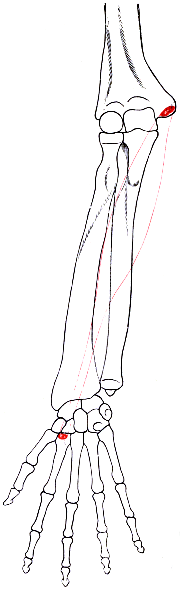
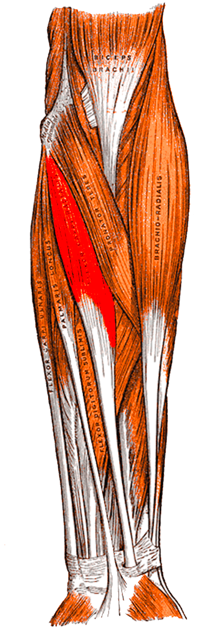
Attachments, Innervation and Relations
Origin
- Medial epicondyle of the humerus through the common flexor origin - FCR is the second most radial of the superficial flexor group after pronator teres.
- Additional fibres from the deep antebrachial fascia and from the intermuscular septa it shares with pronator teres (radially) and palmaris longus (ulnarly).
- The origin lies superficial to the ulnar collateral ligament of the elbow, which is why medial epicondyle debridement and UCL reconstruction both traverse this muscle group.
Muscle belly and musculotendinous junction
- The musculotendinous junction begins on average 15 cm proximal to the radiocarpal joint (range 12 to 17 cm).
- Muscle fibres end on average 8 cm proximal to the wrist (range 6 to 9 cm), so the distal third of the forearm contains pure tendon - the level at which the tendon is harvested or split.
Insertion (three components)
- Base of the second metacarpal - the principal insertion, taking approximately 80 per cent of the tendon.
- Base of the third metacarpal - approximately 20 per cent.
- A small slip to the crest or tuberosity of the trapezium - present in most specimens and the anatomical reason the tendon is intimately applied to the trapezium.
The fibro-osseous tunnel
- The tendon enters a fibro-osseous tunnel at the proximal border of the trapezium.
- The tunnel floor is the roughened groove of the trapezium; the tendon is in direct contact with bone.
- A thick septum separates the FCR tunnel from the carpal canal. The distal border of this septum acts as a pivot point for flexor pollicis longus.
- The synovial sheath extends from origin to insertion of the tendon, so tenosynovitis can extend well proximal to the tunnel.
- Within the tunnel the tendon occupies about 90 per cent of the available cross-sectional space - a functionally unforgiving corridor.
Median nerve, sensory.
- Arises 5 to 8 cm proximal to the wrist crease.
- Runs between FCR and palmaris longus.
- Injury: painful neuroma over the thenar base, hypersensitive scar, intolerable pinch and weight-bearing.
- Avoidance: incise the RADIAL wall of the FCR sheath and retract the tendon ULNARLY.
5 to 10 mm radial to the tendon.
- Safe while the dissection stays inside the tendon sheath.
- The superficial palmar branch crosses the distal extension of the incision.
- Deep palmar arch is 2 to 3 mm distal to the insertion - do not chase length at the metacarpal base.
Action and Biomechanics
Primary actions
- Wrist flexion - the second strongest wrist flexor after flexor carpi ulnaris.
- Radial deviation - acting with extensor carpi radialis longus and brevis; the extensor component of that couple cancels FCR flexion, leaving pure radial deviation.
- Weak forearm pronation - a minor accessory action from its oblique proximal course; not clinically significant in isolation.
- Dynamic stabilisation of the second and third carpometacarpal bases and, through its trapezial slip, of the STT and thumb basal region.
Numbers examiners ask for
- Flexor Carpi Radialis
- Approximately 33 mm
- Comparator
- Finger flexors approximately 70 mm
- Significance
- Adequate for wrist motors, insufficient to power finger flexion without a tenodesis effect
- Flexor Carpi Radialis
- Approximately 1.5 cm
- Comparator
- FCU approximately 1.5 to 2 cm
- Significance
- FCU has the larger cross-section and is the stronger flexor
- Flexor Carpi Radialis
- C6-C7 (median)
- Comparator
- FCU C7-C8 (ulnar)
- Significance
- FCR is the more proximal root pattern - useful in root-level localisation
- Flexor Carpi Radialis
- Approximately 90 per cent
- Comparator
- Carpal tunnel far less crowded
- Significance
- Small osteophytes cause disproportionate stenosis
- Flexor Carpi Radialis
- 15 cm proximal to the radiocarpal joint
- Comparator
- FCU muscular almost to the pisiform
- Significance
- FCR is easier to harvest as pure tendon; FCU harvest carries muscle
Length-tension and transfer suitability
- FCR is a short-excursion, moderate-force motor. Its 33 mm of excursion matches wrist-level work but falls short of the 50 to 70 mm required by finger and thumb flexors, so it cannot be transferred to a finger flexor without accepting a tenodesis-assisted arc.
- It is, however, an excellent motor for extensor digitorum communis in radial nerve palsy. FCR is chosen over FCU in most modern practice because FCU is the principal ulnar deviator and the key wrist stabiliser during power grip - taking it costs more function than taking FCR.
- As a graft, the tendon is long, flat, strong and partly expendable. Harvesting the radial half preserves wrist flexion power while providing 8 to 10 cm of usable graft.
What happens when FCR fails
- Isolated FCR loss (rupture or whole-tendon harvest): a measurable but usually well-tolerated loss of wrist flexion power, with a tendency toward relative ulnar deviation at rest.
- Combined FCR and palmaris longus loss: wrist flexion is carried almost entirely by FCU, and the wrist adopts a flexion-ulnar deviation posture during grip.
- Loss with median neuropathy at the pronator level: FCR weakness accompanies pronator teres weakness, superficialis weakness and thumb and index flexion loss - a global proximal median picture rather than an isolated tendon problem.
Surface Anatomy and Examination
Identifying the tendon
- Ask the patient to flex the wrist against resistance with the fist lightly clenched. Three tendons appear at the volar wrist: from radial to ulnar, FCR, palmaris longus (absent in a significant minority) and FCU.
- FCR is the most radial and the most prominent of the visible tendons; the radial artery pulse is felt immediately radial to it.
- Palmaris longus sits in the midline and is the thinner and more superficial of the pair; FCU is the ulnar-most and inserts into the pisiform, which is palpable as a hard nodule.
- The FCR tendon disappears from view at the distal wrist crease as it enters its tunnel - the surface marking of the tunnel entrance is over the scaphoid tubercle and trapezium, roughly one fingerbreadth distal to the crease.
Isolation test
- Position: forearm supinated and supported, wrist in neutral, fingers relaxed.
- Manoeuvre: the patient flexes and radially deviates the wrist against resistance applied to the base of the thenar eminence.
- What you are grading: visible and palpable tension in the FCR tendon plus wrist flexion power (MRC grading 0 to 5).
- False positives and pitfalls: the long finger flexors substitute if the patient makes a strong fist, and palmaris longus tension can be mistaken for FCR if the examiner palpates too ulnarly. Test with the fingers relaxed and the pulp of the thumb on the tendon itself.
Named clinical tests around the volar radial wrist
- How to perform
- Wrist neutral, fingers relaxed; resist wrist flexion with radial deviation
- Positive finding
- Pain localised over the FCR tendon and tunnel
- What it means
- FCR tendinopathy or stenosing tenosynovitis
- False positives
- Coexisting basal joint arthritis loaded by the same manoeuvre
- How to perform
- Passively extend and ulnar deviate the wrist
- Positive finding
- Reproduction of volar radial pain
- What it means
- Tendon stretch pain, supporting tendinopathy
- False positives
- Radiocarpal synovitis; dorsal impingement
- How to perform
- Axial load plus rotation of the thumb metacarpal
- Positive finding
- Pain and crepitus at the thumb base
- What it means
- Trapeziometacarpal arthritis
- False positives
- Painful STT joint; hypermobile joints in young patients
- How to perform
- Thumb in palm, wrist ulnar deviated
- Positive finding
- Sharp pain over the radial styloid
- What it means
- de Quervain tenosynovitis
- False positives
- Often positive in basal joint arthritis and in normal wrists if forced
- How to perform
- Thumb pressure on the scaphoid tubercle while moving the wrist ulnar to radial
- Positive finding
- Painful dorsal clunk of the proximal pole
- What it means
- Scapholunate instability
- False positives
- Positive in generalised ligamentous laxity - always compare sides
- How to perform
- Direct pressure on the tubercle just distal to the crease
- Positive finding
- Focal tenderness
- What it means
- Scaphoid fracture, STT arthritis or FCR tunnel pathology
- False positives
- The tubercle is tender in many normal wrists if pressed hard
Differential diagnosis of volar radial wrist pain
- Key Discriminator
- Tenderness over the tendon at and just proximal to the crease; pain on resisted flexion-radial deviation
- Confirmatory Finding
- Fluid in the FCR sheath on ultrasound or MRI; relief after image-guided sheath injection
- Key Discriminator
- Grind test positive; pain at the base of the thumb metacarpal
- Confirmatory Finding
- Joint space narrowing and osteophytes on a Robert (true AP thumb) view
- Key Discriminator
- Tenderness just distal to the scaphoid tubercle
- Confirmatory Finding
- STT narrowing and sclerosis; frequently coexists with FCR tendinopathy
- Key Discriminator
- Snuffbox and tubercle tenderness; axial thumb compression pain
- Confirmatory Finding
- CT along the scaphoid axis
- Key Discriminator
- Pain over the radial styloid, positive Eichhoff
- Confirmatory Finding
- Thickened first extensor compartment on ultrasound
- Key Discriminator
- Positive Watson shift with a clunk
- Confirmatory Finding
- Scapholunate widening on a clenched-fist PA view
- Key Discriminator
- Discrete tender swelling that moves with the tendon
- Confirmatory Finding
- Cystic lesion on dynamic ultrasound
The single most useful bedside discriminator: FCR tendinopathy hurts when the tendon is loaded and when it is stretched, and the tenderness follows the tendon proximally. Joint pathology hurts on joint loading, and the tenderness stays over the joint.
Complications
Donor site morbidity after FCR harvest
- Wrist flexion weakness. Whole-tendon harvest produces a measurable reduction in wrist flexion torque; half-tendon harvest is generally well tolerated. Grip strength changes are small but real in manual workers.
- Avoidance: take the radial half only; leave the second metacarpal insertion intact; divide the strip proximally at the musculotendinous junction under direct vision through a counter-incision rather than by blind distal traction.
- Bowstringing and residual tenderness over the harvest site occur when the sheath is left widely open across the crease.
Iatrogenic nerve injury
- Palmar cutaneous branch of the median nerve. The commonest nerve complication of any volar wrist approach. It produces a painful neuroma over the thenar base and a hypersensitive scar that makes pinch and weight-bearing through the hand intolerable.
- Avoidance: ulnar retraction of the tendon within its sheath; incisions in line with the tendon rather than transverse across the crease; if the crease must be crossed, cross it obliquely.
- Treatment: neuroma excision with burial of the stump into pronator quadratus or the distal radius, or targeted muscle reinnervation. Prevention is far superior.
- Median nerve proper. At risk if dissection strays ulnar to the flexor mass; retract the mass as a single block.
Tendon complications
- Adhesions of FCR after a volar approach cause a tender, restricted glide and a click at the crease. Prevented by not repairing the sheath tightly.
- FPL and profundus attrition against prominent volar hardware - the true late complication of the FCR approach, and one to raise at consent.
- Attritional rupture of the harvested remnant is described where a half-tendon harvest is performed in the presence of an unaddressed trapezial osteophyte.
Vascular complications
- Radial artery injury during the approach is unusual but troublesome to correct in a bloody field. The superficial palmar branch should be identified and ligated rather than avulsed.
- Deep palmar arch injury during aggressive distal harvest at the second metacarpal base - remember the arch lies only 2 to 3 mm distal to the insertion.
Approach-specific carpal complications
- Ulnar carpal translation after unrepaired division of the radioscaphocapitate ligament in the extended volar scaphoid approach.
- Loss of volar tilt correction if the pronator quadratus and brachioradialis deforming forces are not addressed during distal radius fixation.
Clinical Relevance
FCR tendinopathy and stenosing tenosynovitis
- Mechanism: the tendon fills roughly 90 per cent of a rigid fibro-osseous tunnel and lies in direct contact with the trapezium. Any reduction in tunnel volume - trapezial or STT osteophytes, thumb basal joint arthritis, a distal radius malunion with volar prominence, a ganglion, or post-traumatic scarring - produces mechanical abrasion and stenosis.
- Presentation: volar radial wrist pain aggravated by gripping, wringing and repetitive wrist flexion. Swelling and crepitus may be felt over the tendon. Frequently mislabelled as a wrist sprain or attributed solely to the thumb base.
- Associations: scaphotrapeziotrapezoid arthritis, thumb carpometacarpal arthritis, scaphoid nonunion with a volar prominence, rheumatoid disease, and prominent volar distal radius hardware.
- Imaging: ultrasound shows sheath fluid, tendon thickening and hypoechoic change, and is dynamic. MRI demonstrates peritendinous fluid, tendon signal change and the causative spur or arthritic joint. Plain radiographs including a Robert view and an STT view identify the culprit osteophyte.
- Management ladder: activity modification with a thumb-spica or wrist splint, then an image-guided injection into the sheath, not into the tendon, and finally surgical release of the tunnel roof with excision of the offending osteophyte. Where basal joint arthritis is the driver, treating the joint often resolves the tendon problem.
FCR rupture
- Uncommon and almost always attritional rather than traumatic: chronic abrasion against a trapezial osteophyte, a scaphoid nonunion prominence, or prominent volar plate hardware.
- Presentation is often painless loss of the tendon contour after a period of pain that then settles - the pain stops when the tendon parts.
- Functional deficit is modest and reconstruction is rarely required, but the underlying bony cause must be addressed or the neighbouring tendons follow.
Volar radial wrist pain in a patient over 50 is almost reflexively attributed to the thumb basal joint. Palpate along the tendon, not just over the joint: if the tenderness extends 2 to 3 cm proximal to the wrist crease and worsens on resisted flexion-radial deviation, the tendon is involved. Treating only the joint in that patient leaves half the pain behind.
Surgical Relevance
The FCR (modified Henry) approach to the volar distal radius
The default approach for volar plating of distal radius fractures.
Incision. Longitudinal, directly over the palpable FCR tendon, typically 6 to 8 cm long, ending at or just proximal to the wrist crease. Extend distally with an oblique limb across the crease only if carpal exposure is needed.
Superficial dissection.
- Incise the radial wall of the FCR tendon sheath.
- Retract the FCR tendon ULNARWARD. This is the key manoeuvre: it interposes the tendon between the operative field and the palmar cutaneous branch of the median nerve and the median nerve itself, and keeps the dissection radial to those structures.
- Incise the floor of the FCR sheath. The flexor pollicis longus muscle belly is then encountered and retracted ulnarly with the rest of the flexor mass.
Deep dissection.
- The pronator quadratus is exposed. Elevate it as an L-shaped flap with the vertical limb along the radial border of the radius and the horizontal limb along its distal edge, preserving a cuff for later repair.
- The radial artery lies radial and is protected by intact soft tissue; the brachioradialis insertion may be released to correct radial-sided deformity, which also relaxes the deforming force on the radial styloid fragment.
Distal limit - the watershed line.
- The watershed line is the most volar transverse ridge of the distal radius, just proximal to the volar rim, where the flexor tendons lie closest to bone.
- Plates placed distal to or directly on the volar rim (Soong grade 2) are associated with flexor tendon attrition and rupture, with flexor pollicis longus rupturing first because it lies most radial and most directly on the plate.
- Always confirm plate position on a true lateral and consider early removal of a prominent plate in a symptomatic patient.
Closure. Repair pronator quadratus over the plate where the tissue allows. Do NOT repair the FCR sheath tightly - a snug repair reproduces the stenosis of FCR tendinopathy.
- Retract the FCR tendon ULNARWARD, never radially. Ulnar retraction shields the palmar cutaneous branch of the median nerve and the median nerve; radial retraction exposes the radial artery and gains nothing.
- Respect the watershed line. A volar plate sitting on or distal to the volar rim (Soong grade 2) puts flexor pollicis longus and then the index profundus in direct contact with metal. Check the true lateral before you leave theatre.
RAP-FUMVolar Wrist Structures, Radial to Ulnar
Hook:Order at the wrist crease: radial artery, FCR, palmar cutaneous branch, palmaris longus, median nerve, then FDS, FCU and the ulnar bundle.
Guidelines, Registries & Global Practice
Anatomical variation across populations
- The three-part insertion pattern described by Bishop and colleagues (trapezial slip plus second and third metacarpal bases) is consistent across published cadaveric series, though the proportion carried by the third metacarpal varies.
- Flexor carpi radialis brevis, an accessory muscle from the distal volar radius to the second or third metacarpal or the trapezium, is reported at low single-figure prevalence in cadaveric series and is encountered incidentally during volar plating.
- Unlike palmaris longus, agenesis of FCR is exceptionally rare; a surgeon planning an FCR-based reconstruction can rely on the tendon being present, which is one reason it is favoured over palmaris longus for structural ligament reconstruction.
Differences in described technique between sources
- Position on the FCR Interval
- Describes the FCR approach as the standard volar exposure for distal radius fixation, with ulnar retraction of the tendon, L-shaped pronator quadratus elevation and repair, and explicit warning about plate position relative to the watershed line.
- Position on the FCR Interval
- Emphasises palmar cutaneous branch protection and reports flexor tendon irritation as the principal late complication of volar plating, recommending removal of prominent implants in symptomatic patients.
- Position on the FCR Interval
- Supports the volar FCR approach as the default for displaced distal radius fractures requiring fixation, with routine documentation of plate position and counselling on tendon rupture risk.
- Position on the FCR Interval
- Trapeziectomy with or without LRTI remains standard; randomised comparisons have not shown clear superiority of LRTI over simple trapeziectomy, so FCR harvest is a surgeon-preference step rather than a mandate.
The LRTI controversy worth knowing
- Randomised and systematic-review evidence comparing simple trapeziectomy, trapeziectomy with ligament reconstruction, and trapeziectomy with LRTI has repeatedly failed to demonstrate a consistent, clinically important advantage for the more complex procedures, while showing higher complication rates with the additional soft-tissue work.
- The practical position: FCR harvest for LRTI is defensible and widely practised, but a candidate who states it is proven superior to trapeziectomy alone will be challenged. State the evidence honestly and justify your choice by patient factors.
High- versus limited-resource practice
- Well-resourced settings: ultrasound-guided sheath injection, MRI for tendinopathy, arthroscopic-assisted carpal ligament reconstruction, and low-profile volar plates specifically designed to sit proximal to the watershed line.
- Limited-resource settings: the FCR approach requires no special equipment and remains fully reproducible; clinical diagnosis of FCR tendinopathy with a landmark-guided sheath injection is reliable, and simple trapeziectomy is an equipment-light, evidence-supported alternative to LRTI.
Registry and outcome signals
- Institutional and national series of volar locked plating consistently report flexor tendon irritation as the leading late soft-tissue complication, with rupture rates well under 5 per cent but concentrated in prominent implants. This is the outcome signal that has driven plate redesign toward lower-profile distal edges.
MCQ Practice Points
Q: Where does the FCR tendon run distally? A: In its own fibro-osseous tunnel entered at the proximal border of the trapezium, separated from the carpal canal by a thick septum. It is NOT a carpal tunnel structure and is not decompressed by carpal tunnel release.
Q: How much of the tunnel does the tendon occupy? A: About 90 per cent, in direct contact with the trapezium - which is why a small osteophyte causes disproportionate stenosing tendinopathy.
Q: Nerve and root value of FCR? A: Median nerve, C6-C7, entering the proximal third of the muscle. Compare with FCU, which is ulnar nerve C7-C8.
Q: Where does FCR insert? A: Base of the second metacarpal (approximately 80 per cent), base of the third metacarpal (approximately 20 per cent), plus a slip to the trapezial crest.
Q: How far distal to the FCR insertion is the deep palmar arch? A: Only 2 to 3 mm. This defines the limit of safe distal dissection during graft harvest.
Q: Which way is the FCR tendon retracted in the volar approach and why? A: Ulnarward - to shield the palmar cutaneous branch of the median nerve and the median nerve itself, and to keep the radial artery safely radial.
Q: At what level does the FCR muscle become tendon? A: The musculotendinous junction begins about 15 cm proximal to the radiocarpal joint; muscle fibres end about 8 cm proximal to the wrist.
Q: How much of the tendon is taken for LRTI? A: The radial half, left attached distally at the second metacarpal base. The ulnar half preserves wrist flexion power.
Q: In radial nerve palsy, why is FCR preferred over FCU as the donor to EDC? A: FCU is the principal ulnar deviator and the key wrist stabiliser during power grip; transferring it costs more function than transferring FCR.
Q: Which ligament must be repaired after the volar scaphoid approach? A: The radioscaphocapitate ligament. Unrepaired division allows ulnar carpal translation.
Exam Viva Scenarios
Practise clinical reasoning and management decisions out loud
“A 58-year-old woman has an unstable extra-articular distal radius fracture. You plan volar locked plating. Take me through your approach and the structures you must protect.”
“A 62-year-old keen gardener has six months of volar radial wrist pain. Radiographs show thumb carpometacarpal and scaphotrapeziotrapezoid narrowing with osteophytes. A basal joint injection helped for three weeks only. What is going on and what do you do?”
“Four months after volar plating of a distal radius fracture through an FCR approach, a 41-year-old carpenter cannot bear weight through the heel of his hand. The fracture has united and the plate looks well positioned. He points to a spot at the proximal thenar eminence where light touch is unbearable. Diagnosis and management?”
Anatomy
- Origin: medial epicondyle via common flexor origin
- Insert: 2nd MC base 80%, 3rd MC 20%, trapezial crest slip
- Nerve: median, C6-C7, proximal third entry
- Own fibro-osseous tunnel over the trapezium, 90% occupied
- MTJ 15 cm proximal to the radiocarpal joint
Relations
- Radial artery 5 to 10 mm radial to the tendon
- Palmar cutaneous branch between FCR and PL
- FPL pivots at the distal border of the FCR septum
- Deep palmar arch 2 to 3 mm distal to the insertion
Approaches
- Modified Henry: retract FCR ULNARLY
- Watershed line - Soong grade 2 risks FPL rupture
- Volar scaphoid: repair the radioscaphocapitate ligament
- Volar radial arthroscopy portal through the FCR sheath
Donor Uses
- LRTI: radial half, distally based, drill hole in thumb MC
- Eaton-Littler volar beak ligament reconstruction
- Modified Brunelli three-ligament tenodesis for SL instability
- FCR to EDC in radial nerve palsy - spare the FCU
Pathology
- Stenosing tendinopathy from trapezial and STT osteophytes
- Attritional rupture over spurs or prominent plates
- FCR sheath ganglion and calcific tendinitis
- Palmar cutaneous neuroma - the classic iatrogenic injury
Evidence Base
Flexor Carpi Radialis Tendinitis. Part I: Operative Anatomy
- Twenty-five adult cadaveric specimens plus sectional, tenographic and histological study
- Musculotendinous portion begins on average 15 cm proximal to the radiocarpal joint; muscle fibres end on average 8 cm proximal to the wrist
- The tendon enters a fibro-osseous tunnel at the proximal border of the trapezium, separated from the carpal canal by a thick septum whose distal border acts as a pivot for flexor pollicis longus
- Within the tunnel the tendon occupies 90 per cent of the available space and lies in direct contact with the trapezium
- Insertion is at three sites: a slip to the trapezial crest, 80 per cent of the remainder to the second metacarpal base and 20 per cent to the third; the deep palmar arch lies 2 to 3 mm distal to the insertion
Surgical Management of Basal Joint Arthritis of the Thumb. Part II: Ligament Reconstruction with Tendon Interposition Arthroplasty
- Twenty-five LRTI procedures using part of the flexor carpi radialis tendon, mean follow-up 2 years
- LRTI improved pinch strength, grip endurance and thumb web space more consistently than silicone implant arthroplasty
- Proximal metacarpal migration averaged only 11 per cent of the arthroplasty space versus nearly 50 per cent loss of height with silicone
- Subluxation averaged 7 per cent of metacarpal base width versus 35 per cent with implants
- Excellent results in 23 of 25 thumbs with no revisions during follow-up
Volar Locking Plate Implant Prominence and Flexor Tendon Rupture
- Two parallel series of volar locked plating (73 and 95 distal radial fractures) with at least six months of follow-up
- Plate prominence graded against a line tangential to the most volar extent of the volar rim: grade 0 not volar to the line, grade 1 volar but proximal to the rim, grade 2 on or distal to the rim
- Three flexor tendon ruptures occurred in the group with 63 per cent grade 2 prominence; two of the three had grade 2 plates
- No ruptures occurred in the second group, which contained no grade 2 plates
Fracture of the Distal Radius: Risk Factors for Complications After Locked Volar Plate Fixation
- 594 patients with volar locked plate fixation reviewed for early complications and 321 for late complications
- Tendon irritation was the most common late complication (14 patients), including one flexor pollicis longus rupture
- Plate-attributed events accounted for 26 of 47 total complications
- Major plate-related complications (intra-articular screws and tendon rupture) occurred in 1.3 per cent early and under 1 per cent late
Anatomical Variations of the Median Nerve in the Carpal Tunnel
- 246 operatively explored carpal tunnels with 29 median nerve variations identified
- Four groups defined: thenar branch course, accessory distal branches, high division, and accessory branches proximal to the tunnel
- In 100 cadaver hands the thenar branch was extraligamentous in 46 per cent, subligamentous in 31 per cent and transligamentous in 23 per cent
- The findings emphasise approaching the median nerve from the ULNAR side when opening the carpal tunnel
Proximal Median Nerve Compression: Pronator Syndrome
- Review of proximal median nerve compression, its anatomical sites and clinical features
- Electrodiagnostic studies are frequently negative, so diagnosis rests on anatomical knowledge and clinical pattern
- Emphasises that clinicians need a thorough understanding of median nerve anatomy and of the possible sites of compression to make a reliable diagnosis
- Non-operative management first; surgical release yields satisfactory outcomes in retrospective series