A location, not a diagnosis — the anatomy under your thumb tells you which of six structures is talking
- Radial wrist pain after a fall on the outstretched hand is a scaphoid fracture until MRI or repeat imaging says otherwise — normal initial radiographs do not exclude it.
- Finkelstein's / Eichhoff's test is positive in both de Quervain's and thumb CMC arthritis; the grind test separates them.
- Tenderness 4-6 cm proximal to the radial styloid with squeaky crepitus on wrist flexion-extension is intersection syndrome, not de Quervain's.
- Purely sensory radial-dorsal pain with a Tinel's sign over the brachioradialis tendon and NO tenderness of the 1st compartment is Wartenberg's syndrome (superficial radial nerve).
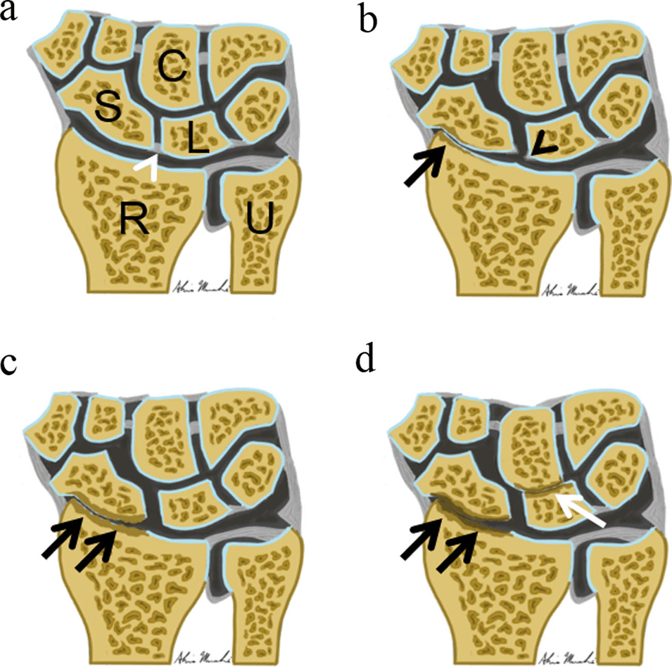
- A scapholunate gap greater than 3 mm on a clenched-fist PA view plus a scapholunate angle greater than 60 degrees on lateral means SL dissociation — the precursor of SLAC wrist.
- In a child or adolescent gymnast, radial wrist pain with widened irregular distal radial physis is a stress physeal injury (gymnast's wrist), not a soft-tissue strain.
- “Say the anatomy out loud: first extensor compartment, anatomical snuffbox floor, scaphoid tubercle, trapeziometacarpal joint, radial styloid, distal radial physis.
- “A 55-year-old woman with radial pain and a positive grind test has CMC arthritis; a 55-year-old woman post-partum or with new lifting has de Quervain's.
- “Non-union of an old scaphoid fracture presents as painless-then-painful radial wrist arthritis — always ask about a remote injury.
- “The 'ganglion that isn't there' — occult dorsal wrist ganglion causes dorsoradial pain reproduced by full extension; ultrasound confirms.
The correct sentence is "no fracture is visible on today's radiographs". With snuffbox and tubercle tenderness, immobilise and re-image or obtain MRI within 7-14 days. Missed scaphoid fracture is a leading hand litigation source.
A positive Eichhoff's test simply loads the radial column. Thumb CMC arthritis, scaphoid non-union and radial styloid impingement all hurt on ulnar deviation with the thumb clasped. Palpate at the styloid versus the CMC joint.
Steroid injected into a superficial radial nerve or subcutaneously causes fat atrophy and depigmentation over the styloid. Test light touch in the dorsal first web space before you offer injection.
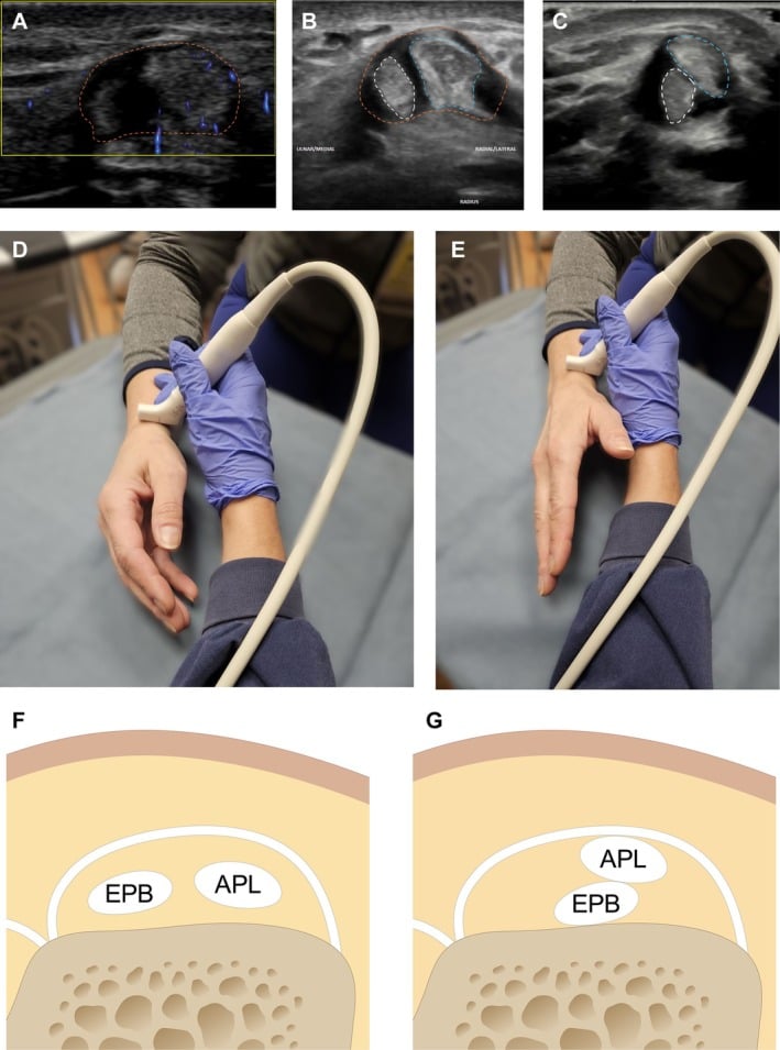
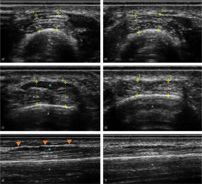
Around 40-60 per cent of first compartments have a septum isolating extensor pollicis brevis. Injecting only the abductor pollicis longus sheath explains most injection failures — and it is why release must open the EPB compartment.
Recognising the Pattern
Definition. Radial-sided wrist pain is pain localised to the region bounded by the radial styloid proximally, the thumb metacarpal base distally, the first extensor compartment volarly-radially and the anatomical snuffbox dorsally. It is a topographical pattern, not a diagnosis — the differential is generated by asking which structure in that box is tender.
Confirming the pattern is genuine. Ask the patient to point with one fingertip. Diffuse whole-wrist pain, night pain waking from sleep, or pain that migrates is not a radial-column pattern and should redirect you to inflammatory arthropathy, CRPS or referred cervical pain. Reproducibility matters: the same point must be tender on repeat palpation, and the pain should be provoked by a loading manoeuvre specific to that structure.
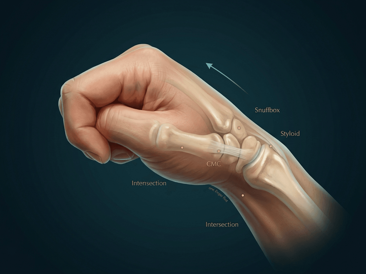
- First extensor compartment over the radial styloid — APL and EPB; tenderness plus a positive Eichhoff's/Finkelstein's test.
- Anatomical snuffbox floor and scaphoid tubercle (volar, distal to the distal wrist crease with the wrist ulnarly deviated) — scaphoid.
- Trapeziometacarpal joint, one centimetre distal to the styloid at the thumb metacarpal base — CMC arthritis; grind test.
- The muscle belly crossing 4-6 cm proximal to Lister's tubercle — intersection syndrome.
"This patient has radial-sided wrist pain. I would localise it to the first extensor compartment, the scaphoid, the trapeziometacarpal joint or the radioscaphoid articulation. My examination is directed at separating these: point tenderness, Eichhoff's test, the grind test, scaphoid shift and a Tinel's over the superficial radial nerve. I would then obtain PA, lateral, oblique and scaphoid views."
- Referred C6 radiculopathy — pain extends proximally into the forearm, sensory change in the thumb, Spurling's positive, no local tenderness.
- Wartenberg's syndrome — burning dorsoradial pain with a Tinel's over the nerve as it emerges between brachioradialis and ECRL; may coexist with de Quervain's ("cheiralgia paraesthetica").
- Thumb MCP ulnar collateral ligament injury — patients report "wrist" pain but tenderness is distal, at the thumb MCP.
- Distal radius stress reaction in gymnasts — pain worse on axial loading, not on tendon loading.
- Referred cardiac or thoracic outlet pain — vanishingly rare but exertional and non-positional.
Next Investigation
Radiographs in de Quervain's are for excluding CMC arthritis in the older patient, not for diagnosing the tendinopathy. Say that out loud rather than "radiographs are normal".
The Differential
- Typical age / setting
- 15-40, fall on outstretched hand, contact sport
- Discriminating feature
- Tenderness in snuffbox AND over the volar scaphoid tubercle; pain on axial thumb compression
- What confirms it
- Scaphoid series (PA in ulnar deviation, lateral, two obliques); MRI within 7-14 days if radiographs normal
- Typical age / setting
- 25-50, remote untreated injury
- Discriminating feature
- Painless interval of months to years, then radial-column arthritic pain and loss of extension
- What confirms it
- Radiographs show sclerosis and cyst at the fracture line; CT along the scaphoid axis grades humpback deformity
- Typical age / setting
- 30-60, dorsal wrist injury or degenerative
- Discriminating feature
- Dorsoradial pain, positive scaphoid shift (Watson) with a painful clunk on release
- What confirms it
- Clenched-fist PA gap greater than 3 mm; lateral SL angle greater than 60 degrees; radial styloid beaking in SLAC I
- Typical age / setting
- Any age, diabetes, immunosuppression, penetrating injury
- Discriminating feature
- Rest pain, erythema, refusal of any passive motion, fever
- What confirms it
- Aspiration with cell count and culture; raised CRP; urgent surgical washout
- Typical age / setting
- Any age, fall; osteoporotic females over 60
- Discriminating feature
- Tenderness over the styloid itself, not distal to it; swelling and ecchymosis proximal to the snuffbox
- What confirms it
- PA and lateral radiographs; CT if intra-articular step-off suspected
- Typical age / setting
- 30-50, post-partum women, new repetitive lifting
- Discriminating feature
- Tenderness AT the styloid over the first compartment with palpable thickening; positive Eichhoff's/Finkelstein's
- What confirms it
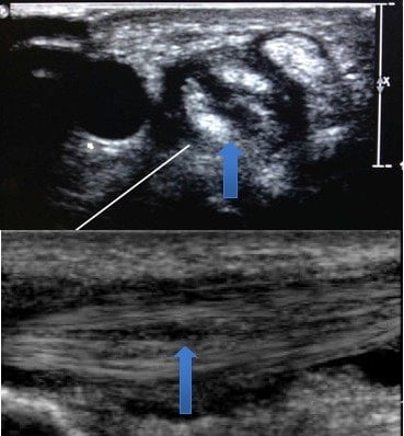
- Clinical; ultrasound shows sheath thickening and can identify an EPB septum before injection
- Typical age / setting
- Women over 50, adduction deformity, weak pinch
- Discriminating feature
- Tenderness one centimetre DISTAL to the styloid at the metacarpal base; positive grind test with crepitus
- What confirms it
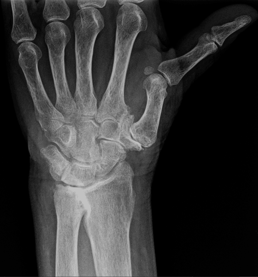
- Robert's (true AP thumb) and lateral views showing joint space loss, subluxation, osteophytes
- Typical age / setting
- 20-40, rowers, weight-lifters, heavy shovelling
- Discriminating feature
- Tenderness and squeaky crepitus 4-6 cm PROXIMAL to the radial styloid where APL/EPB cross ECRL/ECRB
- What confirms it
- Clinical; MRI shows peritendinous oedema at the crossover point
- Typical age / setting
- 20-50, tight watch strap, handcuffs, prior surgery
- Discriminating feature
- Purely sensory: burning and numbness in the dorsal first web, Tinel's over the nerve, NO tendon tenderness
- What confirms it
- Positive Tinel's plus relief with a diagnostic local anaesthetic block; nerve conduction may be normal
- Typical age / setting
- 40-70, post-traumatic or after radial styloidectomy failure
- Discriminating feature
- Pain only at end-range radial deviation, reproduced by forced radial deviation with the wrist loaded
- What confirms it
- Radiographs showing radioscaphoid joint narrowing and styloid beaking
- Typical age / setting
- 20-40, women more than men
- Discriminating feature
- Pain reproduced at full wrist extension, mass appears on volar flexion; may be palpable only when flexed
- What confirms it
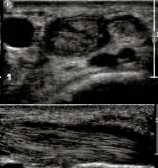
- Ultrasound — cystic, anechoic, arising from the scapholunate interval
- Typical age / setting
- 20-50, steroid use, alcohol, idiopathic
- Discriminating feature
- Insidious radial pain with NO history of injury; proximal pole sclerosis without a fracture line
- What confirms it
- MRI showing proximal pole low signal on T1 with absent enhancement
- Typical age / setting
- 9-16, high-load gymnastics, no acute event
- Discriminating feature
- Dorsoradial pain on axial loading and weight-bearing through the hand, not on tendon loading
- What confirms it
- Radiographs: widened, irregular, sclerotic distal radial physis; compare with the opposite side
- Typical age / setting
- 40-60, often with scaphotrapezial arthritis
- Discriminating feature
- Tenderness along the VOLAR FCR tendon proximal to the trapezial ridge, pain on resisted wrist flexion
- What confirms it
- Ultrasound showing FCR sheath effusion; radiographs for scaphotrapeziotrapezoid arthritis
Narrowing It Down
- 1Step 1 — Was there trauma, and what mechanism?
Ask the mechanism before touching the wrist, and ask specifically about a fall on the outstretched hand however trivial it sounded.
A fall on the outstretched hand aged 15-40 puts scaphoid fracture at the top and it stays there until excluded. A high-energy dorsiflexion injury with dorsal pain raises scapholunate dissociation. No trauma at all shifts you to tendinopathy, arthritis, nerve or Preiser's.
- 2Step 2 — Where exactly is the point of maximal tenderness?
Have the patient put ONE FINGER on the worst spot, then confirm it yourself against the named landmarks - styloid, snuffbox floor, volar tubercle, metacarpal base, four to six centimetres proximal.
At the radial styloid over the tendons equals de Quervain's. In the snuffbox AND at the volar tubercle equals scaphoid. One centimetre distal at the metacarpal base equals CMC arthritis. Four to six centimetres proximal equals intersection syndrome. Volar over flexor carpi radialis equals FCR tendinopathy. This single step does more work than every provocative test that follows.
- 3Step 3 — Which provocative test is positive?
Perform Eichhoff or Finkelstein, the grind test and the scaphoid shift - and interpret each against its known accuracy rather than as a yes-or-no.
Eichhoff plus styloid tenderness supports de Quervain's, but Eichhoff is ALSO positive in CMC arthritis and scaphoid nonunion because it simply loads the radial column - the grind test is what separates them, and the two conditions frequently coexist. A positive Watson shift with a painful clunk points to scapholunate instability, but its sensitivity against arthroscopy is only 0.50 and its specificity 0.62, so a negative test settles nothing.
- 4Step 4 — Is there a neurological component?
Test sensation in the dorsal first web, tap over the superficial radial nerve where it emerges from under brachioradialis, and screen the neck.
Burning, paraesthesiae and numbness in the dorsal first web with a positive Tinel indicate Wartenberg's - and the first compartment is NOT tender, which is the discriminator. Sensory symptoms extending above the wrist with neck-provoked pain indicate C6 radiculopathy. Neither responds to a first-compartment injection.
- 5Step 5 — What is the age and skeletal maturity?
Establish whether the physes are open, and in an adult look for thumb adduction and weak key pinch.
Open physes in a loading athlete means distal radial physeal stress injury - gymnast's wrist. Over 50 with adduction deformity and weak key pinch means CMC arthritis. Aged 20-40 with insidious pain and no trauma, consider Preiser's or an occult ganglion.
- 6Step 6 — Are there systemic or red-flag features?
Take the temperature, look at the skin, test passive motion, and ask about other joints and morning stiffness.
Fever, erythema, rest pain and refusal of passive motion mean septic arthritis or pyogenic tenosynovitis - aspirate and wash out, do not inject. Symmetrical polyarticular morning stiffness with radial pain means inflammatory arthropathy; check serology and examine the ulnar side too.
- 7Step 7 — Does the pain change with wrist position or grip load?
Load the wrist two different ways - through the tendon (resisted thumb extension) and through the joint (axial grip) - and see which reproduces it.
Pain only at end-range radial deviation suggests radial styloid impingement or radioscaphoid arthritis. Pain at full extension with a mass appearing on flexion suggests an occult dorsal ganglion. Pain on axial grip loading rather than tendon loading points to bone or joint, not tendon - and that distinction is what stops a tendon injection being given for an arthritic joint.
MCQ Practice Points
Q: Which single physical sign best separates thumb CMC arthritis from de Quervain's tenosynovitis, given both may have a positive Eichhoff's test?
A: The grind test — axial compression with rotation of the thumb metacarpal reproducing pain and crepitus at the trapeziometacarpal joint. Point tenderness location (at the styloid versus one centimetre distal at the metacarpal base) is the other key discriminator. Eichhoff separates nothing here because it simply loads the whole radial column, which is also why it is positive in scaphoid nonunion and radial styloid impingement.
Q: A patient has snuffbox tenderness after a fall and normal radiographs. What is the appropriate next step?
A: Immobilise and obtain MRI, or repeat radiographs at 10-14 days where MRI is unavailable. Then read the report rather than the conclusion. MRI is the practical reference standard and Dean's eight-centre cohort found occult fractures in 12.9 per cent with a delayed or nonunion rate above 6 per cent despite casting — these are not trivial. But Bulstra found that among suspected fractures only 6 per cent of scans showed a definite or possible fracture while 29 per cent showed nonspecific signal, and 69 per cent of waist signal changes were classified as distracting. A systematic review then found that nearly half of published studies never define what counts as a fracture on MRI, and a third of those that do accept diffuse marrow oedema alone. So the correct answer includes a caveat: a linear bicortical transverse line on multiple planes is a fracture; isolated diffuse marrow oedema is not the same finding, and the two should not lead to the same treatment.
Q: Why do some de Quervain's injections fail despite correct technique?
A: A septum within the first extensor compartment isolates the extensor pollicis brevis subsheath in a large proportion of wrists. Injectate entering only the abductor pollicis longus sheath leaves extensor pollicis brevis untreated. Ultrasound guidance, and at surgery deliberate identification and release of the EPB compartment, address this.
Q: What radiographic measurements confirm scapholunate dissociation?
A: A scapholunate gap greater than 3 mm on a clenched-fist PA view compared with the contralateral side, and a scapholunate angle greater than 60 degrees on the true lateral, with a cortical ring sign from the flexed scaphoid. Do not let a negative Watson scaphoid shift stop you asking for these: against arthroscopy that test has a sensitivity of only 0.50, and 0.61 even in patients referred specifically for suspected scapholunate injury.
Q: A 13-year-old elite gymnast has radial wrist pain on weight-bearing with no acute injury. What is the diagnosis and finding?
A: Distal radial physeal stress injury (gymnast's wrist) — widening, irregularity and metaphyseal sclerosis of the distal radial physis. Untreated it can cause premature physeal closure with relative ulnar overgrowth and positive ulnar variance, converting a self-limiting overuse problem into a permanent deformity that loads the ulnar side of the wrist.
Exam Viva Scenarios
Practise clinical reasoning and management decisions out loud
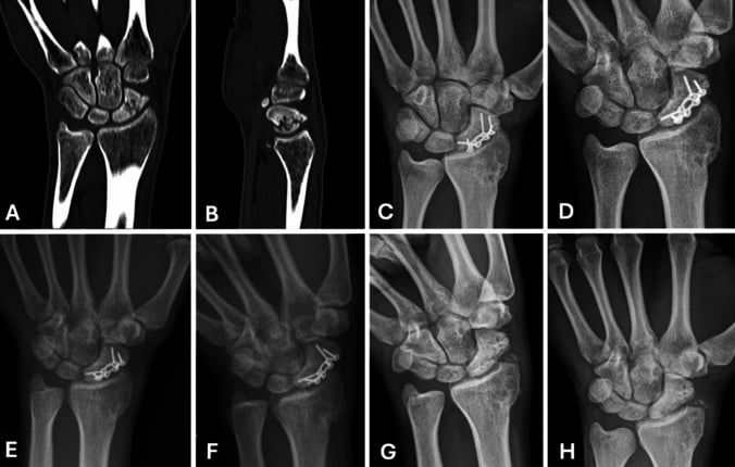
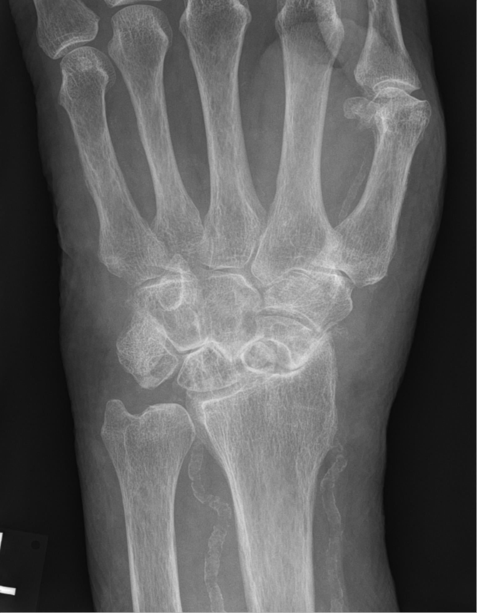
“You are shown this radiograph of the wrist of a 24-year-old man who fell on his outstretched hand during a tackle four days ago. He has pain in the anatomical snuffbox. The scaphoid views are reported as normal.”
“You are shown this radiograph of a 54-year-old woman with three months of radial wrist pain, worse when lifting a kettle. Eichhoff's test is positive.”
“You are shown a wrist radiograph reported as normal in a 27-year-old competitive rower with two weeks of radial forearm pain and audible crepitus. He was previously injected in the first extensor compartment without benefit.”
Palpate in this order
- First extensor compartment at the styloid — de Quervain's
- Snuffbox floor plus volar scaphoid tubercle — scaphoid fracture
- Thumb metacarpal base, one centimetre distal — CMC arthritis
- Four to six centimetres proximal, crossover point — intersection syndrome
- Volar FCR tendon at the trapezial ridge — FCR tendinopathy
Must-not-miss
- Occult scaphoid fracture — normal radiographs do not exclude
- Scaphoid non-union progressing to SNAC wrist
- Scapholunate dissociation progressing to SLAC wrist
- Septic arthritis or pyogenic tenosynovitis
- Undisplaced radial styloid or distal radius fracture
Provocative tests
- Eichhoff's / Finkelstein's — first compartment (not specific)
- Grind test — trapeziometacarpal joint
- Watson scaphoid shift — scapholunate instability
- Tinel's over the superficial radial nerve — Wartenberg's
- Axial thumb compression — scaphoid
Imaging decisions
- PA, lateral, oblique plus scaphoid views after any trauma
- Clenched-fist PA with contralateral comparison for SL gap
- MRI for occult fracture, Preiser's, occult ganglion
- CT along the scaphoid axis for non-union and humpback deformity
- Ultrasound to confirm sheath thickening, find the EPB septum and guide injection
- No imaging needed in classic de Quervain's, intersection syndrome or Wartenberg's
Phrases that score
- 'No fracture is visible on today's radiographs' — never 'no fracture'
- 'Eichhoff's is positive but not specific; the grind test separates them'
- 'Tenderness proximal to the styloid moves me to intersection syndrome'
- 'I would test dorsal first web sensation before offering injection'
Evidence Base
Routine MRI Among Patients With a Suspected Scaphoid Fracture Risks Overdiagnosis
- 256 MRI scans for clinically suspected scaphoid fracture with normal radiographs, imaged a median of 8 days after injury
- Only 6 per cent showed a definite (2 per cent) or possible (5 per cent) scaphoid fracture
- 29 PER CENT showed nonspecific signal changes - 14 per cent at the waist and 15 per cent elsewhere
- Of 51 patients with any scaphoid waist signal change, 69 PER CENT were classified as having distracting and potentially misleading findings rather than a fracture
- Definitions were explicit: a DEFINITE fracture required a linear, focal, BICORTICAL signal abnormality with adjacent oedema, relatively transverse, visible on more than one cut in multiple planes; reliability kappa 0.62
- The authors propose two alternatives: more selective MRI guided by a clinical prediction rule, and shared decision-making about the small risk of symptomatic nonunion
The Rate of Nonunion in the MRI-Detected Occult Scaphoid Fracture
- Multicentre cohort across EIGHT UK centres: 1,989 patients had acute MRI for suspected scaphoid fracture with normal radiographs
- 256 patients (12.9 PER CENT) had a previously occult scaphoid fracture
- Six had early surgery (2.3 per cent); of the 250 treated in cast, there were 16 delayed or nonunions - 6.3 PER CENT
- Nine nonunions (3.5 per cent), of which seven required surgery; seven delayed unions (2.7 per cent), five of which healed with further casting
- Every fracture treated surgically united, with one complication (a prominent screw requiring removal)
- The authors conclude MRI-detected fractures are NOT universally benign and that this supports early MRI
Variation in Definitions of Scaphoid Fracture on MRI Scans for Suspected Fracture: A Systematic Review
- 58 studies of patients with suspected scaphoid fracture examined for how each defined a fracture on MRI
- NEARLY HALF REPORTED NO DEFINITION OR MEASUREMENT OF FRACTURE AT ALL
- Among the 33 that did, 88 per cent described a linear fracture shape, 39 per cent reported the extent across the scaphoid, and NONE specified the orientation of the fracture line
- Of the 25 studies describing shape or extent: 48 per cent required a linear signal THROUGH THE CORTEX, 20 per cent accepted a linear INTRAMEDULLARY signal alone, and 32 PER CENT accepted a DIFFUSE INTRAMEDULLARY SIGNAL alone
- The authors call for a consensus definition before the reliability, diagnostic performance, harms and benefits of MRI can be assessed
Relevance of the Scaphoid Shift Test for the Investigation of Scapholunate Ligament Injuries
- 447 patients who had both a scaphoid shift (Watson) test and wrist arthroscopy, with the clinical result correlated against the arthroscopic findings
- Across the whole cohort the SENSITIVITY WAS 0.50 - the test missed half the ligament injuries
- In the subgroup specifically referred for suspected scapholunate injury sensitivity rose to 0.61, but SPECIFICITY WAS ONLY 0.62
- For the more severe lesions (Geissler grades 3 and 4) sensitivity improved to 0.66
- The authors conclude an isolated scaphoid shift test is of limited value and that A NEGATIVE TEST MUST NOT STOP FURTHER INVESTIGATION