The Independent Flexor and the Surgeon's Spare Part
- Two heads: a humero-ulnar head from the medial epicondyle, ulnar collateral ligament and coronoid, and a radial head from the oblique line of the radius, bridged by the fibrous FDS (sublimis) arcade.
- The median nerve and the ulnar artery pass BENEATH that arcade - it is the most distal of the four classical pronator syndrome compression points.
- Each digit has its own muscle belly and its own tendon, so FDS produces INDEPENDENT proximal interphalangeal flexion; the index belly is the most independent and the little finger the most variable.
- The tendon splits at the proximal phalanx, the slips rotate and decussate dorsal to profundus at Camper chiasm, and insert on the volar middle phalanx.
- In the carpal tunnel the middle and ring FDS tendons lie SUPERFICIAL (volar) to the index and little FDS - the middle finger tendon is the one closest to the median nerve.
- “Test FDS by holding all the OTHER fingers in full extension: this blocks the common profundus belly, so any residual proximal interphalangeal flexion must be FDS.
- “The small finger FDS is functionally independent in only about 58 per cent of hands (Austin). Apparent ABSENCE depends entirely on how you test: 35 per cent of fingers by the standard test but only 4 per cent by the modified test that leaves the ring finger free (Stein) - and every dissected cadaver hand had the tendon present.
- “Camper chiasm is where the FDS slips decussate dorsal to profundus; the vinculum longum to profundus arises from this region, which is why chiasm damage jeopardises profundus nutrition.
- “Harvest an FDS slip and leave one slip attached distally: complete FDS loss in a lax finger produces a swan neck deformity.
Overview
Flexor digitorum superficialis (FDS), historically the sublimis, is the largest muscle of the superficial flexor compartment and the only long flexor with a genuinely independent belly for each digit. That independence is what allows a pianist to flex one proximal interphalangeal joint while the others stay straight, it is the basis of the standard bedside FDS test, and it is why an FDS tendon can be borrowed for a transfer without paralysing the neighbouring fingers - a claim flexor digitorum profundus can never make.
Anatomically it sits in a stratum of its own, deep to the wrist flexors and superficial to profundus and flexor pollicis longus. Two features dominate the examination. First, its proximal fibrous arcade roofs the median nerve and the ulnar artery and is the most distal of the classical pronator syndrome compression sites. Second, its distal architecture in zone II - the split around profundus, the decussation at Camper chiasm, the vincular blood supply and the pulley system - is the anatomy on which the whole of flexor tendon surgery rests.
Two tendons must reach the fingertip through a canal barely wide enough for one. Evolution solved this by making the superficialis open around the profundus:
- At the level of the proximal phalanx the FDS tendon flattens and divides into two slips. Each slip rotates through roughly 180 degrees, passes dorsal to the profundus, and the two decussate at Camper chiasm before partially reuniting and inserting on the volar surface of the proximal half of the middle phalanx on both sides.
- Mechanical consequence: FDS is the sole flexor of the proximal interphalangeal joint and the primary volar restraint against PIP hyperextension. Lose it in a lax finger and the PIP joint drifts into hyperextension - the swan neck deformity.
- Nutritional consequence: the decussation is where the vinculum longum profundus takes origin. Damage or excise the chiasm carelessly and the profundus loses a segmental blood supply.
- Surgical consequence: in zone II the two tendons occupy a single fibro-osseous canal. Bulk is the enemy. Every technical decision in zone II repair - core suture calibre, epitendinous suture, whether to repair one or both FDS slips, whether to vent a pulley - is a compromise between repair strength and the volume that must glide.
- Excursion: FDS requires roughly 64 mm of excursion for full digital motion; profundus needs about 70 mm. Wrist tenodesis contributes several millimetres of that arc, which is why a repaired tendon is protected in wrist flexion.
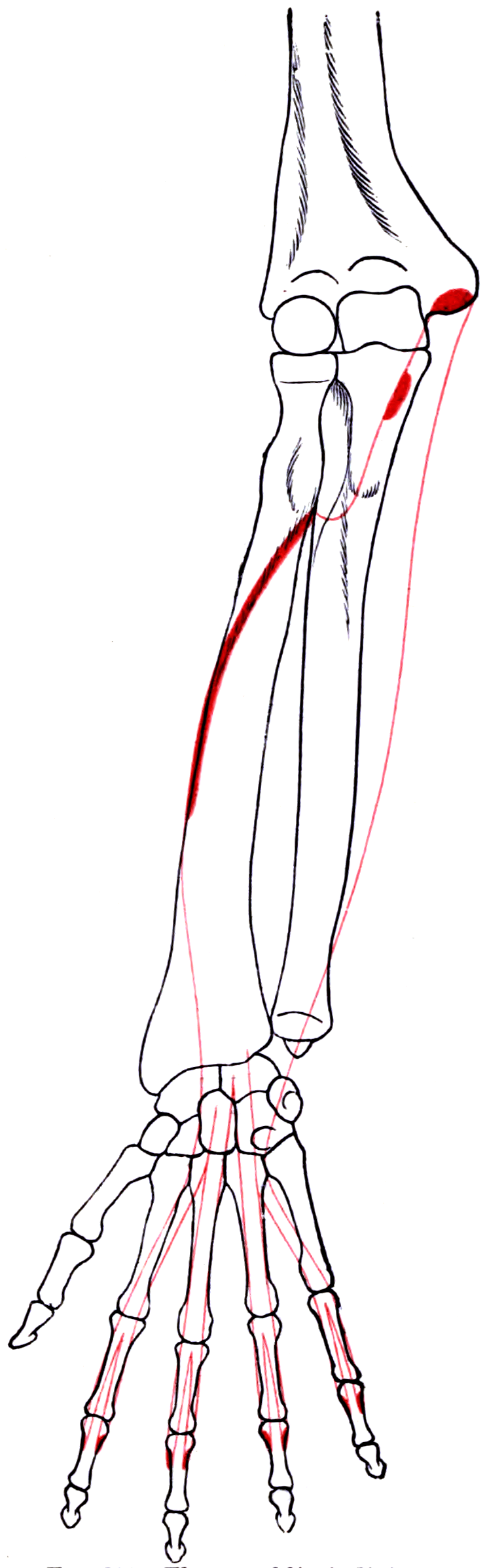
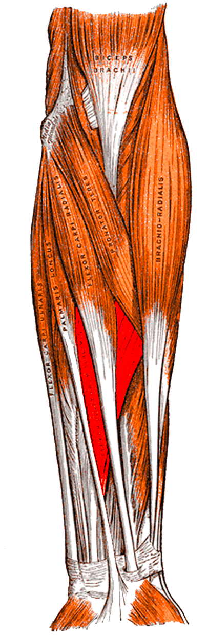
Attachments, Innervation and Relations
Origin - two heads
Humero-ulnar head
- Medial epicondyle of the humerus via the common flexor origin.
- The ulnar collateral ligament of the elbow.
- The medial (sublime) tubercle of the coronoid process of the ulna.
Radial head
- The oblique line and proximal volar shaft of the radius, between the radial tuberosity and the insertion of pronator teres.
The FDS (sublimis) arcade
- The two heads are joined by a fibrous arch - the sublimis bridge or FDS arcade - running obliquely between the coronoid and the radius.
- The median nerve and the ulnar artery pass DEEP to this arcade to enter the deep forearm. It is the fourth and most distal of the four classical pronator syndrome compression points.
The two strata of muscle bellies
- Superficial (volar) stratum: the bellies and tendons to the middle and ring fingers.
- Deep (dorsal) stratum: the bellies and tendons to the index and little fingers.
- The index belly is the most anatomically distinct, arising largely from the radial head - the basis of truly independent index proximal interphalangeal flexion.
- The little finger belly is the most variable, often deriving fibres from, or interconnected with, the ring finger tendon or the small finger profundus.
Arrangement in the carpal tunnel
- Nine tendons cross the carpal tunnel: four FDS, four FDP and flexor pollicis longus.
- The middle and ring FDS lie superficial (volar); the index and little FDS lie deep, in the same two-stratum arrangement as in the forearm.
- The middle finger FDS is therefore the tendon lying directly deep to the median nerve - and the tendon a surgeon meets first if the dissection strays deep during carpal tunnel release.
Insertion
- At the level of the proximal phalanx the tendon flattens and splits into two slips.
- The slips rotate approximately 180 degrees, pass dorsal to the profundus tendon, and decussate at Camper chiasm.
- They partially reunite and insert onto the volar surface of the proximal half of the middle phalanx, on both its radial and ulnar sides.
Volar-lateral to the sheath.
- Both bundles lie immediately lateral to the tendon sheath in the digit.
- Risk: division during a poorly planned midlateral or Bruner incision, or during retrieval of a retracted tendon.
- Avoidance: raise full-thickness flaps, keep the dissection on the sheath, and identify both bundles before opening the sheath.
The profundus lifeline.
- The vinculum longum profundus arises from FDS at the chiasm.
- Risk: excising FDS slips indiscriminately during transfer harvest or repair devascularises the profundus segment.
- Avoidance: harvest FDS proximal to the chiasm where possible and preserve the vincular attachments.
Action and Biomechanics
Primary actions
- Flexion of the proximal interphalangeal joint - its defining and near-exclusive action. FDS is the only tendon that flexes the PIP joint without simultaneously flexing the distal interphalangeal joint.
- Flexion of the metacarpophalangeal joint - secondary, as it crosses that joint.
- Weak wrist flexion - it crosses the wrist and contributes, particularly when the fingers are extended.
- Volar restraint of the PIP joint - a static role that becomes obvious only when it is lost.
Numbers examiners ask for
- Flexor Digitorum Superficialis
- Approximately 64 mm
- Comparator
- FDP approximately 70 mm
- Significance
- Both need long-excursion donors; wrist motors at 33 mm cannot substitute
- Flexor Digitorum Superficialis
- Separate belly per digit
- Comparator
- FDP has one common belly to the ulnar three
- Significance
- FDS harvest does not weaken neighbouring digits; FDP injury causes quadriga
- Flexor Digitorum Superficialis
- Approximately 58 per cent
- Comparator
- Common pattern 21 per cent, absent pattern 21 per cent
- Significance
- The standard test over-diagnoses absence; use the modified test
- Flexor Digitorum Superficialis
- Volar proximal half of the middle phalanx
- Comparator
- FDP on the distal phalanx base
- Significance
- FDS is the sole PIP flexor
- Flexor Digitorum Superficialis
- C7, C8, T1
- Comparator
- FCR C6-C7
- Significance
- FDS is the most distal main-trunk median muscle in the forearm
The FDS-FDP interplay
- Both flex the PIP joint; only profundus flexes the DIP joint. Therefore blocking the DIP joint or eliminating profundus action is the only way to isolate FDS clinically.
- Profundus is the workhorse of grip; FDS contributes disproportionately to precision and to sustained grip strength, and its independence gives the hand its capacity for graded, digit-specific flexion.
- Losing FDS alone: grip is measurably weaker but functional. The specific loss is independent PIP flexion and volar PIP support.
- Losing FDP alone: the finger can still flex at the PIP but the fingertip cannot be brought to the palm, and pulp pinch is lost.
- Swan neck mechanics: with FDS gone, the volar plate becomes the sole PIP restraint. In a lax or rheumatoid finger the PIP hyperextends, the lateral bands subluxate dorsally, and the DIP flexes - the classic swan neck. This is the single most important reason to leave one FDS slip attached distally when harvesting.
Length-tension and transfer suitability
- FDS is a long-excursion, moderate-force motor with roughly 64 mm of amplitude - the ideal donor for another long-excursion task: opposition, thumb extension, finger extension, or profundus reconstruction.
- Its independence means one digit's FDS can be borrowed while the remaining fingers keep normal superficialis function.
- The ring finger FDS is the classic donor. The index FDS is preferentially preserved because independent index flexion is essential to precision pinch, and the little finger FDS is unreliable because it is functionally absent or non-independent in a large minority of hands.
Surface Anatomy and Examination
Palpation and identification
- The FDS tendons are not individually palpable at the wrist, but the mass of the superficialis is felt as the firm bulk immediately deep to palmaris longus during resisted finger flexion.
- At the wrist, remember the stratum rule: middle and ring FDS lie volar, index and little FDS lie deep. The median nerve lies volar and radial, immediately deep to palmaris longus and just superficial to the middle finger FDS.
- In the digit, the FDS insertion can be localised to the volar proximal half of the middle phalanx, and zone II ends there.
The standard FDS test
- Hold ALL the other fingers in full extension at the metacarpophalangeal, proximal and distal interphalangeal joints, flat on the table or against your hand.
- Ask the patient to flex the tested finger.
- Because the profundus tendons to the ulnar three digits share one common muscle belly, holding the other fingers extended prevents the profundus from shortening. Any residual proximal interphalangeal flexion must therefore come from FDS.
- A normal result is isolated PIP flexion with the DIP joint left floppy and extended - the classic appearance of intact FDS.
The modified FDS test for the little finger
- The little finger FDS is functionally independent in only about 58 per cent of hands; a common pattern (linked with the ring finger) occurs in about 21 per cent and an absent pattern in about 21 per cent, with right-left asymmetry in about a quarter of people.
- Modification: hold the index and middle fingers extended but leave the RING finger free. This permits the interconnected ring-little superficialis unit to act and reveals a little finger FDS that the standard test reports as absent.
- Published prospective testing found absent small finger FDS in 35 per cent of fingers on standard testing but only 4 per cent on modified testing - an enormous difference that turns an unnecessary hand surgery referral into a normal examination.
Named clinical tests around FDS
- How to perform
- Hold all other fingers fully extended; ask the patient to flex the test finger
- Positive finding
- Isolated PIP flexion with a floppy DIP
- What it means
- Intact FDS to that digit
- False positives
- A lax finger may allow apparent PIP flexion from FDP if a neighbouring digit escapes the block
- How to perform
- Hold index and middle extended, leave the RING finger free; ask for little finger PIP flexion
- Positive finding
- PIP flexion of the little finger
- What it means
- Functional small finger FDS present via the ring-little interconnection
- False positives
- None of significance - this is the more accurate test
- How to perform
- Block the PIP joint in extension; ask for DIP flexion
- Positive finding
- DIP flexion
- What it means
- Intact FDP to that digit
- False positives
- Apparent motion from tenodesis if the wrist is allowed to extend
- How to perform
- Elbow extended, forearm neutral; resist PIP flexion of the middle finger
- Positive finding
- Pain and paraesthesiae in the median territory
- What it means
- Median nerve compression at the FDS (sublimis) arcade
- False positives
- Positive in medial epicondylitis and in normal people if resistance is excessive
- How to perform
- Resist forearm pronation from full extension
- Positive finding
- Median paraesthesiae
- What it means
- Compression between the two heads of pronator teres
- False positives
- Frequently equivocal - pronator syndrome tests have poor specificity
- How to perform
- Ask the patient to make a circle with thumb and index
- Positive finding
- Square pinch - IP and DIP hyperextend
- What it means
- Anterior interosseous nerve palsy - FPL and index FDP, with FDS intact
- False positives
- Attritional FPL rupture and tendon adhesions mimic it
- How to perform
- Passively extend then flex the wrist
- Positive finding
- Fingers flex on wrist extension, extend on wrist flexion
- What it means
- Flexor tendon continuity - a normal cascade
- False positives
- Preserved in a partial laceration and in a patient with intact FDS but divided FDP
Assessing a suspected zone II laceration
- Examine before anaesthetising the digit and test each tendon separately.
- Loss of the normal resting cascade - the injured finger sitting more extended than its neighbours - is the earliest sign.
- Test FDS and FDP independently as above; a divided FDS with an intact FDP retains DIP flexion and often near-normal composite flexion, so an isolated FDS division is genuinely easy to miss.
- Partial lacerations may retain full active motion but produce pain on resisted flexion and a palpable or audible triggering as the flap of tendon catches at a pulley.
- Wound position matters: a laceration sustained with the fingers flexed lies distal to the tendon injury when the finger is examined extended. Always explore the tendon over its full excursion.
Complications
Donor site morbidity after FDS harvest
- Swan neck deformity of the donor finger from loss of the volar restraint at the proximal interphalangeal joint. Highest risk in hyperlax and rheumatoid patients.
- Avoidance: leave one slip attached distally or perform a formal FDS tenodesis to the proximal phalanx.
- Loss of independent proximal interphalangeal flexion in the donor digit - functionally minor in the ring finger, significant if the index is taken.
- Reduced grip strength - measurable but usually well tolerated after a single-digit harvest.
- Proximal interphalangeal flexion contracture if the harvested tendon stump scars within the sheath.
Zone II repair complications
- Rupture - most commonly in the first three weeks, precipitated by an uncontrolled grasp. A gap of more than 3 mm at the repair predicts it.
- Adhesions - the dominant cause of a poor result. Repair between two tendons in one canal, sheath handling and prolonged immobilisation all contribute. Managed initially with therapy; tenolysis is considered at 3 to 6 months once motion has plateaued, the scar is supple and the joints are passively full.
- Bowstringing from excessive pulley loss.
- Joint contracture, most often a PIP flexion contracture from a protected posture.
- Quadriga - if a profundus repair is over-shortened or a tendon is advanced too far, the common profundus belly cannot extend the other digits fully. Note that quadriga is a profundus phenomenon; FDS independence protects it from this.
- Lumbrical plus finger - paradoxical interphalangeal extension when the profundus is divided distally and its proximal stump pulls through the lumbrical.
Iatrogenic injury
- Digital nerve division during sheath exposure - keep the neurovascular bundles in a full-thickness flap.
- Vincular damage at Camper chiasm during harvest or repair, compromising profundus nutrition.
- Median nerve injury in carpal tunnel release when the plane strays; the middle finger FDS is the orientation landmark deep to the nerve.
- Recurrent motor branch injury if the transverse carpal ligament is divided too radially - always divide at its ulnar margin.
Transfer-specific complications
- Wrong pulley position in opponensplasty - a pulley placed too radially or too distally gives palmar abduction without pronation, so the patient can lift the thumb but cannot rotate the pulp to meet the fingers.
- Over-tensioning a lasso producing a fixed metacarpophalangeal flexion posture with an extension lag.
- Failure to test the Bouvier manoeuvre before a lasso procedure, so a fixed interphalangeal contracture is treated with a dynamic operation that cannot correct it.
Clinical Relevance
The Verdan zones
- Boundaries
- Distal to the FDS insertion on the middle phalanx
- Contents and significance
- FDP only - the zone of jersey finger and of distal FDP repairs
- Boundaries
- From the A1 pulley / distal palmar crease to the FDS insertion
- Contents and significance
- Both FDS and FDP within the digital sheath - the classic no man's land
- Boundaries
- From the distal edge of the carpal tunnel to the A1 pulley
- Contents and significance
- The lumbrical origin zone in the palm; good prognosis for repair
- Boundaries
- Within the carpal tunnel
- Contents and significance
- Nine tendons and the median nerve; repairs risk adhesion between tendons
- Boundaries
- Proximal to the carpal tunnel
- Contents and significance
- Forearm - multiple structures; the spaghetti wrist territory
Why zone II is difficult
- Two tendons in one canal. FDS and FDP must glide independently within a rigid fibro-osseous sheath. Repair bulk, adhesion between the two repairs, and adhesion to the sheath all conspire against motion.
- Camper chiasm. The FDS slips wrap around profundus, so the tendon relationships change over just a few millimetres of tendon length - a repair at the chiasm is technically demanding and prone to a bulky, catching result.
- The vinculum longum profundus arises here, so damage carries a nutritional cost as well as a mechanical one.
- The pulleys are unforgiving. A repair that will not pass under A4 must be trimmed, or the pulley vented.
Principles of zone II repair
- Strong core suture: a four- to six-strand core repair with adequate purchase (approximately 7 to 10 mm from the cut end), placed volarly to spare the dorsal vincular supply, combined with a circumferential epitendinous suture which adds strength, smooths the repair and resists gapping.
- Slight repair-site shortening (a few per cent) is acceptable and reduces gapping; a gap of more than 3 mm predicts rupture and adhesion.
- Pulley venting. Contemporary practice accepts partial or complete venting of A4 and of the distal part of A2 to allow a bulky repair to glide, provided a sufficient length of the pulley system is retained overall. Published series have shown venting is needed in the majority of zone II repairs and does not lead to clinically relevant bowstringing.
- The FDS decision. Repair FDS where it can be done without bulk - this preserves PIP volar restraint and prevents late swan neck deformity, and the FDS repair also preserves the vinculum longum profundus origin. Where both slips cannot be repaired without crowding the canal, repair one slip and excise the other.
- Early active mobilisation under supervision is now standard, and repair strength must be chosen with that regimen in mind.
A patient sent with "absent flexor digitorum superficialis to the little finger" after a minor laceration very often has entirely normal anatomy. Repeat the test with the ring finger left free: prospective testing found apparently absent small finger FDS in 35 per cent of fingers by the standard method and only 4 per cent by the modified method. Test both hands - asymmetry is normal in about a quarter of people.
Surgical Relevance
Exposure of the flexor sheath in the digit
- Bruner (volar zig-zag) incision: the apices of the zig-zag must reach the mid-lateral line at the flexion creases; angles that are too acute produce tip necrosis. Full-thickness flaps are raised down to the sheath, keeping the neurovascular bundles in the flap.
- Midlateral incision: placed at the dorsal limit of the flexion creases, dorsal to the neurovascular bundle. It leaves a less conspicuous scar and avoids a scar directly over the repair, but exposure of the volar sheath is more limited.
- Sheath windows: open the sheath between pulleys, through the cruciate segments, to preserve annular pulley substance.
Retrieval of a retracted tendon
- A divided FDP with an intact vinculum stays close to the injury; a divided tendon with ruptured vincula retracts to the palm.
- Retrieve atraumatically: flex the wrist and fingers and milk the tendon distally; if that fails, make a separate palmar incision at the A1 pulley and pass the tendon distally through the sheath using a small catheter or a suture shuttle.
- Never grasp the tendon repeatedly with toothed instruments - each bite damages the gliding surface and guarantees adhesion.
Proximal forearm release for pronator syndrome
- Anterior approach with a lazy-S incision crossing the antecubital fossa obliquely.
- Identify and protect the lateral antebrachial cutaneous nerve at the lateral edge of the biceps tendon.
- Release in sequence: lacertus fibrosus, then the superficial head of pronator teres (or its deep fascia), then the FDS arcade, tracing the median nerve beneath the arch.
- Look proximally for a supracondylar process and ligament of Struthers - a bony spur on radiographs is the clue.
- Take the ring finger, spare the index, distrust the little. The index provides independent precision flexion, and the little finger superficialis is functionally absent or non-independent in a large minority of hands.
- Leave a slip in a lax finger. Complete FDS loss removes the volar restraint of the proximal interphalangeal joint and produces a swan neck deformity that is harder to treat than the problem you were solving.
- Never harvest an FDS in a high median nerve palsy. It is denervated. Use extensor indicis proprius or an abductor digiti minimi (Huber) transfer instead.
SLAPMedian Nerve Compression Sites, Proximal to Distal
Hook:Release all four at one operation - leaving one unreleased is the commonest cause of failure.
Guidelines, Registries & Global Practice
Anatomical variation across populations
- The best-documented variation is the small finger FDS, functionally independent in roughly 58 per cent, with a common pattern in 21 per cent and an absent clinical pattern in 21 per cent, and asymmetry between hands in about a quarter of individuals. Cadaveric dissection shows the tendon is anatomically present in essentially every hand - the variation is in interconnections and functional independence, not in existence.
- Accessory FDS bellies extending distally into the carpal tunnel are described across populations and are a recognised cause of carpal tunnel syndrome in young manual workers.
- Pulley morphology varies most in the A1 and cruciate pulleys; A2 and A4 are consistent, which is why they anchor the surgical vocabulary.
Differences in described technique between sources
- Position on FDS-related practice
- Endorses multi-strand core repair with an epitendinous suture and controlled venting of A4 and the distal A2, replacing the historical absolute prohibition on dividing those pulleys.
- Position on FDS-related practice
- Emphasises early supervised motion after flexor repair and the diagnosis of partial lacerations by pain on resisted flexion rather than by loss of active motion.
- Position on FDS-related practice
- Recommend specialist referral for all suspected zone II flexor injuries and formal hand therapy access as a prerequisite for early active mobilisation protocols.
- Position on FDS-related practice
- One tendon one function, adequate excursion, expendable donor, straight line of pull, synergism and supple joints - the framework used to justify FDS as a donor and to reject it in high median palsy.
Evidence positions worth stating
- Pulley venting: prospective data show venting is required in the majority of zone II repairs, and multi-strand repair series with deliberate venting report excellent or good outcomes in over 80 per cent with no ruptures. The defensible position is controlled, documented venting rather than reflex preservation.
- Repair of FDS in zone II: there is no high-level randomised evidence mandating repair of both slips. Repairing at least one slip preserves the volar restraint of the PIP joint and the origin of the vinculum longum profundus; excising a slip is accepted where canal volume demands it.
- Timing of tenolysis: widely accepted at 3 to 6 months after repair, once active motion has plateaued, the scar is supple and the joints have full passive range - not before.
High- versus limited-resource practice
- Well-resourced settings: microsurgical repair, high-resolution ultrasound for partial lacerations and anomalous muscle bellies, formal hand therapy with early active protocols, and staged reconstruction with silicone rods where required.
- Limited-resource settings: zone II repair remains achievable with basic instruments and loupes, but the availability of supervised hand therapy is the true determinant of outcome, not the suture technique. Where therapy is not available, a more protective regimen and a robust repair should be chosen, and the patient counselled accordingly. FDS transfers require no implants and are among the most transferable reconstructive techniques between health systems.
MCQ Practice Points
Q: What passes deep to the FDS arcade? A: The median nerve and the ulnar artery. The arcade is the most distal of the four classical pronator syndrome compression points.
Q: What is Camper chiasm? A: The decussation of the two FDS slips dorsal to the profundus tendon at the proximal phalanx, before they insert on the volar middle phalanx. The vinculum longum profundus arises from this region.
Q: Which FDS tendons lie most superficial in the carpal tunnel? A: Middle and ring. Index and little lie deep. The middle finger FDS lies immediately deep to the median nerve.
Q: Why does holding the other fingers extended isolate FDS? A: Because the profundus tendons to the ulnar three digits share ONE common muscle belly, so blocking the other digits prevents profundus shortening.
Q: How often is the small finger FDS functionally independent? A: About 58 per cent, with a common pattern in 21 per cent and an absent pattern on standard testing in 21 per cent. Use the modified test that leaves the ring finger free.
Q: Define zone II. A: From the A1 pulley at the distal palmar crease to the FDS insertion on the middle phalanx - the zone containing both tendons within the digital sheath.
Q: Which annular pulleys overlie the joints? A: The odd-numbered ones - A1 over the MCP, A3 over the PIP and A5 over the DIP. A2 and A4 overlie the proximal and middle phalanges.
Q: Why is the core suture placed volarly within the tendon? A: Because the dorsal surface carries the vincular blood supply. Volar placement spares perfusion.
Q: What deformity follows complete FDS loss in a lax finger? A: Swan neck - PIP hyperextension with dorsal lateral band subluxation and DIP flexion. Prevent it by leaving one slip attached distally.
Q: Why can FDS not be used for opponensplasty in a high median nerve palsy? A: Because it is denervated. Use extensor indicis proprius or an abductor digiti minimi (Huber) transfer instead.
Q: A finger still triggers after an adequate A1 pulley release. What is the usual cause and the salvage? A: A catching FDS slip. Excision of one slip (usually the ulnar slip) decompresses the canal without creating a swan neck.
Exam Viva Scenarios
Practise clinical reasoning and management decisions out loud
“A 19-year-old is referred from the emergency department with a small volar laceration over the base of the little finger. They have documented an absent flexor digitorum superficialis to the little finger and referred for exploration. The finger flexes fully. What do you do?”
“A 44-year-old machine operator has aching volar forearm pain and tingling in the thumb, index and middle fingers. The symptoms are worse at work and do not wake him at night. Nerve conduction studies are normal. Carpal tunnel injection gave no relief. What is your diagnosis and how do you confirm it?”
“Two patients need opposition restored. Patient A has a low median nerve palsy after a wrist laceration repaired two years ago with no recovery of thenar function. Patient B has a high median nerve palsy after a proximal forearm injury. What transfer do you offer each, and why?”
Anatomy
- Humero-ulnar head: medial epicondyle, UCL, coronoid
- Radial head: oblique line of the radius
- FDS arcade bridges them - median nerve and ulnar artery pass beneath
- Nerve: median, C7-C8-T1; last main-trunk forearm branch before the AIN
- Insert: volar proximal half of the middle phalanx
Architecture
- Superficial stratum: middle and ring. Deep stratum: index and little
- Independent belly per digit - no quadriga
- Camper chiasm: slips rotate and decussate dorsal to FDP
- Vinculum longum profundus arises from FDS at the chiasm
Zones and Pulleys
- Zone II: A1 pulley to the FDS insertion
- Five annular, three cruciate, plus the palmar aponeurosis pulley
- Odd pulleys over joints, even pulleys over phalanges
- Venting A4 and distal A2 is accepted practice
Testing
- Block ALL other fingers - isolates FDS from the common FDP belly
- Small finger: independent 58%, common 21%, absent 21%
- Modified test leaves the ring finger free
- Resisted middle finger PIP flexion tests the sublimis arcade
Donor Uses
- Bunnell opponensplasty - FCU pulley at the pisiform
- Royle-Thompson - distal ulnar border of the TCL as pulley
- Zancolli lasso around A1 for claw correction
- Pulley reconstruction, FDS to EPL, FDS to adductor pollicis
- NOT available in high median nerve palsy
Evidence Base
Variations of the Flexor Digitorum Superficialis of the Small Finger
- Fifty normal subjects tested clinically with standard and modified superficialis flexion tests, plus 40 cadaver hands dissected
- An FDS-independent pattern was found 58 per cent of the time, an FDS-common pattern 21 per cent and an FDS-absent pattern 21 per cent
- Right and left hands were asymmetric in 26 per cent of subjects
- All cadaver hands had an FDS tendon present in the palm and finger despite the clinical variability
- Interconnections between the small finger FDS and either the ring FDS or the small finger FDP explain the functional variation
Clinical Evaluation of Flexor Tendon Function in the Small Finger
- One hundred subjects (200 hands) prospectively tested for small finger FDS function by standard and modified methods
- Standard testing showed absent FDS in 70 of 200 small fingers (35 per cent)
- Modified testing showed absent FDS in only 8 fingers (4 per cent)
- Small finger FDS function was frequently unilateral
Anatomy of the Finger Flexor Tendon Sheath and Pulley System
- Sixty-one fresh human cadaver fingers dissected under the operating microscope
- A flexor synovial sheath with visceral and parietal elements extends from the metacarpal neck to the distal interphalangeal joint
- The retinacular system comprises the palmar aponeurosis pulley, five annular pulleys and three cruciform pulleys
- Significant morphological variation exists in the first annular and the cruciform pulleys
- The palmar aponeurosis pulley, described by Manske and Lesker, is a substantive addition to the system
Anatomy and Function of the Palmar Aponeurosis Pulley
- The transverse fibres of the palmar aponeurosis are anchored by vertical septa to the transverse metacarpal ligament
- This forms a constant and substantial retinacular pulley over the flexor tendons proximal to A1
- Its position, breaking strength and demonstrated function justify inclusion in the pulley system
Venting or Partial Lateral Release of the A2 and A4 Pulleys After Repair of Zone 2 Flexor Tendon Injuries
- 126 consecutive zone 2 flexor tendon injuries prospectively studied
- Venting of one or other pulley was required in 81 repairs (64 per cent)
- The A4 pulley was vented between 10 and 100 per cent of its length in 71 fingers (56 per cent)
- The distal edge of A2 was vented by 4 to 10 mm in 10 fingers (8 per cent)
Outcomes of the Six-Strand M-Tang Repair for Zone 2 Primary Flexor Tendon Repair in 54 Fingers
- Fifty-four fingers with complete zone 2 flexor divisions repaired with a six-strand core technique and early motion
- Excellent or good results in 83 per cent of fingers followed beyond one year
- No repair ruptures occurred in the series
- Outcomes were significantly better in patients younger than 37, in male patients, and where the A2 pulley had been vented
Proximal Median Nerve Compression: Pronator Syndrome
- Review of proximal median nerve compression including the flexor digitorum superficialis arcade
- Symptoms frequently overlap with carpal tunnel syndrome, making the clinical distinction difficult
- Electrodiagnostic studies are often negative, so diagnosis depends on anatomical knowledge and clinical pattern
- All patients should initially be managed non-operatively; surgical intervention has been shown to result in satisfactory outcomes, with most outcome data from retrospective case series