One Tendon, One Joint, No Substitute
- Origin from the middle third of the volar radius and adjacent interosseous membrane, with a variable accessory head (Gantzer muscle) from the coronoid or medial epicondyle.
- Insertion on the volar base of the distal phalanx of the thumb - it is the ONLY flexor of the thumb interphalangeal joint.
- Innervated by the anterior interosseous nerve, C7-C8, a purely motor branch - so an isolated FPL palsy has NO sensory loss.
- At the wrist it is the most radial and deepest tendon of the carpal tunnel, lying within a few millimetres of the volar rim of the distal radius.
- It is the FIRST tendon to rupture over a prominent volar plate and the first in the rheumatoid Mannerfelt lesion over a scaphoid osteophyte.
- “The OK sign (Kiloh-Nevin) is abnormal in anterior interosseous palsy: a square pinch instead of a round circle, because FPL and index profundus cannot flex the terminal joints.
- “The Linburg-Comstock anomaly is an FPL to index profundus tendon slip, present clinically in about 31 per cent of individuals in at least one hand.
- “A patient who cannot flex the thumb IP joint 12 months after volar plating of a distal radius fracture has ruptured the FPL until proven otherwise - image the plate position on a true lateral.
- “The radial digital nerve of the thumb crosses obliquely OVER the FPL sheath at the A1 pulley - the classic iatrogenic injury of trigger thumb release.
Overview
Flexor pollicis longus (FPL) is the deep flexor of the thumb and the only tendon that flexes the thumb interphalangeal joint. Every other thumb flexor - flexor pollicis brevis, the thenar group, adductor pollicis - acts proximal to that joint. There is no synergist, no partial substitute and no tenodesis workaround: if FPL fails, terminal thumb flexion is gone, and with it the strong tip pinch on which handwriting, buttoning and lifting a small object all depend.
Two properties dominate its surgical relevance. First, it is supplied by the anterior interosseous nerve, a purely motor branch, so an FPL palsy comes with no sensory loss at all - the anatomical fact behind the OK sign. Second, its anatomical position is exposed: it lies against the volar rim of the distal radius at the watershed line, it pivots at the distal border of the flexor carpi radialis septum, and it runs across the volar carpal bones in the carpal tunnel. Anything hard and prominent at those points - a plate edge, a scaphoid osteophyte, a trapezial spur - abrades it, and FPL is the tendon that goes first.
Attritional flexor rupture at the wrist follows a stereotyped sequence, and flexor pollicis longus is invariably the first casualty. Three anatomical reasons converge:
- It lies most radial and deepest in the carpal tunnel, closest to the volar surface of the scaphoid and trapezium and closest to the volar rim of the distal radius at the watershed line - the exact place a volar plate edge sits.
- It pivots. The distal border of the septum separating the flexor carpi radialis tunnel from the carpal canal acts as a pulley for FPL. The tendon therefore does not slide flat over the bone; it is dragged around a fixed point, concentrating pressure on one segment with every thumb movement.
- It works constantly. The thumb flexes in almost every prehensile task, so the abrading segment sees more cycles than any neighbouring tendon.
- Volar plate rupture: a plate on or distal to the volar rim (Soong grade 2) presents a metal edge to the tendon. Rupture typically occurs 6 to 24 months after fixation, often preceded by months of volar radial wrist pain and crepitus that were dismissed.
- The Mannerfelt lesion: in rheumatoid arthritis, a bony spur within the carpal tunnel - characteristically on the scaphoid - abrades the FPL, which ruptures while the patient assumes the loss of thumb flexion is "just the arthritis".
loss of thumb interphalangeal flexion in a patient with either a volar plate or rheumatoid disease is a tendon rupture until proven otherwise, and the causative bony prominence must be removed at the same operation or the reconstruction will fail in the same way.
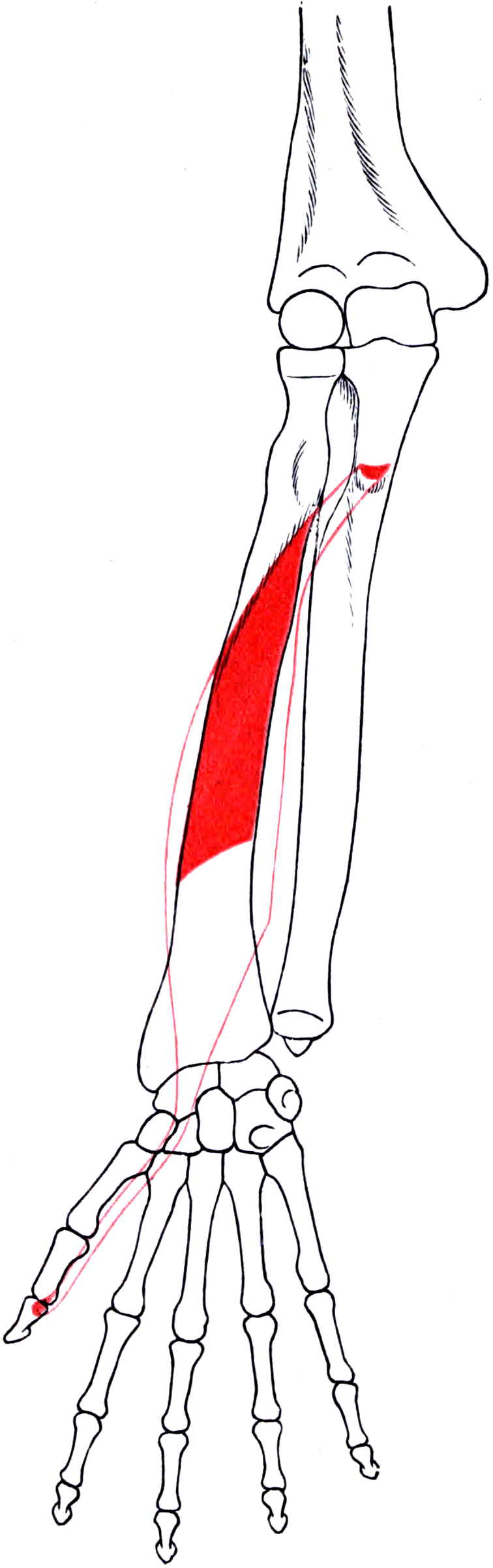
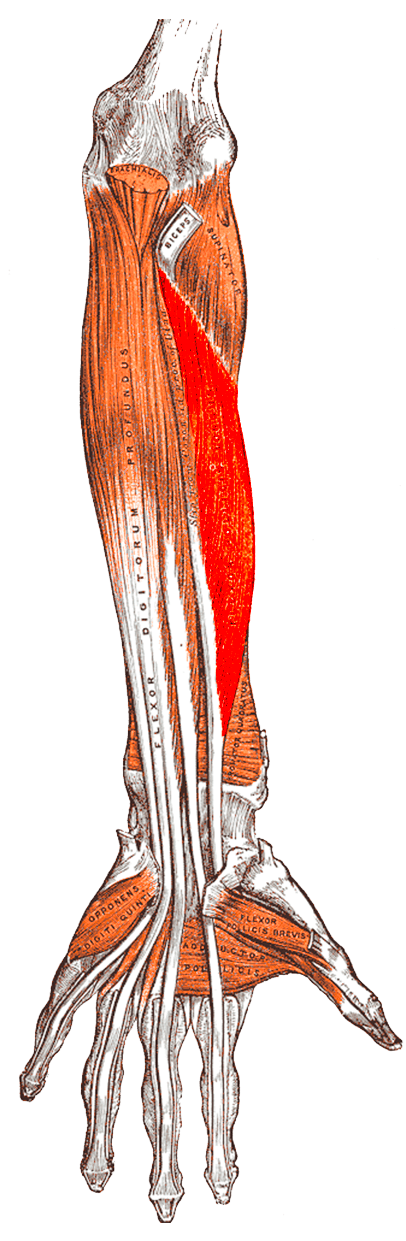
Attachments, Innervation and Relations
Origin
- The middle third of the volar (anterior) surface of the radius, from just distal to the radial tuberosity and oblique line down to the proximal border of pronator quadratus.
- The adjacent interosseous membrane.
- A variable accessory head - the Gantzer muscle - arising from the medial (coronoid) process of the ulna or from the medial epicondyle via the common flexor origin. It is present in a substantial proportion of forearms in cadaveric series, is usually innervated by the anterior interosseous nerve, and is a recognised potential compressor of that nerve.
Course
- Descends on the radial side of the deep flexor compartment, radial to flexor digitorum profundus, with the anterior interosseous nerve and artery in the interval between the two muscles on the interosseous membrane.
- Becomes tendinous in the distal forearm and passes deep to pronator quadratus and then into the carpal tunnel.
- Within the carpal tunnel it is the most radial and the deepest of the nine tendons, surrounded by its own synovial sheath - the radial bursa.
- It pivots at the distal border of the septum that separates the flexor carpi radialis tunnel from the carpal canal.
- It then passes between the two heads of flexor pollicis brevis and through the thumb flexor sheath under the A1, oblique and A2 pulleys.
Insertion
- The volar base of the distal phalanx of the thumb.
The thumb pulley system - different from the fingers
- A1 pulley at the metacarpophalangeal joint.
- Oblique pulley over the proximal phalanx, running proximal-ulnar to distal-radial, continuous with the insertion of adductor pollicis.
- A2 pulley at the interphalangeal joint.
- A variable variable annular pulley (Av) between A1 and the oblique pulley is described.
- The oblique pulley is the biomechanically critical one. Losing it causes bowstringing and loss of interphalangeal flexion power; A1 can be divided freely - which is exactly what a trigger thumb release does.
Crosses OVER the FPL sheath at A1.
- Runs obliquely from ulnar-proximal to radial-distal across the sheath at the metacarpophalangeal level, lying superficially and near the midline.
- Injury: numbness of the radial border of the thumb and a painful neuroma in the pinch surface - functionally disabling.
- Avoidance: open release with a longitudinal or transverse incision, blunt dissection to the sheath, and direct visualisation of A1 before dividing it. Avoid percutaneous release in the thumb.
Between FPL and FDP on the interosseous membrane.
- Runs with the anterior interosseous artery in the interval between the two deep flexors.
- Injury: loss of thumb IP and index DIP flexion with NO sensory deficit.
- Avoidance: identify the interval before deep dissection; beware a Gantzer accessory head crossing the nerve.
Action and Biomechanics
Primary actions
- Flexion of the thumb interphalangeal joint - its unique and irreplaceable action.
- Flexion of the thumb metacarpophalangeal and carpometacarpal joints - secondary, since it crosses both.
- Assists wrist flexion - minor.
- Provides the terminal component of tip pinch, the force that presses the thumb pulp against the index pulp.
Numbers examiners ask for
- Flexor Pollicis Longus
- Approximately 50 mm
- Comparator
- FDP 70 mm, wrist motors 33 mm
- Significance
- Needs a long-excursion donor - FDS or brachioradialis with release
- Flexor Pollicis Longus
- AIN, C7-C8, purely motor
- Comparator
- FDS is main-trunk median
- Significance
- No sensory loss in an isolated AIN palsy
- Flexor Pollicis Longus
- Most radial and deepest
- Comparator
- FDS middle finger most volar
- Significance
- Explains attritional rupture against the radius and carpal bones
- Flexor Pollicis Longus
- 31 per cent clinically in at least one hand, 14 per cent bilateral
- Comparator
- 25 per cent in cadaver dissection, 6 per cent bilateral
- Significance
- A common normal variant, occasionally symptomatic
- Flexor Pollicis Longus
- The OBLIQUE pulley
- Comparator
- A1 may be divided freely
- Significance
- Losing the oblique pulley causes bowstringing and weak IP flexion
- Flexor Pollicis Longus
- Typically 6 to 24 months
- Comparator
- -
- Significance
- A late complication, often preceded by months of pain and crepitus
The thumb flexion chain
- FPL flexes the interphalangeal joint. Flexor pollicis brevis flexes the metacarpophalangeal joint. Opponens pollicis and abductor pollicis brevis position the thumb. Loss of FPL therefore produces a thumb that can be positioned and can grip in a coarse way but cannot pinch precisely at the tip.
- The compensatory pattern: a patient without FPL substitutes with flexor pollicis brevis and adductor pollicis, producing a pulp-to-side or key pinch with a hyperextended interphalangeal joint - an appearance that is easy to mistake for normal function unless the interphalangeal joint is watched specifically.
- Interphalangeal joint stability matters as much as motion. In a low-demand patient, a stable interphalangeal joint that resists collapse during pinch may be more valuable than a weakly mobile one - the argument for arthrodesis as a legitimate primary reconstruction.
What happens when FPL fails
- Complete loss: no active interphalangeal flexion; on attempted tip pinch the interphalangeal joint hyperextends and pinch force falls substantially.
- Adhesion after repair: limited active but full passive interphalangeal flexion; managed with therapy and, if it plateaus, tenolysis.
- Over-tight repair or graft: a fixed interphalangeal flexion posture that interferes with grasp of large objects and with placing the hand flat.
- Linburg-Comstock: the thumb flexes normally but not independently - attempting isolated interphalangeal flexion drags the index distal interphalangeal joint into flexion with it.
Surface Anatomy and Examination
Palpation and identification
- The FPL tendon is palpable in the thenar crease during resisted thumb interphalangeal flexion, and in the distal forearm just radial to the midline deep to the superficial flexors.
- The tendon defect in a rupture is often palpable: an absent tendon cord in the thenar crease on attempted flexion, sometimes with a proximal bunched mass.
- The thumb A1 pulley is at the level of the metacarpophalangeal flexion crease, which lies roughly at the level of the proximal edge of the sesamoids - not at the more distal digital crease. Marking the A1 pulley at the wrong crease is a common cause of an inadequate trigger thumb release.
The FPL isolation test
- Stabilise the thumb metacarpophalangeal joint in extension between the examiner's finger and thumb.
- Ask the patient to bend the tip of the thumb.
- Any interphalangeal flexion must come from FPL - it is the only tendon crossing that joint.
- Grade against resistance and compare with the other side.
Distinguishing palsy from rupture at the bedside
- Passive tenodesis test: passively extend the wrist and observe the thumb. In an intact tendon with a nerve palsy, the thumb interphalangeal joint flexes passively as the wrist extends. In a rupture, that passive flexion is absent.
- Palpation: feel for tendon continuity in the thenar crease and for a tender, thickened segment at the volar wrist.
- Ultrasound is definitive, dynamic and immediately available in most units - it shows the tendon ends, the gap and the retraction level, and identifies the offending bony or metal prominence.
Named clinical tests around FPL
- How to perform
- Block the thumb MCP joint in extension; ask for IP flexion
- Positive finding
- Absent or weak IP flexion
- What it means
- FPL rupture, laceration or AIN palsy
- False positives
- Pain inhibition; a fixed IP contracture; an arthrodesed joint
- How to perform
- Ask the patient to make a circle with the thumb and index
- Positive finding
- A square pinch - thumb IP and index DIP hyperextend, pulps meet along their length
- What it means
- Anterior interosseous palsy affecting FPL and index FDP
- False positives
- FPL rupture alone gives an abnormal thumb but a normal index
- How to perform
- Passively extend the wrist and watch the thumb
- Positive finding
- Thumb IP flexes passively
- What it means
- Tendon continuity preserved - the lesion is neurological
- False positives
- Very stiff joints mask the effect
- How to perform
- Hold the index finger extended and ask for isolated thumb IP flexion
- Positive finding
- Involuntary index DIP flexion, or inability to flex the thumb IP independently, with or without pain
- What it means
- FPL to index FDP tendon interconnection
- False positives
- Some voluntary co-contraction occurs in normal people - look for the fixed, obligatory pattern
- How to perform
- Resist pronation with the elbow flexed to minimise pronator teres
- Positive finding
- Weakness
- What it means
- Pronator quadratus weakness, supporting an AIN lesion
- False positives
- Difficult to grade reliably; pain limits effort
- How to perform
- Ask for repeated full thumb flexion and extension
- Positive finding
- Catching or locking at the MCP level with a palpable nodule
- What it means
- Stenosing tenosynovitis at the A1 pulley
- False positives
- A locked IP joint from other pathology
The examination after a distal radius plate
Every review appointment after volar plating should include: active thumb interphalangeal flexion, palpation over the watershed line for tenderness and crepitus, and a true lateral radiograph assessing plate position relative to the volar rim. A patient reporting volar radial wrist pain and crepitus is describing pre-rupture attrition - the moment when plate removal prevents the rupture.
Complications
Complications of FPL rupture and its treatment
- Recurrent rupture where the causative plate or bony spur is not removed - the single most important preventable failure.
- Progression to index profundus rupture over an unaddressed carpal spur in rheumatoid disease.
- Adhesion of a graft or transfer at either suture line, producing full passive but limited active interphalangeal flexion. Managed with therapy, with tenolysis considered at 3 to 6 months once motion has plateaued.
- Incorrect tension: an over-tight transfer produces a fixed interphalangeal flexion posture that prevents the hand lying flat and impairs grasp of large objects; an under-tensioned transfer produces a weak, lag-prone thumb.
- Donor morbidity after ring finger superficialis harvest: swan neck deformity in a lax finger, loss of independent proximal interphalangeal flexion, and a modest grip strength reduction.
Iatrogenic injury
- Radial digital nerve of the thumb during trigger thumb release. It crosses obliquely over the FPL sheath at the A1 pulley, lying superficially near the midline. Injury produces numbness of the pinch surface and a painful neuroma - a disabling complication after a minor operation.
- Avoidance: open release with direct visualisation; avoid percutaneous technique in the thumb.
- Division of the oblique pulley at trigger thumb release or during zone II repair, producing bowstringing and weak interphalangeal flexion.
- Anterior interosseous nerve injury during deep forearm dissection or fracture fixation; it lies between FPL and FDP on the interosseous membrane.
- FPL laceration during volar plating of the distal radius, or during percutaneous fixation of a scaphoid or thumb metacarpal from the volar side.
Rehabilitation complications
- Interphalangeal joint stiffness - the thumb interphalangeal joint stiffens readily after prolonged immobilisation.
- Rupture of a repair in the first three weeks, usually from an uncontrolled grasp or from a strong pinch during the protected phase.
- First web contracture if the thumb is splinted in adduction rather than palmar abduction.
The counselling point at the index operation
Every patient receiving a volar distal radius plate should be told that flexor tendon irritation and, rarely, rupture is a recognised late complication, that volar wrist pain or crepitus should be reported, and that plate removal may be advised if the implant is prominent. That single sentence at consent converts a late rupture from a complaint into an anticipated and managed event.
Clinical Relevance
Rupture over a prominent volar plate
- Mechanism: the plate edge sits at or distal to the watershed line - the most volar transverse ridge of the distal radius, just proximal to the volar rim - where the flexor tendons lie closest to bone. FPL, being most radial and deepest, contacts the plate first.
- The Soong grading of plate prominence on a true lateral radiograph, using a line tangential to the most volar extent of the volar rim parallel to the volar cortex of the shaft:
- Grade 0: the plate does not extend volar to the line.
- Grade 1: the plate is volar to the line but proximal to the volar rim.
- Grade 2: the plate lies directly on or distal to the volar rim.
- Grade 2 prominence carries the tendon risk. In the original comparative series, three flexor tendon ruptures occurred in a group where 63 per cent of plates were grade 2, and none in a group with no grade 2 plates.
- Timeline: rupture typically 6 to 24 months after fixation, usually preceded by volar radial wrist pain and crepitus.
- Prevention: place the plate proximal to the watershed line, repair pronator quadratus over it where possible, and remove a prominent plate in a symptomatic patient before the tendon goes.
- Treatment of the established rupture: remove the plate at the same operation as the tendon reconstruction - leaving the offending metal in place guarantees a repeat.
The Mannerfelt lesion
- Attritional rupture of FPL within the carpal tunnel over a bony spur, classically arising from the scaphoid (the scaphotrapezial region), in rheumatoid arthritis.
- The mechanism is the same as the plate: a hard prominence in a confined space, with the most radial and deepest tendon taking the abrasion.
- Presentation: painless loss of thumb interphalangeal flexion in a rheumatoid patient, often attributed by the patient to their arthritis. Frequently there is preceding volar wrist swelling from tenosynovitis.
- Progression: if untreated, the index profundus is the next tendon to rupture over the same spur.
- Treatment: flexor tenosynovectomy, excision of the offending spur and repair of the volar capsule over it, plus reconstruction of the tendon. Removing the spur is not optional.
Other causes of rupture
- Scaphoid nonunion with a volar prominence or a trapezial osteophyte, producing the same attrition in a non-rheumatoid patient.
- Closed traumatic rupture in climbers and weightlifters, and after repeated corticosteroid injection for trigger thumb.
- Chronic tenosynovitis of any inflammatory or infective cause, including atypical mycobacterial infection, which weakens the tendon before it parts.
Both an anterior interosseous palsy and a flexor pollicis longus rupture present as a thumb that cannot bend at the tip. Passively extend the wrist. With an intact tendon the thumb interphalangeal joint flexes passively - the tenodesis effect. With a rupture it does not. Then check the index: an AIN palsy takes the index profundus too, a rupture does not. Two seconds of examination, two entirely different operations.
Surgical Relevance
Exposure of the FPL in the forearm
- Volar (Henry) approach: the interval between brachioradialis (with the radial artery) laterally and the flexor carpi radialis medially. Deep dissection exposes flexor pollicis longus lying on the radius, and pronator quadratus distally.
- The anterior interosseous nerve and artery lie in the interval between FPL and flexor digitorum profundus on the interosseous membrane. Identify this interval before any deep dissection.
- The FPL origin can be elevated subperiosteally from the radius to expose the middle third of the bone - the standard exposure for a radial shaft plate through Henry.
Exposure at the wrist
- The FCR approach exposes the volar distal radius; FPL is encountered deep and is retracted ulnarly with the flexor mass after the floor of the FCR sheath is opened.
- The watershed line is the distal limit for plate placement. Confirm on a true lateral.
- Carpal tunnel extension: where FPL must be retrieved or a Mannerfelt lesion addressed, the carpal tunnel is opened with an incision in line with the radial border of the ring finger, staying ulnar to the palmar cutaneous branch of the median nerve and dividing the transverse carpal ligament at its ulnar margin.
Exposure in the thumb
- Trigger thumb release: a transverse or short longitudinal incision at the level of the metacarpophalangeal flexion crease, at the proximal edge of the sesamoids. Spread bluntly down to the sheath. Identify the radial digital nerve, which crosses obliquely over the sheath, before dividing A1 under direct vision. Preserve the oblique pulley.
- Zone II FPL repair: a Bruner-type incision along the volar thumb, opening the sheath between pulleys, with the same nerve-protection discipline.
An FPL that ruptured over a Soong grade 2 volar plate or a rheumatoid scaphoid spur ruptured for a mechanical reason that has not gone away. Remove the plate. Excise the spur and cover the raw bone with capsule or soft tissue. Then reconstruct. A transfer or graft laid across the same prominence will fail in exactly the same way - and the next tendon to go will be the index profundus.
FIPThe Three AIN Muscles
Hook:Purely motor - an anterior interosseous palsy has NO sensory loss at all.
Guidelines, Registries & Global Practice
Anatomical variation across populations
- The Linburg-Comstock anomaly is one of the best-quantified upper limb variants: clinically detectable in about 31 per cent of individuals in at least one hand and bilaterally in 14 per cent, with lower rates on cadaveric dissection (25 per cent and 6 per cent), the discrepancy reflecting that a functional tether is easier to detect clinically than a discrete anatomical slip is to find at dissection.
- The Gantzer accessory head is reported in a substantial proportion of forearms across cadaveric series, with variable origin from the coronoid or medial epicondyle.
- Congenital absence or hypoplasia of FPL occurs in thumb hypoplasia and in some congenital clasped thumb presentations, and must be excluded before attributing a fixed flexed or extended thumb to a pulley problem.
Differences in described technique between sources
- Position on FPL-related practice
- Explicit warning to keep the volar plate proximal to the watershed line and to repair pronator quadratus over the implant where the tissue allows.
- Position on FPL-related practice
- Identifies flexor tendon irritation as the principal late complication of volar plating and supports removal of a prominent implant in a symptomatic patient before rupture occurs.
- Position on FPL-related practice
- Recommend routine documentation of plate position on the postoperative lateral and specific consent regarding late flexor tendon rupture.
- Position on FPL-related practice
- Loss of thumb interphalangeal flexion in rheumatoid disease should prompt urgent hand surgical assessment for the Mannerfelt lesion, since prompt spur excision prevents sequential rupture of the index profundus.
Evidence positions worth stating
- Volar plate prominence: the Soong grading system and its association with flexor rupture is the strongest available evidence linking a modifiable technical factor to a specific tendon complication. No randomised trial exists, nor is one likely; the observational association is nonetheless strong enough to have changed implant design.
- Prophylactic plate removal: there is no high-level evidence mandating removal of an asymptomatic grade 2 plate. The defensible position is removal in a symptomatic patient with volar wrist pain or crepitus, and a discussion of removal in an asymptomatic patient with a grade 2 plate, particularly a young or high-demand one.
- Anterior interosseous palsy: the balance of contemporary opinion is that most spontaneous cases are neuralgic amyotrophy rather than mechanical compression, and that expectant management with a period of observation of at least three to six months is appropriate before considering exploration in a spontaneous palsy without a structural cause.
- Reconstruction choice: no randomised comparison exists between superficialis transfer, bridge grafting and arthrodesis for chronic FPL loss. The decision is made on joint suppleness, bed quality, patient demand and access to therapy - and an examiner will accept arthrodesis readily provided the reasoning is stated.
High- versus limited-resource practice
- Well-resourced settings: ultrasound and MRI to confirm rupture and localise retraction, low-profile plate designs specifically shaped to sit proximal to the watershed line, formal hand therapy for transfer rehabilitation, and two-stage grafting where the bed is scarred.
- Limited-resource settings: the diagnosis is entirely clinical - block the metacarpophalangeal joint and watch the tip, then passively extend the wrist to test tenodesis. Interphalangeal arthrodesis is an excellent, equipment-light, therapy-independent reconstruction that gives a strong stable pinch and is often the most appropriate operation for a manual worker where supervised therapy is unavailable. Where a superficialis transfer is performed, the outcome depends far more on the availability of therapy than on the surgical technique.
MCQ Practice Points
Q: Which tendon flexes the thumb interphalangeal joint? A: Flexor pollicis longus, and only flexor pollicis longus. There is no synergist.
Q: What supplies FPL and what is the sensory consequence of its palsy? A: The anterior interosseous nerve, C7-C8 - a purely motor branch, so there is NO sensory loss.
Q: What is the Kiloh-Nevin sign? A: The abnormal OK sign - a square pinch instead of a circle, because FPL and index profundus cannot flex the terminal joints in anterior interosseous palsy.
Q: How do you distinguish an AIN palsy from an FPL rupture at the bedside? A: Passively extend the wrist. An intact tendon flexes the thumb IP passively (tenodesis effect); a ruptured one does not. Also, the index profundus is weak in a palsy and normal in a rupture.
Q: Where does FPL lie in the carpal tunnel? A: Most radial and deepest of the nine tendons, in its own sheath, the radial bursa.
Q: What is a Soong grade 2 plate? A: A volar plate lying directly on or distal to the volar rim of the distal radius - the configuration associated with flexor tendon rupture.
Q: What is the Mannerfelt lesion? A: Attritional rupture of FPL in the carpal tunnel over a bony spur, classically on the scaphoid, in rheumatoid arthritis. The index profundus ruptures next if the spur is not excised.
Q: Prevalence of the Linburg-Comstock anomaly? A: Clinically in at least one extremity of about 31 per cent and bilaterally in 14 per cent; on cadaveric dissection 25 per cent and 6 per cent.
Q: Which thumb pulley is biomechanically most important? A: The oblique pulley, continuous with the adductor pollicis insertion. A1 may be divided freely; the oblique pulley must be preserved.
Q: Which nerve is at risk during trigger thumb release? A: The radial digital nerve of the thumb, which crosses obliquely over the FPL sheath at the A1 pulley, lying superficially near the midline.
Q: What is the Gantzer muscle? A: An accessory head of FPL from the coronoid process or medial epicondyle, a recognised cause of anterior interosseous nerve compression.
Exam Viva Scenarios
Practise clinical reasoning and management decisions out loud
“Fourteen months after volar plating of a distal radius fracture, a 61-year-old woman notices she can no longer bend the tip of her thumb. She had been complaining of volar wrist discomfort for several months. What has happened and what do you do?”
“A 29-year-old carpenter reports that when he tries to hold a screw between thumb and index, the index fingertip curls involuntarily and his forearm aches. He has full strength and full sensation. What is going on?”
“A 47-year-old plasterer sustained a deep volar wrist laceration eight months ago that was repaired locally. He has no active thumb interphalangeal flexion. The interphalangeal joint is passively supple, sensation is intact, the scar is soft, and the flexor pollicis longus is discontinuous on ultrasound with the proximal end retracted into the forearm. What are his options and what would you offer?”
Anatomy
- Origin: middle third volar radius and interosseous membrane
- Accessory Gantzer head from the coronoid or medial epicondyle
- Insert: volar base of the thumb distal phalanx
- Nerve: anterior interosseous (median), C7-C8, purely motor
- Excursion approximately 50 mm
Position and Risk
- Most radial and deepest tendon in the carpal tunnel
- Pivots at the distal border of the FCR septum
- Lies against the watershed line of the distal radius
- First tendon to rupture over a plate or a carpal spur
Testing
- Block the MCP joint, test IP flexion
- OK sign (Kiloh-Nevin) for AIN palsy
- Passive wrist extension tenodesis separates palsy from rupture
- Linburg-Comstock: hold index extended, test independent thumb flexion
Pathology
- Soong grade 2 plate - rupture at 6 to 24 months
- Mannerfelt lesion - rheumatoid scaphoid spur
- Linburg-Comstock - 31% clinical, 14% bilateral
- Trigger thumb - preserve the oblique pulley
Reconstruction
- Acute: direct repair after atraumatic retrieval
- Chronic: FDS ring transfer, leave a slip distally
- Scarred bed: two-stage silicone rod grafting
- High median palsy: brachioradialis to FPL
- IP arthrodesis at 15-20 degrees - a positive choice
Evidence Base
Volar Locking Plate Implant Prominence and Flexor Tendon Rupture
- Two parallel retrospective series of volar locked plating (73 and 95 distal radial fractures) with at least six months of follow-up
- Prominence graded against a line tangential to the most volar extent of the volar rim: grade 0 not volar to the line, grade 1 volar but proximal to the rim, grade 2 on or distal to the rim
- Three flexor tendon ruptures (4 per cent) occurred in group 1, where 63 per cent of plates were grade 2; two of the three ruptures had grade 2 plates
- No ruptures and no grade 2 plates occurred in group 2, which used a lower-profile design
- The authors conclude that surgeons should avoid implant prominence at the watershed line regardless of plate choice
Attrition Ruptures of Flexor Tendons in Rheumatoid Arthritis Caused by Bony Spurs in the Carpal Tunnel
- A clinical and radiological study of attrition ruptures of the flexor tendons in rheumatoid arthritis, establishing bony spurs within the carpal tunnel as the cause - the lesion that now carries the author's name
- Indexed for rheumatoid arthritis and exostoses as causes, and for spontaneous rupture and operative treatment of the resulting tendon injury
- Established teaching derived from this lesion, rather than a figure quoted here: flexor pollicis longus is characteristically the first tendon to rupture, being the most radial and deepest tendon in the canal
- Established teaching: the index flexor digitorum profundus is the next tendon at risk over the same prominence if the spur is not excised
Anomalous Tendon Slips from the Flexor Pollicis Longus to the Flexor Digitorum Profundus
- Clinical examination of 194 patients suggested the anomaly is present in at least one extremity of 31 per cent and in both extremities of 14 per cent
- Dissection of 43 cadavers demonstrated the anomaly in at least one extremity of 25 per cent and in both of 6 per cent
- Four cases were described in which the anomaly was probably responsible for chronic tenosynovitis
- Surgical excision of the anomalous slip was performed in all symptomatic cases
Linburg-Comstock Syndrome: A Case Report
- A carpenter presented with disability flexing the thumb and index independently while handling screws
- Dynamic high-resolution ultrasound and three-dimensional CT of the distal forearm confirmed the tendinous connection
- Surgical removal of the connection between the FPL and index FDP tendons was performed
- The patient returned to work without difficulty
Flexor Carpi Radialis Tendinitis. Part I: Operative Anatomy
- Twenty-five cadaveric specimens with sectional, tenographic and histological analysis of the flexor carpi radialis tunnel and adjacent structures
- A thick septum separates the FCR tunnel from the carpal canal
- The DISTAL BORDER of that septum functions as a pivot point for the flexor pollicis longus tendon
- The FCR tendon lies within a few millimetres of the distal radius, scaphoid tubercle, scaphotrapezial joint and thumb carpometacarpal joint
Fracture of the Distal Radius: Risk Factors for Complications After Locked Volar Plate Fixation
- 594 patients reviewed for early complications and 321 for late complications after volar locked plating
- Tendon irritation was the most common late complication in 14 patients, including one flexor pollicis longus rupture
- Twenty-six of 47 total complications were attributed to the plate
- Major plate-related complications occurred in 1.3 per cent early and under 1 per cent late