Diffusely reduced bone density — a radiographic appearance, not a diagnosis
- Radiographs are insensitive AND the reader is unreliable: 30-50% of bone mineral must be lost before osteopenia is visible, and reading blinded wrist films against DXA orthopaedic surgeons achieved only 61% sensitivity and 61% specificity. A normal-looking radiograph does not exclude osteoporosis, and an abnormal-looking one does not establish it.
- Osteopenia is a descriptive term for the radiograph; osteoporosis is a DXA/histological diagnosis; osteomalacia is defective mineralisation of normal osteoid volume.
- Osteoporosis = normal bone chemistry. Any abnormal calcium, phosphate, alkaline phosphatase or PTH means the diagnosis is NOT simple osteoporosis.
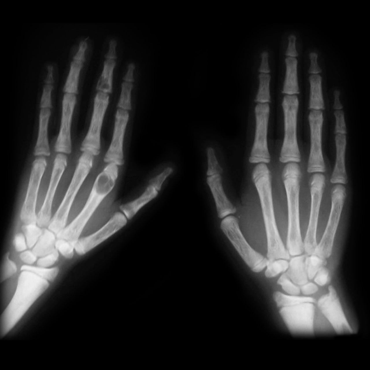
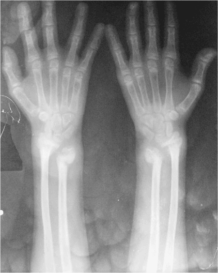
- Subperiosteal resorption of the radial side of the middle phalanges of the index and middle fingers is pathognomonic of hyperparathyroidism.
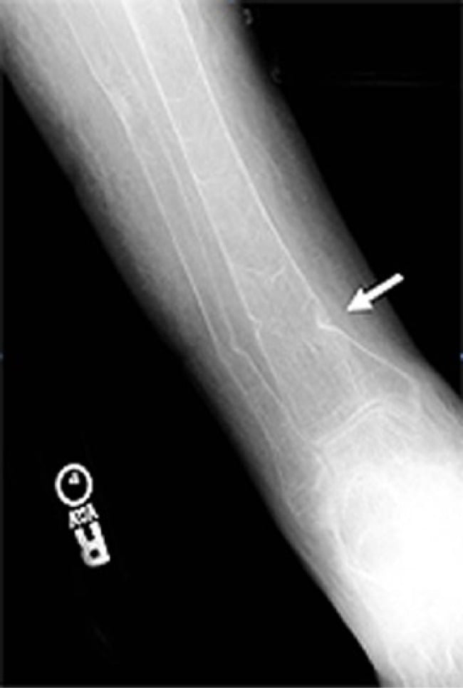
- Looser zones (pseudofractures) — lucent bands perpendicular to the cortex at the medial femoral neck, pubic rami, scapular axillary border and ribs — indicate osteomalacia.
- Diffuse osteopenia with a raised ESR, anaemia and normal ALP in a patient over 50 is myeloma until proven otherwise.
- Regional (not generalised) osteopenia has a different differential: disuse, CRPS, transient osteoporosis of the hip, RSD, inflammatory arthritis.
- “Alkaline phosphatase is the single most useful discriminator: normal in osteoporosis and myeloma, raised in osteomalacia, Paget's and metastases.
- “'Increased vertebral radiodensity relative to soft tissue' — technical overexposure is the commonest mimic of osteopenia. Always check exposure before calling it.
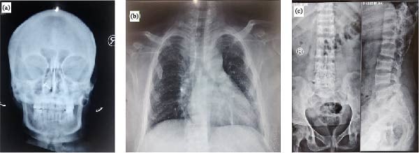
- “Vertebral picture-frame appearance = osteoporosis (accentuated cortex against lost trabeculae). Rugger-jersey spine = renal osteodystrophy.
- “Ward's triangle, principal compressive and tensile trabeculae — the Singh index describes the order of trabecular loss in the femoral neck.
Overexposure blackens bone and mimics demineralisation. Confirm by checking that the soft tissue shadows and the cortex of an adjacent normal bone are appropriately penetrated. Digital radiography with post-processing makes visual density assessment even less reliable.
Osteoporosis is a diagnosis of exclusion on plain film. Missing a raised calcium (myeloma, hyperparathyroidism) or a low phosphate with raised ALP (osteomalacia) is the classic viva failure. Always say "I would check bone chemistry".
An osteoporotic-looking vertebral collapse in a patient with back pain, anaemia and a raised ESR. Pedicle destruction, posterior vertebral body cortex convexity, and a paraspinal soft tissue mass all point away from benign osteoporotic collapse.
Periarticular osteopenia of the hands is inflammatory arthritis, not metabolic bone disease. Osteopenia confined to one limb is disuse or CRPS. Only comment on 'generalised' osteopenia if multiple regions are involved.
Recognising the Pattern
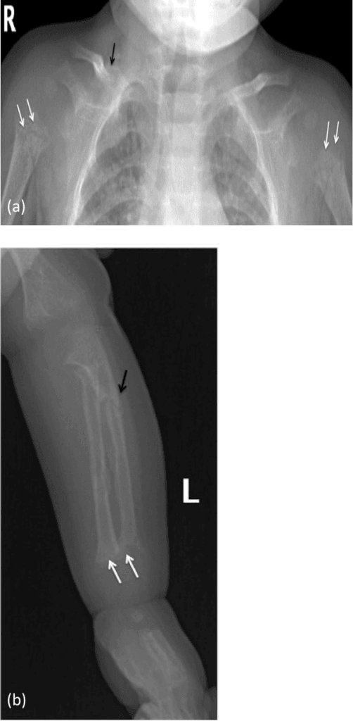
Definition. Generalised osteopenia is a diffuse reduction in radiographic bone density affecting the whole skeleton, with thinning of cortices and loss of trabeculae. It is a radiographic descriptor. The underlying histology may be osteoporosis (reduced quantity of normally mineralised bone), osteomalacia (normal or increased osteoid that is undermineralised), marrow replacement, or increased resorption.
Confirming it is genuinely present. Bone density cannot be measured on a radiograph, so use structural signs rather than greyness:
- Cortical thinning. Measure the second metacarpal: combined cortical thickness less than 50% of the total diaphyseal width at mid-shaft is abnormal (the Barnett–Nordin index).
- Trabecular dropout. Loss of the secondary (non-weight-bearing) trabeculae first, leaving the principal compressive and tensile groups standing out — the femoral neck Singh index.
- Accentuated cortical outline. The vertebral endplates and anterior cortex appear as a "picture frame" or "empty box" because the surrounding trabeculae have vanished.
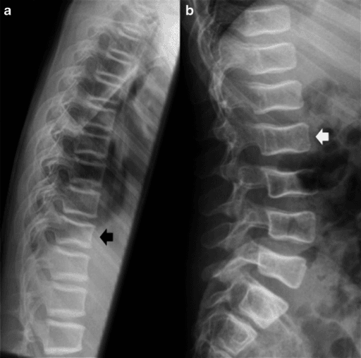
- Vertebral body changes. Increased biconcavity ("codfish" vertebrae), anterior wedging, or frank compression fractures.
- Exposure check. Compare bone density with the adjacent soft tissue shadow on the same film; if the soft tissues are also dark, suspect technique.
How to say it out loud. "This is a plain radiograph of the [lumbar spine] in a [skeletally mature] patient. There is diffusely reduced bone density with cortical thinning and loss of secondary trabeculae, giving the vertebral bodies a picture-frame appearance. I can see biconcave endplate deformity at L2 and L3. The technical factors appear adequate, so I believe this represents genuine generalised osteopenia rather than overexposure. I would want to know the patient's age, sex, drug history and renal function, and I would examine the hands, the femoral necks and the pubic rami for discriminating features."
Mimics — false positives:
- Overexposed or digitally over-processed film.
- Obesity or large body habitus causing scatter and apparent low density.
- Osteomalacia producing a "coarsened, unsharp" texture that is actually increased osteoid, not less bone.
- Diffuse marrow infiltration (leukaemia, myeloma) which reduces density without true osteoporosis.
- Regional osteopenia from disuse or inflammatory arthritis being over-read as generalised.
Next Investigation
Bone Density Loss: HOMESCauses of Generalised Osteopenia
The Differential
- Typical age / setting
- Over 50, back pain, anaemia, fatigue
- Discriminating feature
- Diffuse osteopenia with NORMAL alkaline phosphatase; punched-out lytic lesions without sclerotic rim; vertebral collapse with pedicle involvement
- What confirms it
- Serum and urine protein electrophoresis, serum free light chains, marrow aspirate; whole-body MRI or low-dose CT skeletal survey
- Typical age / setting
- Over 50, known primary (breast, lung, prostate, renal, thyroid)
- Discriminating feature
- Asymmetric, patchy destruction rather than uniform loss; pedicle erosion ('winking owl'); raised ALP
- What confirms it
- Bone scintigraphy (may be photopenic in myeloma), CT chest/abdomen/pelvis, biopsy
- Typical age / setting
- Women 50-70; incidental hypercalcaemia
- Discriminating feature
- Subperiosteal resorption on the RADIAL side of the middle phalanges of index and middle fingers; acro-osteolysis of terminal tufts; brown tumours; salt-and-pepper skull
- What confirms it
- Raised serum calcium with raised or inappropriately normal PTH; low phosphate; sestamibi scan
- Typical age / setting
- Housebound elderly, malabsorption, dark skin, veiled dressing, gastric surgery
- Discriminating feature
- Looser zones — lucent bands perpendicular to cortex at medial femoral neck, pubic rami, scapular axillary border, ribs; blurred, unsharp trabeculae and cortices
- What confirms it
- Low vitamin D, low or low-normal calcium and phosphate, RAISED alkaline phosphatase, raised PTH
- Typical age / setting
- Women over 55; men over 70
- Discriminating feature
- Sharp, well-defined but thinned cortices; normal bone chemistry throughout; fragility fractures of wrist, vertebra, hip
- What confirms it
- DXA T-score of minus 2.5 or less at spine or hip; normal Ca, PO4, ALP, PTH
- Typical age / setting
- Any age on prednisolone; RA, PMR, transplant, asthma
- Discriminating feature
- Axial-predominant loss with exuberant fracture callus; multiple vertebral fractures at a younger age than expected; associated AVN of femoral head
- What confirms it
- Drug history; DXA; consider vertebral fracture assessment. Normal biochemistry
- Typical age / setting
- Dialysis or CKD stage 4-5, any adult age
- Discriminating feature
- Mixed picture — rugger-jersey spine (endplate sclerosis with lucent centre), subperiosteal resorption, soft tissue and vascular calcification
- What confirms it
- Raised phosphate, raised PTH, raised ALP, low or normal calcium, raised creatinine
- Typical age / setting
- Women 30-60; thyroxine excess or Graves'
- Discriminating feature
- High bone turnover with cortical striation and tunnelling in the metacarpals; weight loss, tremor, AF
- What confirms it
- Suppressed TSH with raised free T4/T3; ALP may be mildly raised
- Typical age / setting
- 20-50, endogenous or exogenous steroid
- Discriminating feature
- Vertebral fractures with dense sclerotic marginal condensation of endplates; central obesity, striae, proximal myopathy
- What confirms it
- 24h urinary free cortisol, overnight dexamethasone suppression, late-night salivary cortisol
- Typical age / setting
- Children and adults; systemic upset
- Discriminating feature
- In children, transverse metaphyseal lucent bands ('leukaemic lines') and periosteal reaction; diffuse osteopenia with marrow replacement on MRI
- What confirms it
- Full blood count with film, marrow aspirate and trephine; MRI shows low T1 marrow signal
- Typical age / setting
- Childhood or young adult; positive family history
- Discriminating feature
- Gracile, thin bones with multiple fractures at different ages, bowing, wormian bones, blue sclerae, dentinogenesis imperfecta
- What confirms it
- Clinical criteria plus COL1A1/COL1A2 genetic testing; normal bone chemistry
- Typical age / setting
- Adults 30-60; flushing, urticaria pigmentosa, diarrhoea
- Discriminating feature
- Osteopenia interspersed with small sclerotic foci; hepatosplenomegaly; symptoms provoked by alcohol or opiates
- What confirms it
- Raised serum tryptase; marrow biopsy with CD117/CD25 mast cell aggregates
- Typical age / setting
- Adolescents and young adults, low BMI, amenorrhoea
- Discriminating feature
- Osteopenia at an age where it should not exist; stress fractures of tibia, metatarsals, femoral neck
- What confirms it
- LH, FSH, oestradiol or testosterone; DXA using Z-score not T-score in the young
- Typical age / setting
- Infants 6-24 months; restricted diet, autism spectrum diet
- Discriminating feature
- Dense white line of Frankel, Trümmerfeld lucent zone, Pelkan spurs, ring-sign epiphyses, subperiosteal haemorrhage
- What confirms it
- Low serum ascorbate; dramatic clinical response to vitamin C within days
Narrowing It Down
- 1Step 1 - Is it real, and is it generalised?
Check exposure and compare bone against soft tissue, then confirm on a second region - hands, pelvis or lateral spine.
Regional loss points to disuse, CRPS, transient osteoporosis of the hip or inflammatory arthritis, and takes you off the metabolic pathway entirely. Proceed only if two or more anatomical regions are affected. Remember that the radiographic impression itself is weak: read blind, orthopaedic surgeons achieve only about 61 per cent sensitivity and specificity against DXA.
- 2Step 2 - How old is the patient?
Let age set the differential before the film does.
Under 20: osteogenesis imperfecta, leukaemia, rickets or osteomalacia, anorexia, chronic illness, steroids. 20 to 50: hypogonadism, Cushing's, steroids, malabsorption, hyperthyroidism, pregnancy-associated. Over 50: postmenopausal osteoporosis is commonest, but myeloma, metastases, hyperparathyroidism and osteomalacia must be excluded. Osteopenia in a young patient is NEVER normal and always needs a cause.
- 3Step 3 - Look at the hands and the phalanges
Get a dedicated hand film and inspect the radial border of the middle phalanges of the index and middle fingers.
Subperiosteal resorption there is hyperparathyroidism. Acro-osteolysis of the terminal tufts, brown tumours and cortical tunnelling reinforce it. Sharp cortices with thinned diaphyses favour simple osteoporosis. This is the single highest-yield film on the pathway.
- 4Step 4 - Is the cortex sharp or blurred?
Judge the QUALITY of the cortical margin, not only its thickness.
Osteoporosis gives thin but crisply defined cortices and sharply marginated trabeculae. Osteomalacia gives an unsharp, ground-glass or coarsened texture, because unmineralised osteoid blurs the interfaces. If it looks fuzzy, hunt specifically for Looser zones at the medial femoral neck, the pubic and ischial rami, the axillary border of the scapula and the ribs.
- 5Step 5 - Is the loss uniform, or is there focal destruction?
Scan deliberately for lesions rather than accepting a global impression.
Punched-out lucencies without a sclerotic rim are myeloma; permeative or moth-eaten destruction is metastasis or lymphoma; a missing pedicle on the AP spine is malignancy until proven otherwise; endplate sclerosis with lucent central bands is the rugger-jersey spine of renal osteodystrophy. Uniform loss with no focal lesion supports a metabolic cause.
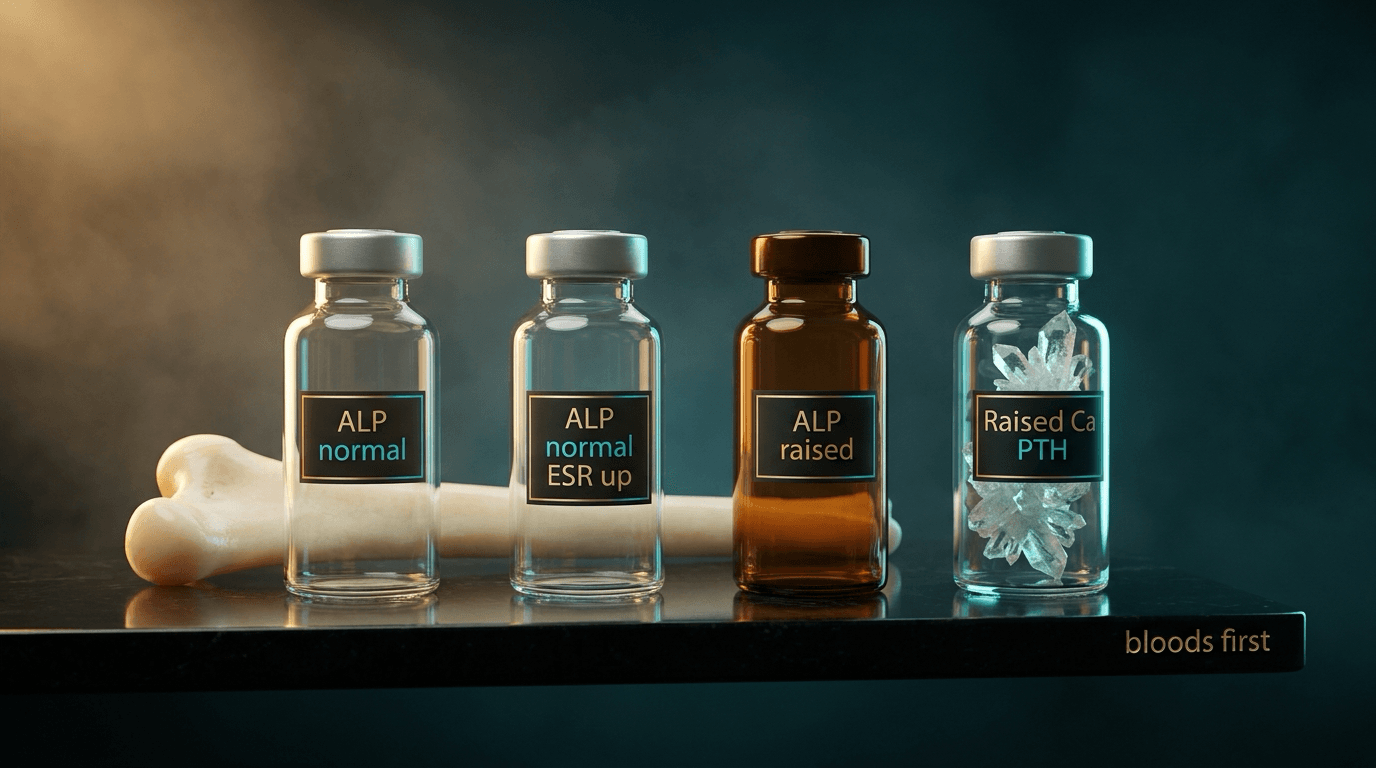
- 6Step 6 - Send the bloods and read the alkaline phosphatase first
Calcium, phosphate, ALP, PTH, 25-hydroxyvitamin D, urea and electrolytes, thyroid function, full blood count, ESR, serum and urine electrophoresis with free light chains, testosterone or oestradiol, and coeliac serology.
Normal ALP with normal calcium and phosphate is osteoporosis or myeloma. Raised ALP with low calcium or phosphate is osteomalacia. Raised calcium with raised PTH is hyperparathyroidism. Raised phosphate with raised creatinine is renal osteodystrophy. The ALP splits the differential faster than any other single result.
- 7Step 7 - Quantify, then stratify fracture risk
DXA of the lumbar spine and proximal femur, reported as a T-score in postmenopausal women and men over 50 and a Z-score in younger patients and children, combined with FRAX or QFracture and lateral spine imaging.
A prevalent vertebral fracture changes the treatment threshold REGARDLESS of T-score. Note also that BMD is not the whole risk: in the FRAX development cohort, ten-year hip fracture probability varied roughly a hundred-fold on clinical risk factors alone, before any BMD was measured.
MCQ Practice Points
Q: How much bone mineral must be lost before osteopenia becomes visible on a plain radiograph, and how good is the reader?
A: Conventionally 30 to 50 per cent - which is why a normal-looking film cannot exclude significant bone loss. The measured version of the same point is more damning: reading blinded wrist radiographs against DXA, orthopaedic surgeons achieved 61 per cent sensitivity and 61 per cent specificity, and visual analogue assessment of porosity, cortical thickness and trabecular number was unreliable for all four parameters (Olschewski 2001). The plain film raises the question; DXA answers it.
Q: Which single biochemical result best separates osteomalacia from osteoporosis?
A: Alkaline phosphatase - raised in osteomalacia, reflecting osteoblastic attempts at mineralisation, and normal in osteoporosis. Calcium and phosphate are also low or low-normal in osteomalacia, with a compensatory rise in PTH.
Q: What radiographic finding is most specific for hyperparathyroidism?
A: Subperiosteal resorption of the radial aspect of the middle phalanges of the index and middle fingers. Supporting signs are terminal tuft acro-osteolysis, distal clavicular resorption, brown tumours, the salt-and-pepper skull and, in renal disease, the rugger-jersey spine.
Q: Why is bone scintigraphy unreliable for multiple myeloma?
A: Myeloma lesions are purely lytic with minimal reactive osteoblastic activity, so they are often photopenic or simply normal on a bone scan. Whole-body low-dose CT or whole-body MRI is now preferred over both scintigraphy and the traditional skeletal survey.
Q: In a 25-year-old woman with amenorrhoea and osteopenia, should DXA be reported as a T-score or a Z-score?
A: Z-score - comparison against age-matched controls. T-scores, which compare against a young adult peak, should be used only in postmenopausal women and men aged 50 or over. A Z-score of minus 2.0 or less is reported as "below the expected range for age".
Q: Which features distinguish a benign osteoporotic vertebral collapse from a malignant one on MRI?
A: Preservation of normal fatty marrow signal within the collapsed vertebra, a band-like pattern of oedema, a fluid sign and a retropulsed bony fragment all favour benign collapse. Complete low T1 marrow replacement, a convex posterior cortex, pedicle involvement and a paravertebral soft tissue mass favour malignancy.
Exam Viva Scenarios
Practise clinical reasoning and management decisions out loud
“You are shown this lateral lumbar spine radiograph of a 68-year-old woman with six weeks of thoracolumbar back pain. There is diffuse reduction in bone density with a wedge compression fracture at T12 and biconcave endplates at L1 and L2. How do you proceed?”
“You are shown this radiograph of both hands in a 58-year-old woman referred with generalised osteopenia found incidentally. There is loss of the cortical margin along the radial aspect of the middle phalanges of the index and middle fingers, with resorption of the terminal tufts.”
“You are shown a pelvic radiograph of a 34-year-old man with bilateral groin pain and difficulty rising from a chair. There is generalised osteopenia with a lucent band crossing the medial cortex of the left femoral neck perpendicular to the cortex, and a similar band in the right inferior pubic ramus.”
Confirm the Pattern
- Check exposure first — overexposure is the commonest mimic
- Second metacarpal combined cortical thickness less than 50% of shaft width is abnormal
- Singh index — order of femoral neck trabecular loss
- Vertebral picture-frame, codfish biconcavity, anterior wedging
- 30-50% mineral loss required before radiographic visibility
Biochemical Fingerprints
- Osteoporosis: Ca normal, PO4 normal, ALP normal, PTH normal
- Osteomalacia: Ca low/normal, PO4 low, ALP RAISED, PTH raised, vitamin D low
- Primary HPT: Ca RAISED, PO4 low, PTH raised, ALP normal or raised
- Renal osteodystrophy: Ca low/normal, PO4 RAISED, PTH raised, creatinine raised
- Myeloma: Ca may be raised, ALP NORMAL, ESR raised, paraprotein present
- Metastases: ALP raised, Ca may be raised, PTH suppressed
Signature Radiographic Signs
- Looser zones at medial femoral neck, pubic rami, scapular border, ribs — osteomalacia
- Subperiosteal resorption radial border middle phalanges index and middle fingers — HPT
- Rugger-jersey spine — renal osteodystrophy
- Punched-out lucencies without sclerotic rim, pedicle sparing early — myeloma
- Winking owl sign (absent pedicle) — metastasis
- Metaphyseal lucent bands in a child — leukaemia
- Frankel line, Trümmerfeld zone, Pelkan spurs — scurvy
- Wormian bones, gracile bones, blue sclerae — osteogenesis imperfecta
Investigation Sequence
- Bone profile, ALP, PTH, 25-OH vitamin D, U&Es, TFTs
- FBC, ESR, serum and urine electrophoresis with free light chains if over 50
- Radiographs of hands and pelvis — cheap and often diagnostic
- DXA spine and hip; Z-score if under 50 or premenopausal
- MRI whole spine if malignant collapse suspected
- Whole-body low-dose CT or MRI, not bone scan, for myeloma
- Transiliac biopsy only for unexplained or discordant cases
Do Not Forget
- Osteopenia in anyone under 50 always needs a cause
- Correct vitamin D before starting a bisphosphonate
- Regional osteopenia is a different differential — disuse, CRPS, transient osteoporosis of the hip, inflammatory arthritis
- A prevalent fragility vertebral fracture warrants treatment regardless of T-score
- Degenerative sclerosis and aortic calcification falsely raise the lumbar DXA score — use the hip
Evidence Base
Assessment of Osteoporosis Using Standard Radiographs of the Wrist
- 38 patients with a unilateral distal radius fracture had DXA of the UNINJURED distal radius within a week of injury, with standard radiographs of both wrists
- Radiographs were read blinded and randomised, on two separate occasions by three orthopaedic surgeons and once by a fourth, who were asked simply to state whether osteoporosis was present
- RADIOGRAPHIC DETERMINATION OF OSTEOPOROSIS HAD A SENSITIVITY OF 61 PER CENT AND A SPECIFICITY OF 61 PER CENT against DXA - barely better than a coin toss in either direction
- Visual analogue assessment was UNRELIABLE for all four parameters tested: porosity, cortical thickness, trabecular thickness and the number of trabeculae in the ultradistal radius
- Intraobserver agreement averaged 81 per cent, but with a kappa of only 0.54 - moderate once chance agreement is accounted for
- The authors conclude that orthopaedic surgeons cannot predict from plain radiographs, with sufficient accuracy, whether a patient is significantly osteoporotic
Assessment of Fracture Risk and Its Application to Screening for Postmenopausal Osteoporosis - Synopsis of a WHO Report
- The WHO Study Group report that gives orthopaedics its diagnostic vocabulary - the densitometric definition of osteoporosis against a young adult reference, from which the T-score threshold derives
- States the central limitation of the technique in its own words: bone mineral measurements have HIGH SPECIFICITY BUT LOWER SENSITIVITY, so their value is greatest in those already identified as higher risk
- Concluded there was little evidence that osteoporosis could usefully be tackled by population-level policy on smoking, exercise and nutrition, and that targeting treatment to susceptible individuals was the appropriate strategy
- Framed screening as SELECTIVE rather than universal, on cost-benefit grounds, with the menopause as an appropriate point to consider it
FRAX and the Assessment of Fracture Probability in Men and Women from the UK
- Built the FRAX tool from clinical risk factors identified in previous meta-analyses - BMI as a continuous variable, prior fracture, parental hip fracture, oral glucocorticoids, rheumatoid arthritis and other secondary causes, current smoking, and alcohol at three or more units daily
- Four models: ten-year probability of hip fracture and of major osteoporotic fracture, each WITH and WITHOUT femoral neck BMD
- WITHOUT ANY BMD AT ALL, ten-year hip fracture probability in women at fixed BMI ranged from 0.2 per cent at age 50 with no risk factors to 22 per cent at age 80 with a parental history - an approximately HUNDRED-FOLD range driven by clinical factors alone
- For major osteoporotic fracture the range was 3.5 to 31 per cent in women and 2.8 to 15 per cent in men; risk factors compounded incrementally
- At any given T-score and age the probabilities were comparable between men and women, except in the elderly where women's were higher because of men's higher competing mortality