PAD — Three Unipennate Adductors
- PAD: Palmar ADduct. Three muscles, all unipennate, each arising from a single metacarpal — the metacarpal of the finger it moves.
- They arise from and act on the index, ring and little fingers only; the middle finger has none, because the reference axis is the middle finger itself.
- All are supplied by the deep motor branch of the ulnar nerve, C8-T1.
- Insertion is predominantly into the lateral band of the extensor hood, with a smaller bony contribution than the dorsal interossei — hence a greater share in interphalangeal extension.
- Their loss, together with the lumbricals and dorsal interossei, produces the intrinsic-minus claw hand; their contracture produces the intrinsic-plus hand.
- “The Bouvier test is the single most decision-changing test in claw hand: block MCP hyperextension and see whether the PIP joints extend.
- “The ulnar paradox — a high ulnar lesion claws less than a low one, because the flexor digitorum profundus is also paralysed and cannot curl the interphalangeal joints.
- “The Finochietto-Bunnell test distinguishes intrinsic tightness from a proximal interphalangeal joint contracture by comparing PIP flexion with the MCP extended versus flexed.
- “Every anti-claw tendon transfer must be routed volar to the deep transverse metacarpal ligament; routed dorsal to it, the transfer worsens the claw.
Overview
The palmar (volar) interossei are three small unipennate muscles lying on the palmar aspect of the intermetacarpal spaces, deep in the palm, palmar to the dorsal interossei. Each arises from a single metacarpal — the metacarpal of the finger it moves — and adducts that finger toward the axis of the middle finger.
They are smaller and weaker than the dorsal interossei, and their adduction function is easy to underestimate. But their contribution to the extensor mechanism is proportionally larger, and it is through them, the dorsal interossei and the lumbricals that the hand converts extrinsic tendon pull into a coordinated grasp. They are also the muscles whose behaviour underlies the two tests that determine what operation a clawed hand needs.
The intrinsics pass palmar to the metacarpophalangeal (MCP) axis and dorsal to the interphalangeal (IP) axes via the lateral bands. Everything follows from that.
Intrinsic-MINUS (the claw hand) — the intrinsics are paralysed:
- The extensor digitorum communis, unopposed, hyperextends the MCP joints. Its force is dissipated at the first joint it crosses and never reaches the IP joints.
- The long flexors, unopposed at the IP joints, flex the PIP and DIP.
- Posture: MCP hyperextension with IP flexion.
- Function: the fingers curl before they open around an object; the patient cannot grasp anything large. Grip is weak; the hand rolls up rather than closing.
Intrinsic-PLUS — the intrinsics are shortened or contracted:
- The MCP joints are pulled into flexion and the IP joints into extension.
- Posture: MCP flexion with IP extension.
- Function: the fingers cannot curl; the patient cannot make a fist. The PIP joints drift into swan-neck deformity.
The safe (protective) position of immobilisation is deliberately intrinsic-plus: wrist 20-30 degrees extended, MCP joints 70-90 degrees flexed, IP joints straight, thumb palmarly abducted. This is chosen because the cam-shaped metacarpal head puts the MCP collateral ligaments at maximal length in flexion, and the IP volar plates at maximal length in extension. Immobilise a hand flat and the MCP collaterals shorten and the IP volar plates contract — the hand stiffens in a useless posture from which it never recovers.
PAD and DABInterossei — Numbers, Actions and Pennation
Hook:Unipennate = one bone of origin = adduction. Bipennate = two bones of origin = abduction. The middle finger is the reference axis, so it has two dorsal interossei and no palmar interosseous.
FWMBThe Four Bedside Signs of Interosseous Loss — and What Each One Actually Tests
Hook:Froment and Wartenberg tell you the ulnar nerve is out; Masse tells you how long it has been out; Bouvier tells you which operation to do. Only Bouvier changes the plan.
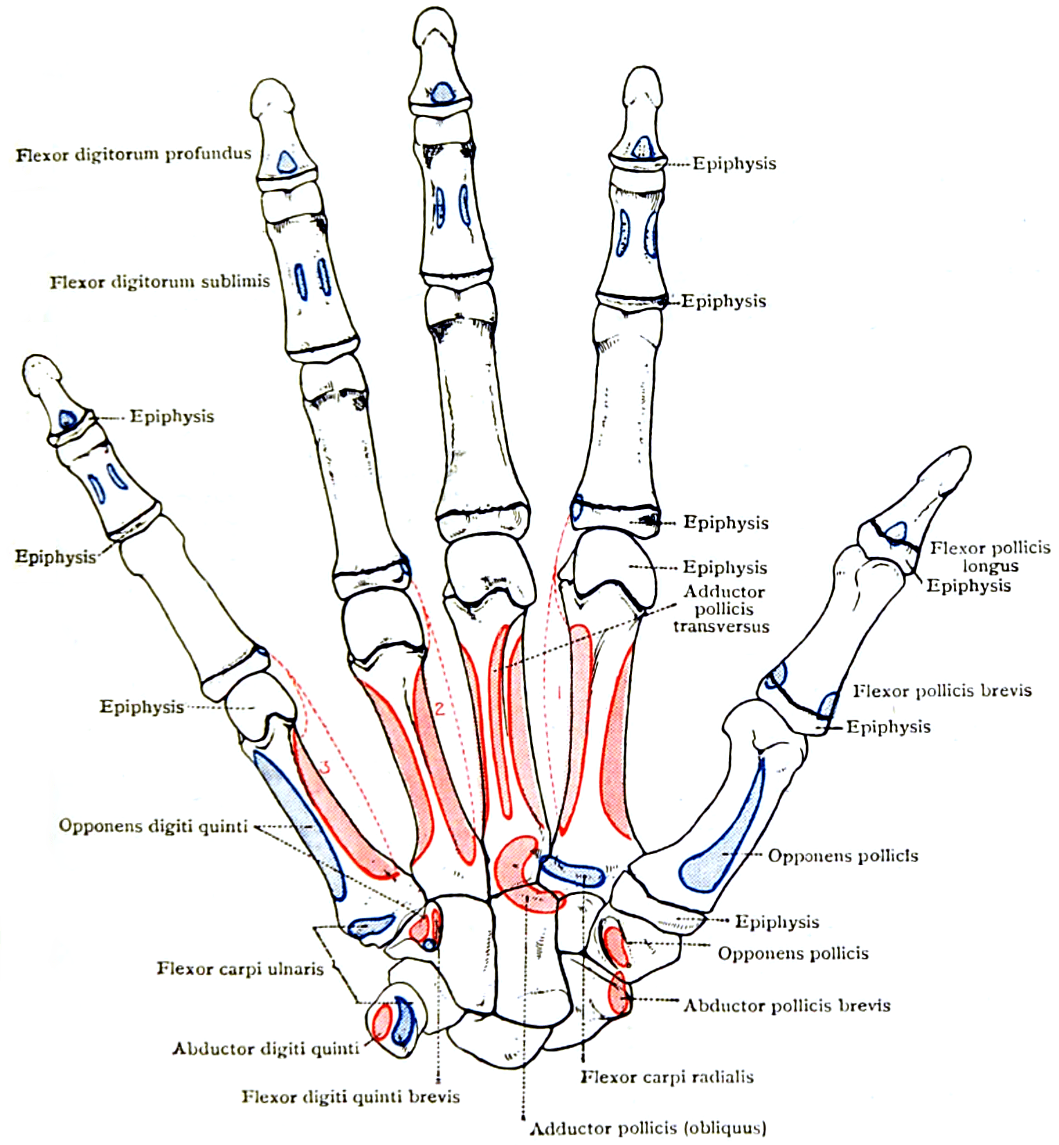
Attachments, Innervation and Relations
General plan
Each palmar interosseous is unipennate, arising from one side of a single metacarpal — the metacarpal of the digit it moves — and running distally to insert into the same side of that digit, adducting it toward the middle finger.
Individual attachments
- Origin
- Ulnar side of the palmar surface of the second metacarpal
- Insertion
- Ulnar base of the index proximal phalanx and the ulnar lateral band
- Action
- Adducts the index toward the middle finger
- Origin
- Radial side of the palmar surface of the fourth metacarpal
- Insertion
- Radial base of the ring finger proximal phalanx and the radial lateral band
- Action
- Adducts the ring finger toward the middle finger
- Origin
- Radial side of the palmar surface of the fifth metacarpal
- Insertion
- Radial base of the little finger proximal phalanx and the radial lateral band
- Action
- Adducts the little finger — the muscle whose loss gives Wartenberg sign
The disputed fourth palmar interosseous
- Many anatomists describe a fourth (or first, in an alternative numbering) palmar interosseous acting on the thumb, arising from the ulnar side of the first metacarpal and inserting into the ulnar sesamoid and the base of the thumb proximal phalanx.
- Where it is described, its function is subsumed within the oblique head of adductor pollicis, with which it is continuous.
- The safe answer in an examination is "three palmar interossei, with a variably described fourth acting on the thumb that is functionally part of adductor pollicis." Numbering the palmar interossei as one to three for index, ring and little is the convention most sources use.
Insertion — proportions matter
- Like the dorsal interossei, each palmar interosseous divides into a medial (bony) tendon to the base of the proximal phalanx and MCP capsule, and a lateral (tendinous) contribution to the lateral band of the extensor hood.
- The palmar interossei have a proportionally greater lateral band contribution and a smaller bony insertion than the dorsal interossei. They therefore contribute relatively more to interphalangeal extension and relatively less to raw MCP flexion power.
- The lumbricals sit at the extreme of this spectrum: they insert exclusively into the radial lateral band with no bony insertion at all, making them the pure IP extensors.
The spectrum, in one line
First dorsal interosseous (mostly bone, powerful abductor and MCP flexor) — other dorsal interossei — palmar interossei — lumbricals (entirely lateral band, pure IP extensors).
Block the MCP and see what happens.
- Passively hold the MCP joints in flexion, preventing hyperextension, and ask the patient to extend the PIP joints.
- They extend fully (positive, correctable claw): the extensor mechanism and lateral bands are intact. An MCP flexion block — static or a simple dynamic transfer — is all that is needed.
- They do not extend (negative, non-correctable): the lateral bands are attenuated or the PIP is contracted. A dynamic transfer inserting into the lateral bands is required, after releasing any fixed contracture.
Volar to the deep transverse metacarpal ligament.
- The intermetacarpal ligament marks the plane of the MCP joint axis.
- A transfer volar to it flexes the MCP joint — which is what an anti-claw transfer must do.
- A transfer dorsal to it extends the MCP joint and makes the claw worse.
- The lumbricals are the anatomical model: they run volar to the ligament, which is exactly why they flex the MCP and extend the IP joints.
Action and Biomechanics
Actions
- Mechanism
- Unipennate pull from a single metacarpal, off-axis at the MCP joint
- Clinical correlate
- Card test between the fingers; Wartenberg sign at the little finger
- Mechanism
- Line of pull passes palmar to the MCP axis of rotation
- Clinical correlate
- Combined with the dorsal interossei and lumbricals, dominates MCP flexion in grip
- Mechanism
- Insertion into the lateral bands, which pass dorsal to the IP axes
- Clinical correlate
- Only effective with MCP hyperextension prevented — the basis of the Bouvier test
- Mechanism
- Bony insertion into the base of the proximal phalanx and accessory collateral ligament
- Clinical correlate
- Loss contributes to ulnar drift in the rheumatoid hand
- Mechanism
- Coordinated adduction narrows the hand around an object
- Clinical correlate
- Loss produces the splayed, weak hand of intrinsic palsy
Position dependence of adduction
- Interosseous abduction and adduction are strongest with the MCP joints extended, because in flexion the cam-shaped metacarpal head tightens the collateral ligaments and locks the joint in the coronal plane.
- Test adduction with the hand flat and the fingers extended. A patient asked to hold a card between flexed fingers is being tested for something else entirely.
- This same cam geometry is the reason the MCP joints must be immobilised in flexion, and the reason a stiff MCP joint after immobilisation in extension is so hard to salvage.
Architecture and force
- The palmar interossei are unipennate with short fibres, giving relatively high force per unit volume but very small excursion (fibre lengths of the order of 1.5-2.5 cm).
- Collectively the interossei generate the great majority of intrinsic force. Within that group the first dorsal interosseous and adductor pollicis stand apart, with cross-sectional areas comparable to extrinsic muscles; the palmar interossei are ordinary members of the interosseous group rather than outliers.
- Implication for reconstruction: you cannot replicate an interosseous with a small local transfer. Anti-claw procedures work by changing the geometry (blocking MCP hyperextension) rather than by replacing intrinsic force.
The ulnar paradox — why the worse injury looks better
- High ulnar lesion (elbow)
- Paralysed
- Low ulnar lesion (wrist)
- Paralysed
- High ulnar lesion (elbow)
- Paralysed
- Low ulnar lesion (wrist)
- Intact
- High ulnar lesion (elbow)
- Absent
- Low ulnar lesion (wrist)
- Present and unopposed
- High ulnar lesion (elbow)
- LESS
- Low ulnar lesion (wrist)
- MORE
- High ulnar lesion (elbow)
- Abnormal
- Low ulnar lesion (wrist)
- Normal
- High ulnar lesion (elbow)
- Clawing WORSENS as the profundus reinnervates before the intrinsics
- Low ulnar lesion (wrist)
- Clawing improves as the intrinsics reinnervate
The paradox in one sentence: the more proximal the lesion, the less the deformity — because the muscle that produces the deformity is itself paralysed. The corollary is the one candidates forget: as a high lesion recovers, the clawing gets worse before it gets better, because the profundus reinnervates first. Warn the patient, or they will believe they are deteriorating.
Synergists and antagonists
- Synergists: dorsal interossei and lumbricals at the MCP and IP joints; adductor pollicis in the pinch couple.
- Antagonists: dorsal interossei in the abduction-adduction plane; extensor digitorum communis, extensor indicis proprius and extensor digiti minimi at the MCP joint; flexor digitorum superficialis and profundus at the IP joints.
Surface Anatomy and Examination
Inspection
- The palmar interossei are deep and not directly visible or palpable. Their wasting is inferred from guttering between the metacarpals on the dorsum, which is largely dorsal interosseous atrophy, and from the overall skeletal appearance of the intrinsic-minus hand.
- Inspect the resting posture: MCP hyperextension with IP flexion in the ring and little fingers is the ulnar claw; in all four fingers it is a combined median and ulnar palsy.
The key clinical tests
- How to perform
- Passively hold the MCP joints in flexion, blocking hyperextension, and ask the patient to actively extend the PIP joints
- Positive finding
- PIP joints extend fully
- What it means
- Correctable claw — extensor mechanism intact. An MCP flexion block suffices
- False positives
- A fixed PIP flexion contracture makes the test uninterpretable — assess passive PIP extension first
- How to perform
- Hold the MCP in extension and passively flex the PIP; repeat with the MCP flexed
- Positive finding
- PIP flexion more restricted with the MCP EXTENDED
- What it means
- Intrinsic tightness — the intrinsics are contracted
- False positives
- Equal restriction in both positions indicates a PIP capsular contracture, not intrinsic tightness
- How to perform
- Compare passive PIP flexion with the wrist and MCP flexed versus extended
- Positive finding
- PIP flexion more restricted with the wrist and MCP FLEXED
- What it means
- Extrinsic extensor tightness — the opposite pattern to intrinsic tightness
- False positives
- Combined intrinsic and extrinsic tightness gives a mixed picture
- How to perform
- Hand flat, fingers adducted; ask the patient to hold them together
- Positive finding
- Little finger drifts into abduction and cannot be adducted
- What it means
- Third palmar interosseous weakness with unopposed extensor digiti minimi
- False positives
- Congenital little finger abduction; extensor digiti minimi subluxation; little finger MCP arthritis
- How to perform
- Place a card between two adjacent extended fingers and pull it out
- Positive finding
- The patient cannot retain the card
- What it means
- Palmar interosseous adduction weakness
- False positives
- Pain from MCP arthritis; a stiff joint that cannot adduct
- How to perform
- Flex the middle finger MCP joint and ask the patient to deviate it both radially and ulnarwards
- Positive finding
- Inability to deviate to both sides
- What it means
- Second and third dorsal interosseous weakness
- False positives
- Middle finger MCP stiffness
- How to perform
- Key pinch on a card; watch the thumb IP joint
- Positive finding
- Thumb IP joint flexes
- What it means
- Adductor pollicis weakness — the terminal deep ulnar muscle
- False positives
- Painful thumb MCP; Riche-Cannieu anastomosis gives false negatives
Sequencing the examination of a clawed hand
- Inspect the resting posture and record which digits claw.
- Assess passive range at the PIP joints. A fixed contracture must be excluded before any dynamic test is interpretable.
- Bouvier test — correctable or not. This is the decision point.
- Finochietto-Bunnell — is there superimposed intrinsic tightness?
- Extrinsic extensor tightness — is the extensor system also tight?
- Motor examination: first dorsal interosseous, abductor digiti minimi, Froment, Jeanne, Wartenberg, Egawa, and flexor digitorum profundus to ring and little.
- Sensation: palmar little and ulnar ring, and dorsal ulnar hand — normal dorsal sensation places the lesion at the wrist.
- Grip and pinch dynamometry, and a functional assessment of what the patient actually cannot do.
Complications
- Mechanism
- Tunnelling through the wrong plane
- Prevention
- Pass the tendon volar to the deep transverse metacarpal ligament under direct vision
- Salvage
- Revision with correct routing — the transfer will not work otherwise
- Mechanism
- Blind passage of a tendon through the web space
- Prevention
- Blunt passer under direct vision; expose the web space if in doubt
- Salvage
- Direct repair; a divided common digital nerve must be repaired
- Mechanism
- Ring FDS harvest leaves the PIP volar plate unsupported, especially in hypermobile patients
- Prevention
- Leave a distal FDS slip attached, or use an extensor donor in hypermobile hands
- Salvage
- PIP volar plate capsulodesis or FDS tenodesis
- Mechanism
- FDS harvested too distally, or FDP tethered by scar
- Prevention
- Harvest at the mid-palm or PIP level under vision
- Salvage
- Release the tether; tenolysis
- Mechanism
- Set with the MCP joints too flexed or the wrist extended
- Prevention
- Tension with the wrist neutral and MCP 60-70 degrees flexed, IP straight
- Salvage
- Lengthening or re-insertion
- Mechanism
- Capsulodesis stretches out in a high-demand hand
- Prevention
- Choose a dynamic transfer for manual workers
- Salvage
- Convert to a dynamic transfer
- Mechanism
- Excision of transverse fibres or the central slip
- Prevention
- Excise only the oblique lateral band fibres; check on the table
- Salvage
- Boutonniere or claw reconstruction — both difficult
- Mechanism
- Uncorrected wrist radial deviation
- Prevention
- Correct the wrist first or at the same sitting
- Salvage
- Revision including wrist correction
- Mechanism
- Deep palmar dissection at the metacarpal bases
- Prevention
- Know the nerve lies on the palmar surface of the interossei with the deep arch
- Salvage
- Direct repair if recognised; late reconstruction
- Mechanism
- Untreated hand compartment syndrome
- Prevention
- Test passive intrinsic stretch in every swollen hand; release all ten compartments
- Salvage
- Intrinsic release plus prolonged splinting; often incomplete
Clinical Relevance
Causes of the intrinsic-minus hand
- Ulnar nerve palsy — ring and little claw (radial two lumbricals are median-innervated and spare the index and middle).
- Combined low median and ulnar palsy — all four digits claw. This is the classic leprosy and combined wrist laceration picture.
- Lower trunk brachial plexus injury and C8-T1 root avulsion.
- Charcot-Marie-Tooth disease and other hereditary neuropathies.
- Compartment syndrome and Volkmann ischaemic contracture — though here the claw is often mixed with intrinsic contracture.
- Leprosy remains the largest global cause.
Why only the ulnar two digits in an ulnar palsy
The first and second lumbricals are median-innervated; the third and fourth are ulnar. All seven interossei are ulnar. In an isolated ulnar palsy the index and middle retain their lumbricals, which insert exclusively into the radial lateral band and are sufficient to prevent clawing in those digits. The ring and little lose both interossei and lumbricals and therefore claw.
Bouvier test and the operation it selects
- Interpretation
- Extensor mechanism intact; only an MCP flexion moment is missing
- Options
- Static: Zancolli lasso, volar plate capsulodesis, bone block, MCP volar tenodesis. Dynamic: any transfer inserted into the A1 or A2 pulley or the proximal phalanx
- Notes
- The simplest adequate operation is the right one
- Interpretation
- Lateral bands attenuated or PIP contracted
- Options
- Dynamic transfer inserting into the LATERAL BANDS: modified Stiles-Bunnell (ring FDS), Brand transfer (ECRL or ECRB with grafts)
- Notes
- Release any fixed PIP contracture first, or the transfer will fail
- Interpretation
- Fixed PIP flexion contracture
- Options
- Serial splinting and therapy first; consider PIP release
- Notes
- Never plan a transfer against a fixed contracture
Anti-claw procedures in detail
- Zancolli lasso: a slip of flexor digitorum superficialis is divided, passed around the A1 pulley and sutured back to itself, creating an active MCP flexion tether without crossing the IP joints. Simple, durable, and the standard in high-volume leprosy programmes.
- Volar plate capsulodesis (Zancolli capsulodesis): the MCP volar plate is advanced proximally and reattached to the metacarpal neck, mechanically preventing hyperextension. Tends to stretch out over time in high-demand hands.
- Bone block and MCP volar tenodesis: described alternatives, less commonly used.
- Advantage: no donor morbidity, no re-education. Disadvantage: no added power.
- Modified Stiles-Bunnell: the ring finger flexor digitorum superficialis is divided distally, split into slips, and routed volar to the deep transverse metacarpal ligament into the lateral bands of the clawing digits. Powerful, but risks swan-neck at the donor PIP joint and quadriga if harvested too distally.
- Brand transfer: extensor carpi radialis longus or brevis is extended with free tendon grafts, routed around the radial side of the wrist, through the intermetacarpal spaces volar to the deep transverse metacarpal ligament, into the lateral bands (Brand EE, extensor to extensor) or into the A1/A2 pulleys. Provides genuine intrinsic power and is the standard where strength is required.
- Flexor carpi radialis with grafts (Brand FF) is the volar variant.
every one of these must run volar to the deep transverse metacarpal ligament.
The staging principle
In a combined median and ulnar palsy the priorities are, in order: sensation and skin, then thumb opposition, then correction of claw, then pinch (adductorplasty and index abduction), then finger flexion power. Do not correct a claw in an insensate hand.
Surgical Relevance
Selecting and performing an anti-claw procedure
- 1Step 1 — Is the hand sensate?
Assess protective sensation and two-point discrimination. Restore or accept sensation before correcting posture.
Never correct a claw in an insensate hand
- 2Step 2 — Are the joints supple?
Assess passive PIP extension and MCP range. Treat any fixed contracture with therapy and serial splinting first.
A transfer cannot move a stiff joint
- 3Step 3 — Bouvier test
Passively block MCP hyperextension and ask for active PIP extension.
Positive: MCP flexion block suffices. Negative: lateral band transfer required
- 4Step 4 — Assess donors
Grade every candidate: ring FDS (needs a working profundus and no hypermobility), extensor carpi radialis longus or brevis, flexor carpi radialis, brachioradialis. Check innervation is intact given the level of the lesion.
Donor availability shapes the plan
- 5Step 5 — Choose static or dynamic, and know what the randomised evidence says
Low-demand hand, positive Bouvier, limited donors: Zancolli lasso or capsulodesis. High-demand hand or negative Bouvier: Brand or modified Stiles-Bunnell transfer. Where BOTH the lasso and the four-tail FDS are options, the one randomised comparison (Chaudhuri 2026, 50 patients) found NO clear superiority overall - but the two differ in what they deliver: the LASSO recovered more grip (129 against 117 per cent, p = 0.013) with a shorter operation and faster return to activity, while the FOUR-TAIL FDS gave numerically better claw correction (excellent in 12 against 7) that did not reach significance.
Simplest adequate operation - then name which axis you are optimising, grip and speed or appearance of correction
- 6Step 6 — Route volar to the intermetacarpal ligament
Pass the transfer through the intermetacarpal space VOLAR to the deep transverse metacarpal ligament, replicating the lumbrical course.
The single non-negotiable technical rule
- 7Step 7 — Insert and tension
Into the lateral bands (negative Bouvier) or the A1 or A2 pulley or proximal phalanx (positive Bouvier). Tension with the wrist neutral and the MCP joints in 60-70 degrees of flexion with the IP joints straight.
Over-tension gives an intrinsic-plus hand
- 8Step 8 — Address pinch separately
Add an adductorplasty and index abduction augmentation if key pinch is deficient.
Claw correction alone does not restore pinch
- 9Step 9 — Immobilise and re-educate
Splint with MCP joints flexed and IP joints extended for 4 weeks, then graded active motion with re-education using the donor's original command.
Rehabilitation determines the result as much as the operation
- Deep motor branch of the ulnar nerve: lies directly on the palmar surface of the interossei with the deep palmar arch, crossing the palm roughly 1-2 cm distal to the distal edge of the transverse carpal ligament, deep to the flexor tendons. It is the structure divided by deep palmar dissection, by fifth carpometacarpal fracture-dislocation surgery, and by careless tunnelling of a transfer through the intermetacarpal space.
- Deep palmar arch: approximately 1 cm proximal to the superficial arch, which lies about 1 cm distal to the transverse carpal ligament at Kaplan's cardinal line.
- Common digital nerves and arteries: lie in the web spaces immediately volar to the deep transverse metacarpal ligament. A transfer being passed volar to that ligament passes directly through their plane — pass the tendon under direct vision with a blunt passer, never blindly.
- Lumbrical canal: the tunnel through which a transfer is passed in the volar route; it contains the lumbrical and communicates proximally with the midpalmar space and distally with the web space. Infection tracks along it.
- Deep transverse metacarpal ligament: the plane that must be respected. Volar equals MCP flexion; dorsal equals MCP extension and a worse claw.
Distal intrinsic release (Littler) — technique
- Indication: established intrinsic tightness confirmed by a positive Finochietto-Bunnell test that has not responded to therapy.
- Approach: dorsolateral incision over the proximal phalanx, or a dorsal midline incision for multiple digits.
- Step: identify the lateral band and the oblique fibres of the intrinsic tendon distal to the MCP joint. Excise a triangle of the oblique fibres, preserving the central slip and the transverse fibres of the extensor hood — the transverse fibres maintain MCP flexion, and the central slip maintains PIP extension.
- Check: repeat the Finochietto-Bunnell test on the table. PIP flexion should now be equal with the MCP extended and flexed.
- Avoid: excising the transverse fibres (loses MCP flexion) or the central slip (produces a boutonniere).
Crossed intrinsic transfer — technique
- Indication: ulnar drift in the rheumatoid hand, at the time of MCP arthroplasty or soft tissue reconstruction.
- Step: divide the ulnar interosseous tendon of the index, middle and ring fingers at the level of the proximal phalanx and transfer each to the radial lateral band of the adjacent ulnar digit, passing volar to the intermetacarpal ligament.
- Do not perform in isolation: the wrist must be corrected, the MCP joints reconstructed, and the extensor tendons centralised, or the drift recurs.
Guidelines, Registries & Global Practice
Anatomical variation across populations
- The existence and numbering of a fourth palmar interosseous acting on the thumb varies between anatomical authorities. Where described, it is functionally continuous with the oblique head of adductor pollicis. Cadaveric series report it inconsistently, and its presence or absence has no clinical consequence.
- Fusion between adjacent interossei and accessory slips are common and functionally silent.
- The Riche-Cannieu anastomosis is reported at high cadaveric frequency worldwide (quoted rates for any communication commonly between 50% and 77%), with complete functional crossover rare. It occasionally explains preserved interosseous function in a documented ulnar division.
- The Martin-Gruber anastomosis is present in roughly 15-20% of limbs across most populations and confounds electrodiagnostic localisation everywhere.
Differences in described practice
- Anti-claw surgery shows the widest international variation of any hand reconstruction. High-volume programmes in South Asia, Brazil and parts of Africa, where leprosy is the dominant cause, favour the Zancolli lasso and Brand transfers for their simplicity, low equipment requirement and reproducibility across large numbers of patients. Practice in well-resourced systems, where post-traumatic palsy dominates, more often uses modified Stiles-Bunnell transfers. There are no randomised comparisons, and the choice is properly driven by Bouvier status, donor availability and demand rather than by geography.
- Static versus dynamic procedures: consensus across sources is that a positive Bouvier test permits a static procedure, and that static procedures tend to stretch out in high-demand hands. The threshold for choosing dynamic over static is not standardised.
- Rheumatoid MCP reconstruction: crossed intrinsic transfer is a widely described adjunct, but the emphasis in current practice — reflected in BOA/BSSH and rheumatology guidance — has shifted toward earlier medical control of disease, so the operation is performed less often than it once was.
Global context
- Leprosy remains the largest global cause of intrinsic paralysis and claw hand, and the reconstructive principles set out here were substantially developed within leprosy programmes. Every procedure described is implant-free and directly transferable to any setting.
- Hand therapy is the rate-limiting resource. The outcome of intrinsic reconstruction depends at least as much on supervised rehabilitation as on the operation, and in settings without hand therapy the simpler static procedures, which require less re-education, are the more reliable choice.
- Compartment syndrome recognition requires no equipment: the passive intrinsic stretch test is free and available everywhere, and its use prevents the ischaemic contracture that is the hardest intrinsic problem to treat.
MCQ Practice Points
Q: How many palmar interossei are there and what do they do? A: Three, all unipennate, and they ADduct the index, ring and little fingers toward the axis of the middle finger.
Q: What is the architectural difference between palmar and dorsal interossei? A: Palmar are unipennate from a single metacarpal; dorsal are bipennate from two adjacent metacarpals.
Q: Which palmar interosseous is responsible for Wartenberg sign? A: The third, adducting the little finger. Its loss leaves extensor digiti minimi unopposed.
Q: What does a positive Bouvier test indicate? A: A correctable claw — the extensor mechanism is intact, so an MCP flexion block will restore IP extension.
Q: Where must an anti-claw transfer be routed? A: Volar to the deep transverse metacarpal ligament. Dorsal to it, the transfer extends the MCP joint and worsens the claw.
Q: PIP flexion is restricted equally with the MCP extended and flexed. What does this indicate? A: A PIP joint capsular contracture, not intrinsic tightness.
Q: Why does a wrist-level ulnar lesion claw more than an elbow-level lesion? A: The flexor digitorum profundus to ring and little is intact in the low lesion and curls the IP joints unopposed.
Q: What is the safe position of hand immobilisation and why? A: Wrist 20-30 degrees extension, MCP 70-90 degrees flexion, IP straight, thumb abducted — the position of maximal collateral ligament and volar plate length.
Q: How does the lumbrical insertion differ from the interosseous insertion? A: Lumbricals insert exclusively into the radial lateral band with no bony insertion; interossei insert into both bone and the lateral band.
Q: How many palmar interosseous compartments are there? A: Three, of the ten compartments of the hand. Two dorsal metacarpal incisions decompress all seven interosseous compartments.
Exam Viva Scenarios
Practise clinical reasoning and management decisions out loud
“A 38-year-old labourer has a low ulnar nerve palsy from a wrist laceration repaired two years ago. The ring and little fingers claw. When you hold the MCP joints flexed, he fully extends the PIP joints. How will you reconstruct him?”
“Two patients: one with an ulnar nerve division at the elbow, one at the wrist. The wrist patient claws dramatically; the elbow patient barely at all. Explain this, and tell me what happens as the elbow patient recovers.”
“A 42-year-old cannot make a fist eight months after a crush injury. Passive PIP flexion is 40 degrees with the MCP held extended and 90 degrees with the MCP held flexed. What is the diagnosis, and how do you manage it?”
Anatomy
- Three muscles, all unipennate, one metacarpal of origin each
- PAD — palmar adduct toward the middle finger axis
- Index, ring and little only; the middle finger has none
- Insert mostly into the lateral band, less into bone than dorsal interossei
Nerve
- Deep motor branch of the ulnar nerve, C8-T1
- The nerve runs on the palmar surface with the deep arch
- Third palmar interosseous loss gives Wartenberg sign
- Interossei dorsal to the intermetacarpal ligament; lumbricals volar
Deformity
- Intrinsic-minus (claw): MCP hyperextension, IP flexion
- Intrinsic-plus: MCP flexion, IP extension — the safe position
- Ulnar paradox: high lesion claws less
- Clawing worsens as a high lesion recovers
Tests and Surgery
- Bouvier: positive means an MCP block suffices
- Finochietto-Bunnell: worse with MCP extended equals intrinsic tightness
- Route every anti-claw transfer VOLAR to the intermetacarpal ligament
- Zancolli lasso for correctable; Brand or Stiles-Bunnell for non-correctable
Evidence Base
The Anatomy of the Dorsal Aponeurosis of the Human Finger and Its Functional Significance
- The anatomical description on which the whole functional account of the interossei and lumbricals rests: the dorsal aponeurosis of the finger and the way the intrinsic muscles enter it
- The interosseous tendon passes VOLAR to the metacarpophalangeal axis and DORSAL to the interphalangeal axes via the lateral band
- That single geometric fact produces the intrinsic action - simultaneous MCP flexion with IP extension - and its loss produces the reciprocal claw
- The oblique retinacular ligament and the sagittal band are described in the same account and are what make the mechanism behave as a linkage rather than a set of independent tendons
Architectural Design of the Human Intrinsic Hand Muscles
- 180 muscles across 20 muscle types measured for length, mass, pennation angle, fibre length and sarcomere length
- THE INTEROSSEI ARE THE ONE GROUP THIS PAPER CHARACTERISES DIRECTLY: relatively high physiological cross-sectional area with a LOW fibre-length-to-muscle-length ratio - adapted for HIGH FORCE and LOW EXCURSION
- The lumbricals are the exact opposite - an extremely high fibre-length ratio, designed for excursion
- First dorsal interosseous, with adductor pollicis, has a cross-sectional area comparable to the EXTRINSIC muscles and much greater than the other intrinsics
- Intrinsic muscle lengths were similar across the group, which the authors read as a space constraint within the hand rather than a functional design