The Muscle Every Volar Plate Goes Through
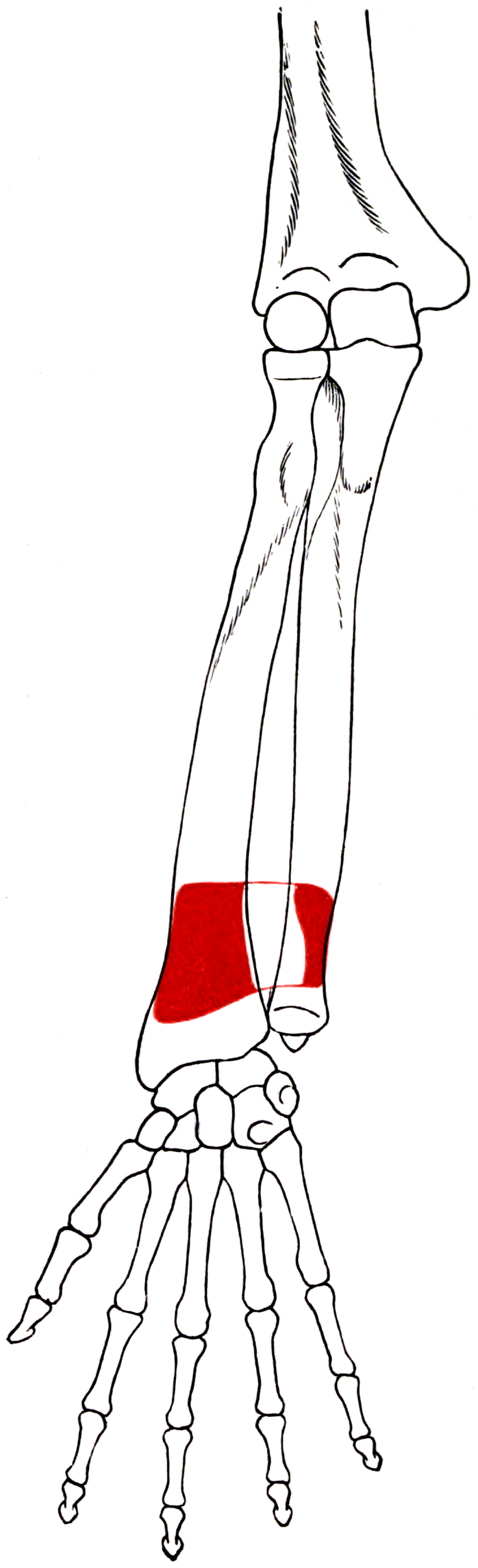
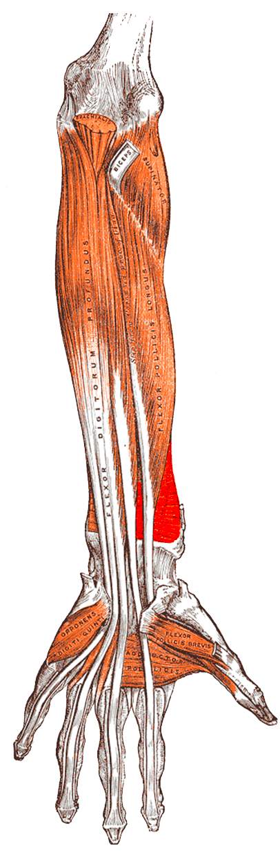
- A flat quadrilateral muscle running TRANSVERSELY across the distal quarter of the forearm, from the anterior surface of the distal ULNA to the anterior surface of the distal RADIUS. It is the deepest muscle of the volar compartment.
- Two heads: a SUPERFICIAL head that pronates, and a DEEP head whose oblique fibres COMPRESS the radius against the ulna and stabilise the distal radioulnar joint.
- Innervated by the ANTERIOR INTEROSSEOUS NERVE (C7, C8) — it is the most distal muscle the nerve supplies, and the nerve continues beyond it as a purely sensory terminal branch to the volar wrist capsule.
- It is elevated in the distal window of the volar Henry approach and in every volar plating of the distal radius; the plate is placed on the bone beneath it.
- Two independent meta-analyses found NO significant functional benefit of pronator quadratus repair after volar plating — no difference in DASH, grip strength, pronation strength or range of motion.
- The repair HOLDS but does not do what it is performed for: radiographic failure in 1 of 24 at three months and every repair intact on ultrasound at nine months, yet the plate was fully covered in only 55 per cent and flexor pollicis longus still touched it in 20 per cent - and in a cadaveric model an anatomic repair over a Soong grade 2 plate RAISED peak FPL-implant pressure by 29 per cent relative. Plate position, not muscle repair, is what protects the tendon.
- “Isolate pronator quadratus clinically by testing resisted pronation with the elbow FULLY FLEXED, which slackens pronator teres and removes its contribution.
- “Soong grading describes volar plate prominence relative to the volar rim: in the original two-group study the series using a plate prominent at the watershed line had a 4 per cent flexor tendon rupture rate, while the series with no grade 2 plates had none.
- “Pronator quadratus interposition after distal ulnar resection did NOT reduce radioulnar convergence in a cadaveric biomechanical study — the flap is not a mechanical solution to instability.
- “The anterior interosseous nerve terminal branch is resected, usually with the posterior interosseous nerve, in wrist denervation for painful arthritis.
Overview
Pronator quadratus is a flat, quadrilateral sheet of muscle lying transversely across the distal quarter of the forearm, deep to every flexor tendon and directly on the bone. Anatomically it is unglamorous. Surgically it matters for one overwhelming reason: it is the tissue a surgeon divides to place a volar distal radius plate, and the distal radius fracture is the commonest fracture treated operatively in the adult upper limb.
Four things follow from its position:
- It is the only soft tissue between a volar plate and the flexor tendons, which is why the question of whether to repair it has generated randomised trials and meta-analyses.
- Its deep head is a distal radioulnar joint stabiliser, compressing the radius against the ulna — so it is not purely a pronator.
- It is the most distal muscle supplied by the anterior interosseous nerve, making it the confirmatory muscle for an anterior interosseous nerve lesion on electromyography.
- It forms the distal window of the volar Henry approach.
This is the concept that makes pronator quadratus surgically important, and it is examined constantly.
The watershed line is the transverse ridge on the volar distal radius marking the most volar prominence of the bone, just proximal to the volar rim of the lunate facet. Distal to it, the flexor tendons — particularly flexor pollicis longus — lie in direct contact with the bone. Any implant placed distal to the watershed line therefore sits directly against the tendons.
Soong grading classifies plate prominence on the lateral radiograph. A line is drawn tangential to the most volar extent of the volar rim, parallel to the volar cortex of the radial shaft:
- Grade 0: the plate does not extend volar to this line.
- Grade 1: the plate lies volar to the line but proximal to the volar rim.
- Grade 2: the plate lies directly on or distal to the volar rim — the most prominent, and the highest risk.
The original data. In two parallel series, Group 1 (73 plated radii, one plate design) had 3 flexor tendon ruptures, a prevalence of 4 per cent; grade 2 prominence was present in 2 of the 3 ruptures and in 46 cases, 63 per cent, overall. Group 2 (95 plated radii, a different, lower-profile design) had no ruptures and no grade 2 plates.
The practical rules:
- Place the plate proximal to the watershed line wherever the fracture allows.
- A prominent plate is a plate that may need removing. Discuss elective removal with a patient who has a Soong grade 2 plate, particularly if there is volar wrist discomfort or crepitus.
- Repair the pronator quadratus over the plate if it will cover it — but do not claim it protects the tendon, because that has now been measured and it did not. The repair itself is durable: Swigart found only 1 of 24 (4 per cent) had failed radiographically at three months, and Hinds found every repair intact on dynamic ultrasound at a mean nine months. What the repair fails to do is the thing it is performed for. In Hinds's series the plate was completely covered in only 55 per cent of patients despite an intact repair, and flexor pollicis longus was still in contact with the plate in 20 per cent. Worse, Kilinc measured the interface directly with a pressure sensor in 16 cadaveric wrists carrying a deliberately Soong-grade-2 plate: anatomic repair raised mean peak FPL-implant pressure from 37.5 to 48.5 per cent (p equals 0.003) — a 29 per cent relative increase, greatest in extension. Their conclusion is explicit that interposition alone may not reliably mitigate flexor tendon loading. So repair it for the reasons that remain (a covered plate, a restored floor to the flexor compartment, no measured harm to function), and treat plate position as the intervention that actually protects the tendon.
- Flexor pollicis longus is the tendon that ruptures, because it lies most radially and most directly over the distal plate edge. Attritional rupture presents as a painless inability to flex the thumb interphalangeal joint, mimicking an anterior interosseous nerve palsy.
FFPThe Deep Volar Forearm — All Anterior Interosseous Nerve
Hook:These three, and nothing else. All are lost in an anterior interosseous nerve palsy, and there is NO sensory deficit.
Attachments, Innervation and Relations
Origin
- The anterior (volar) surface and anteromedial border of the distal quarter of the ULNA.
- The origin extends over roughly the distal 5 to 7 cm of the ulna, beginning about 3 to 4 cm proximal to the ulnar head.
- Some fibres arise from the overlying fascia and the medial intermuscular septum.
Insertion
- The anterior (volar) surface and anterolateral border of the distal quarter of the RADIUS, extending laterally to the anterior border of the radius.
- The distal limit of the muscle lies several millimetres to about 1 cm proximal to the watershed line — which is precisely why a plate placed under the muscle and proximal to the watershed line can be covered by it, whereas a distally placed plate cannot.
Two Heads
- Superficial head — the larger, with transverse fibres running directly from ulna to radius. Its action is pure pronation.
- Deep head — smaller, with oblique fibres running from the ulna distally and laterally to the radius. Its vector has a compressive component drawing the radius toward the ulna, and it is this head that acts as a dynamic stabiliser of the distal radioulnar joint.
- The two heads are separated by a thin fascial plane and by a small interposed fat pad — the pronator quadratus fat pad, which is visible on a lateral wrist radiograph and whose displacement or obliteration is a soft-tissue sign of an occult distal radius fracture.
Bony Landmarks to Quote
- Origin: distal quarter of the anterior ulna, extending over roughly 5 to 7 cm.
- Insertion: distal quarter of the anterior radius, with its distal edge proximal to the watershed line.
- The watershed line itself is the transverse ridge marking the most volar prominence of the distal radius, just proximal to the volar rim of the lunate facet.
Action and Biomechanics
Primary Action
Pronation of the forearm. Pronator quadratus initiates and sustains slow, unresisted pronation and is active throughout the pronation arc regardless of load. Pronator teres is recruited additionally for fast or resisted pronation.
Secondary Action: Distal Radioulnar Joint Stabilisation
- The deep head's oblique fibres produce a compressive vector drawing the radius toward the ulna, tightening the distal radioulnar joint.
- This is a dynamic stabilising function, complementing the static restraints — the triangular fibrocartilage complex (the primary stabiliser, principally the dorsal and volar radioulnar ligaments), the interosseous membrane (particularly the distal oblique bundle), and the extensor carpi ulnaris subsheath.
Pronator Quadratus versus Pronator Teres
- Pronator quadratus
- ANTERIOR INTEROSSEOUS nerve C7, C8
- Pronator teres
- Median nerve main trunk C6, C7
- Pronator quadratus
- Distal quarter, TRANSVERSE, deepest volar muscle
- Pronator teres
- Proximal, OBLIQUE, superficial
- Pronator quadratus
- INITIATES and sustains; active throughout, at all speeds and loads
- Pronator teres
- Recruited for FAST or RESISTED pronation
- Pronator quadratus
- Does NOT cross the elbow — unaffected by elbow position
- Pronator teres
- Crosses the elbow; weak flexor; contribution falls with the elbow extended
- Pronator quadratus
- DRUJ stabilisation via the deep head
- Pronator teres
- None
- Pronator quadratus
- Resisted pronation with the elbow FULLY FLEXED
- Pronator teres
- Resisted pronation with the elbow EXTENDED
- Pronator quadratus
- No — and it is not used as one
- Pronator teres
- YES — the standard donor for wrist extension in radial nerve palsy
The clinical corollary: to test pronator quadratus in isolation, flex the elbow fully. This slackens pronator teres, which crosses the elbow, and removes most of its contribution — leaving pronator quadratus as the muscle being tested. This is the standard bedside method for detecting an anterior interosseous nerve lesion.
What Happens When It Fails
- Isolated pronator quadratus loss (anterior interosseous nerve palsy, or division without repair at surgery) produces measurable but often subtle weakness of pronation, because pronator teres compensates for resisted pronation. Patients notice reduced endurance and reduced power in the fully flexed elbow position specifically.
- Distal radioulnar joint instability may be subtly worsened by loss of the deep head, though the triangular fibrocartilage complex remains the dominant restraint. This is the theoretical argument for repair, and it is one of the few arguments not comprehensively refuted by the meta-analysis data.
- Loss of both pronators (a high median nerve lesion) abolishes active pronation; the patient compensates with shoulder abduction and internal rotation.
Examiners will use pronator quadratus as a way into distal radioulnar joint stability. Structure the answer as static and dynamic restraints.
- Triangular fibrocartilage complex, and within it the dorsal and volar radioulnar ligaments — the true primary stabilisers. The deep fibres (ligamentum subcruentum) inserting at the fovea are the critical component; a foveal tear produces instability, whereas a superficial styloid-based tear may not.
- Bony congruity of the sigmoid notch — shallow or flat notch morphology predisposes to instability.
- Interosseous membrane, particularly the distal oblique bundle, a meaningful secondary restraint when present.
- PRONATOR QUADRATUS deep head — compresses the radius against the ulna.
- Extensor carpi ulnaris and its subsheath — a dynamic and static stabiliser; subsheath disruption produces extensor carpi ulnaris subluxation and contributes to instability.
- Extensor digiti minimi and the ulnocarpal ligaments contribute.
the piano key sign (dorsal ballottement of the ulnar head with the forearm pronated), the radioulnar ballottement or shuck test in neutral, pronation and supination, and comparison with the contralateral side, since asymptomatic laxity is common.
Sauerbier and colleagues showed that after distal ulnar resection, neither pronator quadratus interposition nor extensor and flexor carpi ulnaris tenodesis reduced radioulnar convergence on a dynamic cadaveric loading rig. Soft-tissue procedures do not restore the mechanics of a resected distal ulna — which is a large part of why the Darrach procedure has been superseded by the Sauvé-Kapandji and by distal ulnar replacement in younger patients.
Surface Anatomy and Examination
Palpation
- Pronator quadratus is the deepest muscle of the volar forearm and is not directly palpable. It lies beneath every flexor tendon, and any bulk felt in the distal volar forearm is tendon and fascia, not muscle.
- Its position is defined by bony landmarks: it occupies the distal quarter of the interval between the radius and ulna on the volar side, with its distal edge roughly 2 to 3 cm proximal to the distal wrist crease.
- Its fat pad is a radiographic rather than a clinical landmark.
Clinical Tests and What They Mean
- How to perform
- Elbow flexed as far as possible to slacken pronator teres; forearm supinated; resist pronation
- Positive finding
- Weakness compared with the other side
- What it means
- Pronator quadratus deficit — an anterior interosseous nerve lesion
- False positives
- Pain inhibition after trauma; incomplete elbow flexion leaves pronator teres contributing
- How to perform
- Ask the patient to make a circle with the thumb and index finger
- Positive finding
- A flat, pinched, triangular pinch rather than a round O
- What it means
- AIN palsy — FPL and index FDP cannot flex the terminal joints
- False positives
- Attritional tendon rupture produces an identical sign — use the tenodesis test
- How to perform
- Passively EXTEND the wrist and observe the thumb and index terminal joints
- Positive finding
- Terminal joints flex passively
- What it means
- The tendons are INTACT, so the problem is neurological (AIN palsy)
- False positives
- A stiff or fused interphalangeal joint prevents the response regardless of tendon status
- How to perform
- Test the whole median territory including the thenar SKIN
- Positive finding
- NORMAL sensation throughout
- What it means
- Confirms AIN rather than a proximal median lesion
- False positives
- A concomitant carpal tunnel syndrome confuses the picture
- How to perform
- Forearm pronated, wrist supported; press the ulnar head dorsally and release
- Positive finding
- Ulnar head depresses and springs back; painful
- What it means
- Distal radioulnar joint instability
- False positives
- Asymptomatic bilateral laxity is common — always compare sides
- How to perform
- Stabilise the radius; translate the ulnar head volarly and dorsally in neutral, pronation and supination
- Positive finding
- Increased translation with a soft end-point compared with the other side
- What it means
- TFCC or DRUJ ligamentous insufficiency
- False positives
- Generalised ligamentous laxity
Imaging
- Pronator quadratus fat pad sign: a thin lucent stripe volar to the distal radius on the lateral wrist radiograph, representing the fat between the muscle and the overlying flexor tendons. Displacement, bowing or obliteration of this stripe is a soft-tissue sign of an occult distal radius fracture and should prompt a careful look at the bone or repeat imaging.
- Soong grading is performed on the lateral radiograph with a line tangential to the most volar extent of the volar rim, parallel to the volar cortex of the shaft.
- Ultrasound is excellent for assessing flexor pollicis longus continuity and for detecting tenosynovitis over a prominent plate — the warning sign before a rupture.
- CT is used to assess distal radioulnar joint congruity and sigmoid notch morphology in suspected instability.
The Critical Differential to State Explicitly
A patient who cannot flex the thumb interphalangeal joint after a distal radius volar plate has either:
- an anterior interosseous nerve palsy (tenodesis test positive — the joint flexes on passive wrist extension), or
- an attritional flexor pollicis longus rupture over the plate (tenodesis test negative).
The distinction determines everything: the first is observed, the second requires plate removal and tendon reconstruction.
Complications
Donor and Division Morbidity
- Dividing pronator quadratus without repair produces measurable weakness of pronation with the elbow flexed, but the functional deficit is small because pronator teres compensates. Two meta-analyses found no significant difference in pronation strength between repaired and unrepaired groups.
- The theoretical loss of distal radioulnar joint stability from the deep head is the one argument that survives the outcome data, though it has not been demonstrated clinically. Preserving as much of the deep head as the exposure allows is a reasonable habit.
Adhesions and Stiffness
- The distal forearm and wrist are unforgiving of adhesions. Early active finger, thumb and forearm rotation under hand therapy supervision is the single most important preventive measure after any volar approach.
- Flexor tendon adhesions over an unrepaired pronator quadratus and a bare plate are a recognised cause of poor finger motion after distal radius fixation, and are one of the mechanistic arguments for repair.
Complex Regional Pain Syndrome
- The distal radius fracture is the classic orthopaedic trigger for complex regional pain syndrome. Early motion, avoidance of tight dressings and casts, and adequate analgesia are the practical preventive measures.
Clinical Relevance
The Question
After volar plating of a distal radius fracture, the pronator quadratus has been elevated to place the plate. Should it be repaired over the implant?
- It interposes soft tissue between the plate and the flexor tendons, which should reduce attritional tendon rupture.
- It may restore pronation strength.
- It may preserve distal radioulnar joint stability through the deep head.
- It may reduce adhesions and improve the gliding surface for the flexors.
- Repairing a thin, often macerated muscle over a plate is technically difficult, and the repair frequently fails or dehisces.
- It adds tourniquet time.
- The muscle may be under tension over a prominent plate, and a tight repair can itself restrict rotation.
- The plate, not the muscle, is the variable that determines rupture risk.
The Evidence: Two Independent Meta-Analyses, Same Conclusion
- Included studies
- 6 studies (4 randomised controlled trials, 2 retrospective)
- Patients
- 203 with repair versus 180 without
- Primary outcome
- DASH score
- Result
- NO significant difference in DASH (SMD 0.43, 95 per cent CI minus 0.12 to 0.98, p equals 0.12), grip strength or pronation strength; no benefit in range of motion
- Included studies
- 11 studies (5 randomised controlled trials, 6 retrospective case-control)
- Patients
- Pooled by outcome rather than as a single total
- Primary outcome
- DASH and pronation strength
- Result
- NO significant difference in primary or secondary outcomes at a minimum of 6 months
The one signal worth knowing. Lu and colleagues performed a subgroup analysis by AO fracture type and found a divergence:
- AO type B fractures: pronation strength favoured repair (SMD minus 0.94, 95 per cent CI minus 1.54 to minus 0.34).
- Non-type B fractures: pronation strength favoured NO repair (SMD 0.39, 95 per cent CI 0.07 to 0.70).
This is hypothesis-generating rather than practice-changing, and the authors said so explicitly.
The Honest Position for a Viva
- There is no demonstrated functional benefit to pronator quadratus repair in DASH, grip strength, pronation strength, pain or range of motion.
- Repair remains reasonable on the mechanical rationale of interposing tissue between plate and tendon, provided it does not require tension and does not compromise the fixation or add meaningful time.
- The intervention that actually reduces flexor tendon rupture is plate position, not muscle repair. Keep the plate proximal to the watershed line.
- Say all three. The examiner is testing whether you can hold an evidence-based position while retaining surgical judgement.
Surgical Relevance
The Modified Henry (FCR-Based) Approach to the Distal Radius
The standard approach for volar plating, and the one to be able to describe step by step.
- Incision: longitudinal, 6 to 8 cm, over the flexor carpi radialis tendon, extending proximally from the wrist crease. Do not cross the crease at a right angle.
- Open the flexor carpi radialis sheath and retract the tendon ulnarly. This protects the palmar cutaneous branch of the median nerve, which lies just ulnar to the flexor carpi radialis tendon — retracting the tendon ulnarly takes the branch out of harm's way with it.
- Open the floor of the flexor carpi radialis sheath; the flexor pollicis longus is encountered and retracted ulnarly.
- The radial artery lies radial to the flexor carpi radialis and is protected by staying ulnar to it and by retracting radially only on muscle, not on the vessel.
- Pronator quadratus is now exposed. It is incised along its radial border and distal border in an L shape, and elevated from radial to ulnar as a flap hinged on the ulnar side. This preserves the neurovascular supply, which enters from the deep and ulnar side.
- Place the plate on the bone beneath the muscle, PROXIMAL to the watershed line.
- Repair the pronator quadratus over the plate where the tissue allows, without tension.
Structures at Risk with Distances
- Location
- Just ULNAR to the flexor carpi radialis tendon
- Distance from a landmark
- Arises 5 to 8 cm proximal to the wrist crease; runs superficial to the flexor retinaculum
- Protection
- Retract the flexor carpi radialis tendon ULNARLY, taking the branch with it; open the FCR sheath rather than dissecting around it
- Location
- Directly over the distal plate edge, the most radial flexor tendon
- Distance from a landmark
- Lies on the pronator quadratus; in direct contact with bone distal to the watershed line
- Protection
- Plate proximal to the watershed line; repair pronator quadratus over the plate
- Location
- Radial to the flexor carpi radialis
- Distance from a landmark
- In the distal forearm, immediately medial to the brachioradialis tendon
- Protection
- Stay ulnar to it; retract muscle, not vessel
- Location
- Between flexor digitorum superficialis and flexor digitorum profundus, becoming superficial at the wrist
- Distance from a landmark
- Between the FDS and FCR tendons at the wrist
- Protection
- It is retracted ulnarly with the flexor mass; avoid forceful sustained retraction
- Location
- Deep surface of pronator quadratus, entering from the ulnar side
- Distance from a landmark
- On the interosseous membrane
- Protection
- Elevate the muscle from RADIAL to ULNAR, hinged ulnarly
The Watershed Line Rule, Restated
The plate goes proximal to the watershed line. If the fracture demands more distal support — a volar rim or lunate facet fragment — use a dedicated volar rim plate or a fragment-specific implant designed for that position, accept the higher tendon risk, and plan for early elective removal.
Guidelines, Registries & Global Practice
Anatomical Variation
- An accessory head of pronator quadratus, or a slip extending distally toward the carpus, is described in a minority of limbs and has been implicated in compression of the median or ulnar nerve at the distal forearm and in unexplained volar wrist pain.
- The distal oblique bundle of the interosseous membrane, a discrete thickening functioning as a secondary distal radioulnar joint restraint, is present in only a proportion of people. Its presence or absence is a genuine anatomical variable in the stability equation to which pronator quadratus contributes.
- The extent of the deep head varies, and with it the muscle's contribution to distal radioulnar joint compression.
- Sigmoid notch morphology varies from flat to ski-slope to C-type, and flatter notches are associated with greater instability — an anatomical factor that modulates how much the dynamic stabilisers matter.
Side-by-Side Guidance
- Position relevant to pronator quadratus and distal radius plating
- Supports operative fixation for displaced fractures failing acceptable reduction, without prescribing implant type or soft-tissue technique; notes the limited evidence base for many technical variables.
- Position relevant to pronator quadratus and distal radius plating
- Emphasise timely definitive management, appropriate imaging, early mobilisation and structured follow-up rather than prescribing pronator quadratus handling.
- Position relevant to pronator quadratus and distal radius plating
- Describes the modified Henry approach with L-shaped release and radially based elevation of pronator quadratus, and instructs that the plate be positioned proximal to the watershed line.
- Position relevant to pronator quadratus and distal radius plating
- No functional benefit from pronator quadratus repair; both conclude that further large randomised trials would be needed to demonstrate any difference.
Registry and Outcome Signals
- Distal radius fractures are not captured in national joint registries, and there is no dedicated international distal radius implant registry. The evidence base is therefore randomised trials and cohort studies, which is why the two meta-analyses carry disproportionate weight in this debate.
- Implant removal rates after volar plating vary widely between health systems and between individual surgeons, and are strongly influenced by local thresholds for elective removal rather than by any objective indication. A patient with a Soong grade 2 plate is a patient in whom that threshold should be lower.
- Large national datasets consistently show rising rates of operative fixation of distal radius fractures in older patients, despite trial evidence that many elderly patients do as well non-operatively. This makes the tendon-rupture question more, not less, relevant over time.
High- versus Limited-Resource Practice
- Well-resourced settings: locking plates of multiple designs and profiles, fragment-specific implants for volar rim fragments, routine CT for intra-articular patterns, and a low threshold for elective plate removal.
- Limited-resource settings: a smaller range of implants means the surgeon may not have a low-profile plate available for a given fracture. In that situation the mitigations become more important, not less: place the available plate as proximal as the fracture allows, repair the pronator quadratus over it, counsel the patient explicitly about the symptoms of impending tendon rupture, and arrange follow-up. Non-operative management with a well-moulded cast remains entirely appropriate for many distal radius fractures, particularly in older lower-demand patients, and avoids the problem altogether.
- Ultrasound is widely available and is an excellent substitute for MRI in assessing flexor pollicis longus continuity and detecting tenosynovitis over a prominent plate.
Rehabilitation Consensus
- Early active finger, thumb and forearm motion after volar plating is universally recommended and is the single most important measure against adhesions and stiffness.
- There is no evidence that repairing pronator quadratus alters the rehabilitation protocol, and it should not be used as a reason to delay mobilisation.
- Patients with a prominent plate should be specifically instructed to report volar wrist pain, crepitus or triggering, which are the warning signs that precede attritional rupture.
MCQ Practice Points
Q: Where does pronator quadratus arise and insert? A: From the anterior surface of the distal quarter of the ULNA to the anterior surface of the distal quarter of the RADIUS, running transversely. It is the deepest muscle of the volar compartment.
Q: What do the two heads of pronator quadratus do? A: The superficial head PRONATES. The deep head's oblique fibres COMPRESS the radius against the ulna, stabilising the distal radioulnar joint.
Q: What innervates pronator quadratus and why is that significant? A: The ANTERIOR INTEROSSEOUS NERVE (C7, C8). It is the most distal muscle the nerve supplies, making it the confirmatory site for electromyography in an anterior interosseous nerve lesion.
Q: How do you test pronator quadratus in isolation? A: Resisted pronation with the elbow FULLY FLEXED, which slackens pronator teres and removes most of its contribution.
Q: Does repairing pronator quadratus after volar plating improve outcomes? A: NO. Two independent 2020 meta-analyses found no significant difference in DASH, grip strength, pronation strength or range of motion.
Q: What is the watershed line and why does it matter? A: The transverse ridge marking the most volar prominence of the distal radius. Distal to it the flexor tendons lie directly on bone, so a plate placed distal to it abrades flexor pollicis longus.
Q: What is a Soong grade 2 plate? A: A plate lying directly ON or DISTAL to the volar rim — the most prominent grade. Grade 2 prominence was present in 63 per cent of the series with a 4 per cent flexor rupture rate, and in none of the series with zero ruptures.
Q: Which flexor tendon ruptures over a volar distal radius plate, and how do you distinguish it from a nerve palsy? A: Flexor pollicis longus. Distinguish from an anterior interosseous nerve palsy with the TENODESIS TEST — passive wrist extension flexes the interphalangeal joint if the tendon is intact.
Q: In which direction do you elevate pronator quadratus, and why? A: RADIAL to ULNAR, hinged ulnarly, because the anterior interosseous nerve and artery enter the deep surface from the ulnar side.
Q: How do you protect the palmar cutaneous branch in the volar approach? A: Open the flexor carpi radialis sheath and retract the tendon ULNARLY. The branch lies just ulnar to the tendon and travels with it.
Q: What is the pronator quadratus fat pad sign? A: Displacement, bowing or obliteration of the lucent fat stripe volar to the distal radius on the lateral wrist radiograph — a soft-tissue sign of an occult distal radius fracture.
Q: Does pronator quadratus interposition prevent radioulnar convergence after distal ulnar resection? A: NO. In a dynamic cadaveric study, neither pronator quadratus interposition nor extensor and flexor carpi ulnaris tenodesis reduced convergence.
Exam Viva Scenarios
Practise clinical reasoning and management decisions out loud
“You have just applied a volar locking plate to a distal radius fracture. Your registrar asks whether you are going to repair the pronator quadratus, and what the evidence says. What do you tell them?”
“Fourteen months after volar plating of a distal radius fracture, a 58-year-old woman cannot bend the tip of her thumb. It is not painful. What are the two possibilities and how do you distinguish them?”
“Describe the modified Henry approach to the distal radius for volar plating, and tell me exactly what you do with the pronator quadratus and what you are protecting at each step.”
Anatomy
- Transverse, quadrilateral, DEEPEST muscle of the volar forearm
- Distal quarter of the anterior ULNA to the distal quarter of the anterior RADIUS
- Superficial head pronates; DEEP head compresses the radius against the ulna (DRUJ stabiliser)
- Distal edge lies PROXIMAL to the watershed line
Innervation
- ANTERIOR INTEROSSEOUS NERVE C7, C8 - the MOST DISTAL muscle it supplies
- Nerve and artery enter the DEEP surface from the ULNAR side
- AIN continues beyond it as a purely SENSORY branch to the volar wrist capsule
- Isolate clinically: resisted pronation with the elbow FULLY FLEXED
Volar Plating
- PQ is the ONLY tissue between the plate and the flexor tendons
- L-shaped release; elevate RADIAL to ULNAR, hinged ulnarly
- Plate PROXIMAL to the watershed line
- Open the FCR sheath and retract ULNARLY to protect the palmar cutaneous branch
The Repair Debate
- Shi and Ren 2020: 6 studies, 203 vs 180 - NO difference in DASH, grip or pronation strength
- Lu 2020: 11 studies - no difference; AO type B subgroup favoured repair (hypothesis-generating)
- Repair on mechanical grounds only, and only if TENSION-FREE
- Plate POSITION, not muscle repair, is what determines tendon rupture risk
Tendon Rupture
- FLEXOR POLLICIS LONGUS is the tendon at risk - most radial, over the distal plate edge
- Soong grade 2 = plate on or distal to the volar rim; 4 per cent rupture rate versus zero
- TENODESIS TEST distinguishes rupture from AIN palsy
- Reconstruct with ring FDS to FPL; consider elective plate removal
Evidence Base
Is Pronator Quadratus Repair Necessary to Improve Outcomes After Volar Plate Fixation of Distal Radius Fractures? A Systematic Review and Meta-Analysis
- Six studies met inclusion criteria: four randomised controlled trials and two retrospective studies
- Analysis of 203 patients with pronator quadratus repair versus 180 without
- NO statistically significant difference in DASH scores (SMD 0.43, 95 per cent CI minus 0.12 to 0.98, I squared 85 per cent, p equals 0.12)
- NO difference in grip strength (SMD minus 0.10, 95 per cent CI minus 0.53 to 0.33) or pronation strength (SMD minus 0.02, 95 per cent CI minus 0.82 to 0.78)
- The pooled analysis showed no benefit of repair in improving postoperative range of motion
A Systematic Review and Meta-Analysis of the Pronator Quadratus Repair Following Volar Plating of Distal Radius Fractures
- Eleven studies included: five randomised controlled trials and six retrospective case-control studies
- No significant difference in primary outcomes (DASH and pronation strength) or secondary outcomes (pain, wrist mobility, grip strength) at a minimum of 6 months
- Subgroup analysis by AO fracture type showed pronation strength FAVOURED REPAIR in AO type B fractures (SMD minus 0.94, 95 per cent CI minus 1.54 to minus 0.34)
- In non-type B fractures the subgroup analysis FAVOURED NO REPAIR (SMD 0.39, 95 per cent CI 0.07 to 0.70)
- Registered with PROSPERO; the authors concluded future studies are needed to confirm the relationship between repair and pronation strength across fracture patterns
Volar Locking Plate Implant Prominence and Flexor Tendon Rupture
- Two parallel retrospective series of volar locked plating with at least 6 months of follow-up: 73 radii with one plate design (Group 1) and 95 with a different design (Group 2)
- Prominence graded on the lateral radiograph against a line tangential to the most volar extent of the volar rim: grade 0 not volar to the line, grade 1 volar to the line but proximal to the rim, grade 2 directly on or distal to the rim
- Group 1: three flexor tendon ruptures, a prevalence of 4 per cent; grade 2 prominence in two of the three ruptures and in 46 cases (63 per cent) overall
- Group 2: no flexor tendon ruptures and no grade 2 plates
- The authors concluded that regardless of plate selection, surgeons should avoid implant prominence at the watershed line
Radioulnar Convergence After Distal Ulnar Resection: Mechanical Performance of Two Commonly Used Soft Tissue Stabilizing Procedures
- Seven fresh-frozen cadaveric upper extremities tested on a dynamic computer-controlled device generating forearm rotation with physiological muscle loading
- Four conditions compared: intact, distal ulna resection alone, resection with PRONATOR QUADRATUS INTERPOSITION, and resection with extensor and flexor carpi ulnaris tenodesis
- Distal ulna resection altered kinematics, most predictably creating convergence of the radius toward the ulna, with detectable anteroposterior translations
- Neither pronator quadratus interposition NOR extensor and flexor carpi ulnaris tenodesis reduced the radioulnar convergence created by the resection
- The study provides a biomechanical assessment of two commonly used soft-tissue procedures whose clinical efficacy had been reported but not mechanically tested
The Pronator Teres Syndrome: Compressive Neuropathy of the Median Nerve
- Thirty-nine patients with proximal median nerve compression seen over seven years, with 36 forearms explored in 32 patients
- Typical complaints were aching forearm discomfort, hand weakness and numbness in the thumb and index finger, brought on by cyclic stress
- Electrophysiological testing showed abnormalities in only a few patients and localisation was rarely possible
- Intramuscular tendinous bands in the pronator and indentation of the flexor superficialis muscle belly were found in most explored forearms
- Twenty-eight of 36 operations gave good or excellent results; the causes of failure were inadequate decompression or misdiagnosis
Assessment of Pronator Quadratus Repair Integrity Following Volar Plate Fixation: A Prospective Clinical Cohort Study
- 24 patients, prospective; the pronator quadratus was repaired along its radial and distal borders after volar plate fixation
- RADIOPAQUE HEMOCLIPS were attached on each side of the repair - two radially, two distally - and the distance between them measured at time zero, two weeks, six weeks and three months
- Displacement of 1 cm or more from time zero defined failure
- ONE of 24 repairs (4 per cent) had failed at three months - the repairs withstand the forces of the healing period
- No correlation between the graded severity of pronator quadratus injury and radiographic failure of the repair, and no difference in wrist motion, forearm rotation or grip strength by injury type
Assessment of Pronator Quadratus Repair Integrity Using Dynamic Ultrasonography Following Volar Plate Fixation
- 20 adults who had volar plate fixation with pronator quadratus repair, assessed by BILATERAL DYNAMIC wrist ultrasound at a mean 9 plus or minus 4 months
- ALL 20 repairs were intact - the paper was designed to test the prevailing belief that these repairs often fail, and found the opposite
- The volar plate was COMPLETELY COVERED by the muscle in only 55 PER CENT of patients, and complete coverage was associated with a larger pronator quadratus (p equals 0.026)
- FLEXOR POLLICIS LONGUS WAS IN CONTACT WITH THE PLATE IN 20 PER CENT, with a trend towards greater wrist flexion in those patients (p equals 0.053)
- No difference in range of motion, strength or outcome scores between completely and incompletely covered plates
Anatomic Pronator Quadratus Repair Increases Flexor Pollicis Longus Tendon Pressure After Volar Rim Plating: A Cadaveric Study
- 16 wrists from eight fresh-frozen upper extremities, each plated through a modified volar Henry approach with a 2.4 mm variable-angle volar rim plate placed in a STANDARDISED SOONG GRADE 2 position
- A 0.15 mm piezoresistive sensor was placed between flexor pollicis longus and the distal plate edge to measure interface pressure directly during four active motion cycles
- Anatomic pronator quadratus repair SIGNIFICANTLY INCREASED peak pressure at every wrist position tested
- Mean overall pressure rose from 37.5 to 48.5 per cent (p equals 0.003) - an 11 per cent absolute and 29.3 per cent relative increase
- The rise was greatest in DORSIFLEXION (plus 17.2 per cent, p less than 0.001), then neutral (plus 11.0 per cent, p equals 0.005), then flexion (plus 4.8 per cent, p equals 0.013)
- The authors state that CONTRARY TO PRIOR ASSUMPTIONS, interposition alone may not reliably mitigate flexor tendon loading