The Gateway of the Thoracic Outlet
- The interscalene triangle is bounded by anterior scalene in front, middle scalene behind and the first rib below; it transmits the brachial plexus roots and trunks and the SUBCLAVIAN ARTERY.
- The SUBCLAVIAN VEIN passes ANTERIOR to the anterior scalene, in the costoclavicular space - it does NOT enter the interscalene triangle. This is the single most frequently examined point in the region.
- The phrenic nerve (C3, C4, C5) descends on the ANTERIOR surface of the anterior scalene, running from lateral to medial, deep to the prevertebral fascia.
- The dorsal scapular nerve and the C5-C6 contributions to the long thoracic nerve pierce the MIDDLE scalene; the C7 contribution to the long thoracic nerve passes anterior to it.
- Interscalene block causes ipsilateral hemidiaphragmatic paresis in the great majority of patients with conventional volumes - a relative contraindication in significant respiratory disease.
- “Cervical ribs were present in 2.0 per cent of 3404 consecutive cervical spine CT scans - higher than the figure classically quoted from plain films - were bilateral in 40 per cent, twice as common in women, and are symptomatic in only about 10 per cent. Most thoracic outlet syndrome occurs without a cervical rib.
- “Gilliatt-Sumner hand is the wasting pattern of true neurogenic thoracic outlet syndrome: thenar wasting exceeding hypothenar wasting, from a lower trunk or T1 lesion.
- “Ultrasound work shows the phrenic nerve lies only millimetres from the C5 root at the cricoid, diverging roughly 3 mm for every centimetre caudally - which is why the block cannot reliably spare the diaphragm at the classical level.
- “In the supraclavicular approach, identify the phrenic nerve on the anterior scalene BEFORE dividing the muscle, and remember the thoracic duct arches over the subclavian artery on the LEFT.
Overview
The scalene muscles are three (often four) paired muscles that run from the cervical transverse processes to the first and second ribs, forming the lateral wall of the root of the neck. Anatomically they are lateral vertebral muscles; functionally they are accessory muscles of respiration and lateral flexors of the neck. Surgically, they matter almost entirely because of what passes between them.
The interscalene triangle - anterior scalene in front, middle scalene behind, first rib below - is the first and narrowest of the three compartments through which the neurovascular supply of the upper limb must pass. It is the commonest site of thoracic outlet syndrome, the target of the interscalene brachial plexus block, the field of the supraclavicular approach to the plexus, and the place where three nerves that matter to the shoulder surgeon - the phrenic, the dorsal scapular and the long thoracic - are found in fixed and examinable relationships.
The neurovascular bundle to the arm passes through three sequential narrow spaces, and thoracic outlet syndrome can arise at any of them. Naming all three, in order, is the expected answer:
1. Interscalene triangle (the commonest site)
- Anterior: anterior scalene
- Posterior: middle scalene
- Inferior: first rib
- Contents: the roots and trunks of the brachial plexus and the subclavian artery
- NOT the subclavian vein
2. Costoclavicular space
- Anterosuperior: clavicle and subclavius
- Posteromedial: first rib
- Posterolateral: upper border of the scapula
- Contents: the subclavian artery, the subclavian VEIN and the plexus divisions
- This is where the vein travels - having passed anterior to the anterior scalene - and it is why venous thoracic outlet syndrome (Paget-Schroetter effort thrombosis) is a costoclavicular problem, not an interscalene one.
3. Retropectoralis minor (subcoracoid) space
- Anterior: pectoralis minor
- Posterior: ribs 2 to 4
- Contents: the axillary artery and vein and the cords of the plexus
- Compression here is provoked by hyperabduction (the Wright manoeuvre).
The mechanical reason the interscalene triangle dominates the clinical picture is that it is the only one of the three that narrows with normal physiological movement: the scalenes contract with every inspiration and with every neck movement, and the triangle is at its narrowest with the neck extended and rotated. Everything else in the region - cervical rib, fibrous band, scalene hypertrophy, post-traumatic fibrosis - simply reduces a space that is already the tightest of the three.
VANWhat Passes Where at the Anterior Scalene
Hook:Vein in front, artery and nerves behind, phrenic on the face of the muscle.
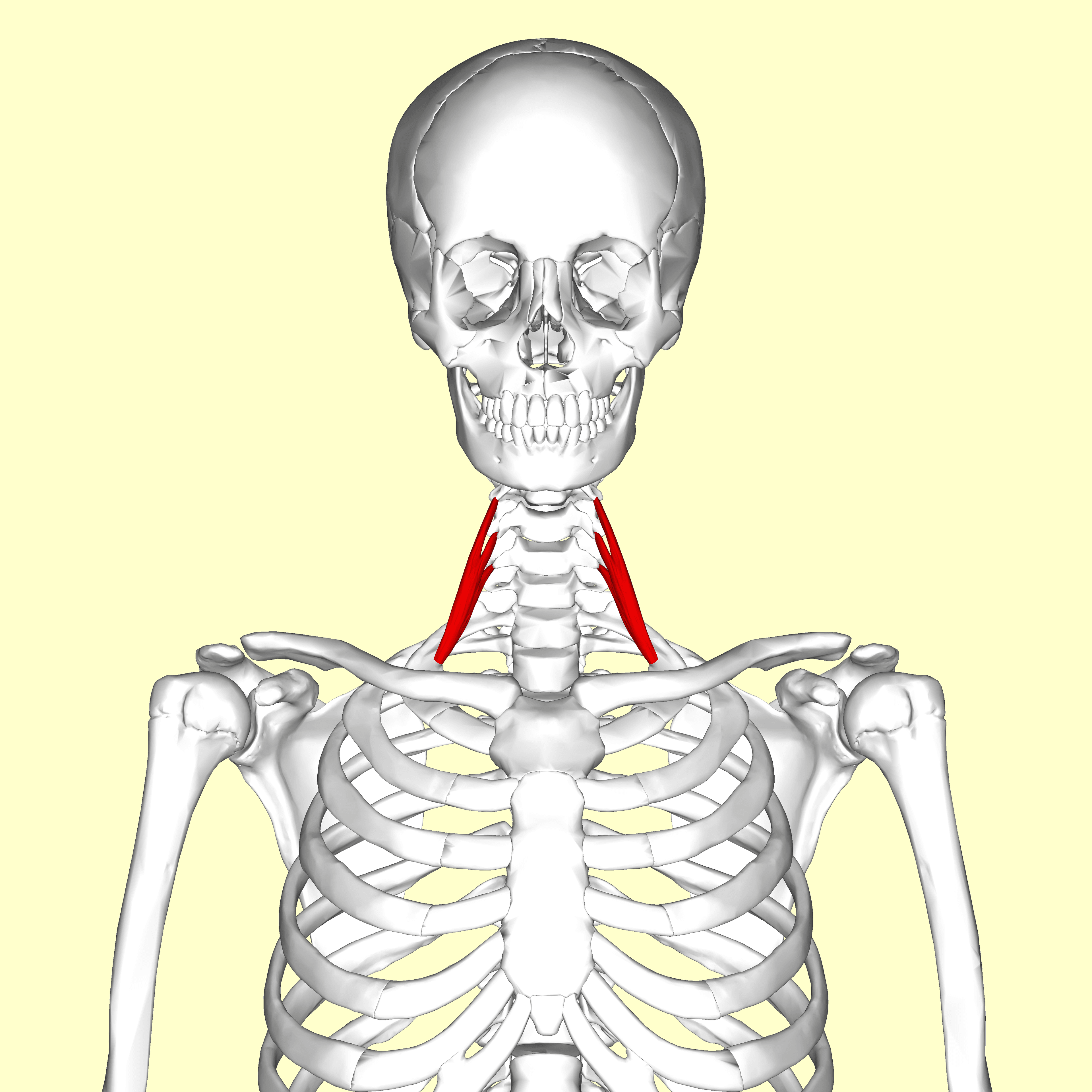
Attachments, Innervation and Relations
Anterior scalene (scalenus anterior)
- Origin: the anterior tubercles of the transverse processes of C3 to C6.
- Insertion: the scalene tubercle of Lisfranc on the inner border of the upper surface of the first rib, and a short ridge running forward from it.
- The tubercle separates two shallow grooves on the first rib: the groove for the subclavian vein anteriorly and the groove for the subclavian artery posteriorly. That single bony landmark encapsulates the whole relationship.
Middle scalene (scalenus medius)
- Origin: the posterior tubercles of the transverse processes of C2 to C7 (frequently described as C1 to C7).
- Insertion: the upper surface of the first rib, behind the groove for the subclavian artery, extending back to the tubercle for serratus anterior.
- It is the largest of the three and is pierced by the dorsal scapular nerve and by the C5 and C6 contributions to the long thoracic nerve.
Posterior scalene (scalenus posterior)
- Origin: the posterior tubercles of the transverse processes of C4 to C6.
- Insertion: the outer surface of the second rib.
- Often blended with middle scalene; occasionally absent.
Scalenus minimus (scalenus pleuralis, Sibson's muscle)
- A variable fourth muscle, present in a substantial minority of people, arising from the transverse process of C6 or C7 and inserting into the inner border of the first rib and into the suprapleural membrane (Sibson's fascia).
- Crucially, it passes between the subclavian artery and the T1 root, splitting the triangle. Where present, it is a recognised anatomical cause of neurogenic thoracic outlet syndrome and must be looked for and excised at scalenectomy.
Inside the interscalene triangle.
- Passes behind the anterior scalene, between it and the middle scalene, with the plexus.
- Grooves the first rib posterior to the scalene tubercle of Lisfranc.
- Compressed in arterial thoracic outlet syndrome, classically by a cervical rib, producing post-stenotic dilatation, aneurysm and distal embolisation.
Outside the triangle, anterior to the muscle.
- Passes in front of the anterior scalene, in the costoclavicular space.
- Grooves the first rib anterior to the scalene tubercle.
- Compressed in venous thoracic outlet syndrome (Paget-Schroetter effort thrombosis) between the clavicle, the subclavius and the first rib - not by the scalenes.
- Practical corollary: a scalenectomy alone does not decompress the vein. Venous thoracic outlet syndrome needs the costoclavicular space opened, usually by first rib resection.
Action and Biomechanics
Actions
- Action
- Ipsilateral lateral flexion of the cervical spine
- Muscle
- All three
- Clinical Relevance
- Restricted contralateral side-bending in scalene shortening
- Action
- Contralateral rotation (anterior scalene)
- Muscle
- Anterior scalene
- Clinical Relevance
- Contributes to the forward-head, rotated posture of chronic thoracic outlet syndrome
- Action
- Elevation of the first and second ribs
- Muscle
- Anterior and middle (rib 1), posterior (rib 2)
- Clinical Relevance
- Active even in quiet inspiration - the scalenes are NOT purely accessory
- Action
- Marked elevation of the upper ribs
- Muscle
- All three
- Clinical Relevance
- Hypertrophy in chronic obstructive airways disease narrows the triangle
- Action
- Cervical spine flexion and stabilisation
- Muscle
- Anterior scalene predominantly
- Clinical Relevance
- Stabilising role during upper limb loading
Why the scalenes narrow their own triangle
- The triangle is narrowest with the neck extended and rotated toward the affected side, and with the arm elevated and the shoulder braced back. Every provocative test for thoracic outlet syndrome exploits one of these positions.
- The scalenes contract with every breath. In a patient with chronic respiratory disease, a habitual upper-chest breathing pattern, or a forward-head posture, the muscles hypertrophy and shorten, and the triangle progressively narrows. This is the mechanism behind non-bony neurogenic thoracic outlet syndrome and the reason postural rehabilitation and breathing retraining are genuinely first-line treatment, not a delaying tactic.
- Post-traumatic fibrosis after a whiplash injury converts a mobile muscle into a fixed fibrous constriction; a substantial proportion of neurogenic thoracic outlet syndrome patients date their symptoms to a neck injury.
The plexus takes the strain
Because the lower trunk (C8, T1) is the most inferior structure and lies directly on the first rib, it is the part of the plexus that is compressed first and hardest - whether by a cervical rib, a fibrous band, or the scalene itself. That single mechanical fact explains the ulnar-side symptoms and the T1-dominant wasting pattern of true neurogenic thoracic outlet syndrome.
Surface Anatomy and Examination
Surface landmarks
- The anterior scalene lies deep to the clavicular head of sternocleidomastoid, immediately above the clavicle. Ask the patient to sniff sharply: the muscle tenses under the finger.
- The interscalene groove - the palpable depression between the anterior and middle scalene - is found by rolling the finger laterally off the posterior border of the clavicular head of sternocleidomastoid at the level of the cricoid cartilage (C6). This is the classical landmark for the interscalene block.
- The subclavian artery pulsation is palpable in the supraclavicular fossa, immediately lateral to the anterior scalene insertion, behind the middle third of the clavicle.
- A cervical rib may be palpable as a hard, fixed, tender fullness in the supraclavicular fossa; a bruit over it suggests arterial involvement.
Provocative tests for thoracic outlet syndrome
The most useful of a poor set.
- Arms abducted to 90 degrees and externally rotated ("stick-up" position), elbows at 90 degrees; the patient opens and closes the fists slowly for three minutes.
- Positive: reproduction of the patient's arm pain, heaviness, paraesthesia or blanching, forcing the patient to lower the arm before three minutes.
- Means: the elevated arm stress test is the most sensitive provocative manoeuvre, but it is positive in a considerable proportion of normal people - it supports, never establishes, the diagnosis.
Classic, and largely of historical value.
- Patient inspires deeply and holds, extends the neck and rotates the head toward the tested side while the examiner palpates the radial pulse.
- Positive: obliteration or marked diminution of the radial pulse, ideally with reproduction of symptoms.
- Means: very little on its own. The test has been shown to be of no clinical value - it is normal in most patients with neurogenic thoracic outlet syndrome and positive in many control volunteers. Only symptom reproduction counts.
Tests the retropectoralis minor space.
- The arm is passively hyperabducted and externally rotated with the head turned away; the radial pulse is monitored.
- Positive: pulse loss with symptom reproduction.
- Means: compression beneath pectoralis minor rather than at the scalenes - relevant because the surgery differs.
Tests the costoclavicular space.
- Shoulders drawn downward and backward, chest thrust forward; the radial pulse is monitored.
- Positive: pulse loss with symptom reproduction.
- Means: narrowing between the clavicle and the first rib - the space relevant to venous thoracic outlet syndrome and to clavicular malunion.
The examination that actually makes the diagnosis
- Tenderness over the anterior scalene with reproduction of the arm symptoms on firm pressure (the "scalene sign") is more useful than any pulse test.
- Neurological examination: look specifically for the Gilliatt-Sumner hand - wasting of abductor pollicis brevis (thenar) greater than the hypothenar and interossei, with sensory loss along the medial forearm in the medial antebrachial cutaneous distribution. This pattern reflects a lower trunk / T1 lesion and defines true neurogenic thoracic outlet syndrome, which is rare.
- Vascular examination: bilateral blood pressures, radial pulses in all provocative positions, hand colour and capillary refill, and auscultation for a supraclavicular bruit.
- Exclude the mimics in every case: cervical radiculopathy (C8-T1), ulnar neuropathy at the elbow or Guyon's canal, carpal tunnel syndrome (which may coexist - the "double crush"), Pancoast tumour, and brachial plexitis.
Pitfalls
- Provocative tests have poor specificity. Positive Adson and Wright tests occur in a large minority of asymptomatic volunteers; treating a positive test rather than a symptomatic patient is the classic error.
- A cervical rib on a radiograph is not a diagnosis. Roughly 90 per cent of cervical ribs are asymptomatic.
- Missing the alternative diagnosis. Apical lung malignancy presenting as lower-plexus pain is the diagnosis that must never be missed - image the apex.
Complications
Phrenic nerve injury
- After a block: expected and usually temporary, but functionally significant in the patient with limited reserve. Persistent palsy is described.
- After surgery: from traction, diathermy or transection during scalenectomy. Present with breathlessness, orthopnoea and a raised hemidiaphragm on an erect chest radiograph; confirm with ultrasound or fluoroscopic sniff testing.
- Avoidance: identify the nerve first; stimulate before dividing; never use monopolar diathermy on the face of the anterior scalene.
Long thoracic and dorsal scapular nerve injury
- Medial scapular winging or a subtle rhomboid wing after scalenectomy, first rib resection or an interscalene block. Both nerves are within the middle scalene; split it under direct vision with a stimulator.
Pneumothorax and chyle leak
- Pneumothorax from breaching Sibson's fascia and the pleural dome during scalenectomy, rib resection or a supraclavicular block. Test with saline and ventilation before closure and obtain a post-operative chest radiograph.
- Chylothorax or a chyle leak after a left-sided supraclavicular dissection, from the thoracic duct arching over the subclavian artery. Manage with ligation if seen, drainage and a low-fat or medium-chain-triglyceride diet if it presents late.
Vascular injury
- Subclavian artery or vein injury, particularly when mobilising a cervical rib that has distorted the anatomy. Obtain proximal control first.
- Vertebral artery puncture during an interscalene injection - causes immediate seizure from a tiny intra-arterial dose; aspirate and inject incrementally.
Neurological injury
- Traction plexopathy from arm positioning during transaxillary rib resection.
- Direct plexus injury during dissection, worst for C8 and T1 which are deepest.
- Horner syndrome from stellate ganglion injury medially, or from local anaesthetic spread.
Recurrence and failure
- Recurrent thoracic outlet syndrome from scar formation around the plexus or an incomplete first rib resection - the posterior rib stump is the classic culprit. Revision surgery is technically harder and has worse outcomes.
- Operating on the wrong diagnosis - the commonest cause of a failed decompression is that the patient had a cervical radiculopathy, an ulnar neuropathy or a myofascial pain syndrome all along.
Clinical Relevance
The three types
- Proportion
- About 95 per cent
- Structure Compressed
- Brachial plexus, chiefly the lower trunk
- Presentation
- Arm pain, ulnar-side paraesthesia, heaviness with overhead use; occupational and postural
- Site
- Interscalene triangle
- Proportion
- A small subset of the above
- Structure Compressed
- Lower trunk / T1, with objective axonal loss
- Presentation
- Thenar wasting greater than hypothenar, medial forearm sensory loss, abnormal EMG
- Site
- Usually a cervical rib or fibrous band
- Proportion
- About 3 to 4 per cent
- Structure Compressed
- Subclavian vein
- Presentation
- Sudden arm swelling, cyanosis, dilated shoulder veins after effort; typically a young athlete
- Site
- Costoclavicular space - ANTERIOR to anterior scalene
- Proportion
- About 1 per cent
- Structure Compressed
- Subclavian artery
- Presentation
- Claudication, coldness, digital ischaemia, embolic infarcts; supraclavicular bruit
- Site
- Interscalene triangle, almost always with a bony anomaly
Anatomical causes
- Cervical rib - complete or incomplete, articulating with or attached by a fibrous band to the first rib.
- Fibrous bands - the most frequent finding at operation, often without any bony abnormality. Several distinct band types are described running from a rudimentary cervical rib, an elongated C7 transverse process, or the scalene fascia to the first rib.
- Elongated C7 transverse process without a true rib.
- Scalenus minimus muscle - passing between the subclavian artery and the T1 root.
- Scalene hypertrophy or fibrosis - post-traumatic, occupational or postural.
- First rib anomalies, clavicular malunion narrowing the costoclavicular space, and healed first rib fracture.
- Anomalous scalene insertions broadening the anterior scalene footprint on the first rib.
Investigation
- Radiographs of the cervical spine and chest - look for a cervical rib, an elongated C7 transverse process, and above all for an apical lung lesion.
- EMG and nerve conduction studies - to confirm a lower trunk lesion in true neurogenic disease and, more often, to exclude the mimics (ulnar neuropathy, C8 radiculopathy, carpal tunnel).
- Duplex ultrasound with provocative positioning for the vascular forms.
- CT or MR angiography and venography where a vascular cause is suspected, with the arm in the provocative position.
- MRI of the brachial plexus to demonstrate bands and to exclude a mass lesion.
- Anterior scalene block with local anaesthetic (or botulinum toxin) - relief predicts benefit from scalenectomy and is one of the more useful selection tools in a field short of them.
Management
- Non-operative for at least 3 to 6 months in neurogenic disease: posture correction, scalene and pectoralis minor stretching, breathing retraining away from an upper-chest pattern, scapular stabilisation, ergonomic and load modification, and weight loss where relevant. The majority improve.
- Surgery for failure of rehabilitation with a convincing clinical picture and a positive scalene block, and urgently for the vascular forms.
- Venous disease - catheter-directed thrombolysis for acute effort thrombosis followed by decompression (usually first rib resection) and anticoagulation.
- Arterial disease - decompression with arterial reconstruction where there is aneurysm or embolisation. This is limb-threatening disease and is not managed conservatively.
Because the phrenic nerve lies on the anterior surface of the anterior scalene, only a few millimetres from the C5 root at the level of the cricoid, an interscalene block placed at the classical level will produce ipsilateral hemidiaphragmatic paresis in the great majority of patients when conventional volumes of local anaesthetic are used. Ultrasound studies show the nerve diverging from the C5 root by only about 3 mm for every centimetre travelled caudally, which is why moving the needle a little lower does not solve the problem.
Practical implications:
- Screen for severe respiratory disease, morbid obesity, contralateral diaphragmatic paralysis and contralateral pneumonectomy before offering the block.
- Use ultrasound guidance with the lowest effective volume, and consider more distal or extrafascial injection, or a superior trunk or suprascapular-plus-axillary nerve block, in the at-risk patient.
- A patient with unexplained breathlessness after shoulder surgery has a phrenic palsy until an erect chest radiograph or ultrasound of diaphragmatic movement says otherwise.
- Persistent phrenic palsy beyond the duration of the local anaesthetic is described and is usually, though not always, temporary.
Surgical Relevance
The supraclavicular approach to the brachial plexus
Indication
Exploration and reconstruction of the supraclavicular brachial plexus (roots, trunks and divisions), scalenectomy and cervical rib excision for thoracic outlet syndrome, and access to the third part of the subclavian artery.
Position and incision
- Supine, with a sandbag between the scapulae, the head turned away, and the table in slight reverse Trendelenburg to reduce venous engorgement.
- Transverse incision approximately 2 cm above and parallel to the clavicle, from the posterior border of sternocleidomastoid extending laterally for 8 to 10 cm.
Layer by layer
- Platysma divided in line with the incision.
- External jugular vein ligated where it crosses.
- Supraclavicular nerves identified and preserved where possible - dividing them gives numbness over the upper chest that patients notice.
- Clavicular head of sternocleidomastoid retracted or partly divided medially; the omohyoid inferior belly divided or retracted.
- Supraclavicular fat pad mobilised laterally to medially as a flap; the transverse cervical artery is encountered crossing the field and is ligated.
- Prevertebral fascia opened. The phrenic nerve is now identified on the anterior surface of the anterior scalene and must be found before anything is divided.
- Anterior scalene divided close to its first rib insertion, having slung and protected the phrenic nerve. Divide it under direct vision; do not simply cut down onto the rib.
- The subclavian artery and the roots and trunks of the plexus are now exposed. The middle scalene may be divided for wider access, protecting the dorsal scapular and long thoracic nerves within it.
Orientation inside the wound
- C5 and C6 are found first, most superficially and laterally; the upper trunk and Erb's point, where the suprascapular nerve leaves, are the reliable landmarks.
- C7 lies deeper; C8 and T1 are deepest, behind and below the subclavian artery, and are the most difficult and most dangerous part of the exposure.
- The pleural dome is immediately below and medial.
Regional anaesthesia and the scalenes
- Interscalene block: needle inserted in the interscalene groove at the level of the cricoid cartilage, ideally under ultrasound guidance with the transducer in the transverse plane identifying the "stoplight sign" of the C5, C6 and C7 roots stacked between the scalenes. Accept the phrenic palsy or choose another technique.
- Supraclavicular block: performed at the level of the trunks and divisions where they cluster lateral to the subclavian artery on the first rib - the "corner pocket". Lower incidence of phrenic palsy than the interscalene block but a higher historical risk of pneumothorax, markedly reduced by ultrasound.
- Alternatives that spare the diaphragm: suprascapular nerve block combined with axillary nerve block, superior trunk block, and low-volume extrafascial injection. These are the techniques to know for the patient with poor respiratory reserve.
Guidelines, Registries & Global Practice
Anatomical variation
- Scalenus minimus is present in a substantial minority of dissections and is a recognised structural cause of neurogenic thoracic outlet symptoms.
- Anomalous insertions of the anterior and middle scalene, with interdigitating fibres crossing the triangle and splitting the plexus, are frequently found at operation and are one reason a purely imaging-based diagnosis is unreliable.
- The C5 root may pass through or anterior to the anterior scalene rather than behind it - a variation that alters both block technique and the safety of scalene division.
- Fibrous bands without any bony abnormality are the commonest structural finding at decompression and are radiolucent.
- Cervical ribs were found in 2.0 per cent of consecutive cervical spine CT scans, were bilateral in 40 per cent, were twice as common in women, and were disproportionately common in African American patients; they were reported by the radiologist in only about a quarter of cases.
Practice framing across bodies
- Position Relevant to the Scalenes and Thoracic Outlet
- Define neurogenic, venous and arterial thoracic outlet syndrome separately, with explicit diagnostic criteria, because the natural history and the operations differ.
- Position Relevant to the Scalenes and Thoracic Outlet
- Venous and arterial thoracic outlet syndrome warrant prompt intervention; neurogenic disease warrants a structured trial of rehabilitation first.
- Position Relevant to the Scalenes and Thoracic Outlet
- Ultrasound guidance is standard for interscalene and supraclavicular blocks; diaphragm-sparing alternatives are recommended for patients with limited respiratory reserve.
- Position Relevant to the Scalenes and Thoracic Outlet
- The phrenic nerve must be identified before division of the anterior scalene, and the long thoracic and dorsal scapular nerves before division of the middle scalene.
- Position Relevant to the Scalenes and Thoracic Outlet
- Posture correction, breathing retraining away from an upper-chest pattern, and scapular stabilisation are first-line for neurogenic thoracic outlet syndrome.
Resource-dependent practice
- Well-resourced settings: ultrasound-guided diagnostic and botulinum scalene blocks, dynamic CT and MR angiography, MR neurography of the plexus, and catheter-directed thrombolysis for effort thrombosis.
- Limited-resource settings: the diagnosis remains clinical, supported by a plain cervical spine and chest radiograph, and by a landmark-guided anterior scalene block. Supraclavicular scalenectomy with band and rib excision requires only standard instruments and good lighting; the critical resource is the surgeon's willingness to identify the phrenic nerve before dividing anything.
Global framing of the condition
- Neurogenic thoracic outlet syndrome remains one of the most contested diagnoses in upper limb practice, with wide variation in reported incidence and operative rates between countries and between individual surgeons. The variation reflects the absence of an objective diagnostic test in the non-specific form, not a difference in disease.
- Vascular thoracic outlet syndrome, by contrast, is objectively defined and treated consistently worldwide - thrombolysis and decompression for venous disease, decompression with arterial reconstruction for arterial disease.
- Interscalene block practice has shifted internationally from high-volume landmark technique to low-volume ultrasound-guided injection and to diaphragm-sparing alternatives, driven specifically by the phrenic nerve anatomy described on this page.
MCQ Practice Points
Q: What forms the interscalene triangle? A: Anterior scalene in front, middle scalene behind, first rib below.
Q: Does the subclavian vein pass through the interscalene triangle? A: No. It passes anterior to the anterior scalene, in the costoclavicular space. Only the subclavian artery and the plexus are in the triangle.
Q: What is the scalene tubercle of Lisfranc? A: The insertion of anterior scalene on the inner border of the first rib, separating the groove for the subclavian vein in front from the groove for the subclavian artery behind.
Q: Where does the phrenic nerve lie relative to the anterior scalene, and in what direction does it run? A: On its anterior surface, deep to the prevertebral fascia, running from lateral to medial as it descends.
Q: Which nerves pierce the middle scalene? A: The dorsal scapular nerve and the C5 and C6 contributions to the long thoracic nerve. The C7 contribution passes anterior to the muscle.
Q: How common is a cervical rib, and how often is it symptomatic? A: 2.0 per cent on cervical spine CT (higher than the 0.5 to 1 per cent classically quoted from plain films), bilateral in 40 per cent, twice as common in women; only about 10 per cent are symptomatic.
Q: Describe the wasting pattern of true neurogenic thoracic outlet syndrome. A: Thenar (abductor pollicis brevis) wasting greater than hypothenar and interosseous wasting, with medial forearm sensory loss - a lower trunk / T1 lesion.
Q: Where is the compression in Paget-Schroetter effort thrombosis? A: The costoclavicular space - between the clavicle with subclavius above and the first rib below. Scalenectomy alone does not decompress it.
Q: Which trunk is characteristically spared by an interscalene block? A: The inferior trunk (C8-T1), so the ulnar border of the hand may remain unblocked.
Q: What structure is specifically at risk in a LEFT supraclavicular dissection? A: The thoracic duct, arching over the subclavian artery to the venous angle - injury causes a chyle leak or chylothorax.
Exam Viva Scenarios
Practise clinical reasoning and management decisions out loud
“Describe the interscalene triangle, its contents, and where the subclavian vein runs. Why does that distinction matter clinically?”
“A 68-year-old with chronic obstructive pulmonary disease has an interscalene block for a cuff repair. In recovery he is breathless, with oxygen saturations of 88 per cent on air. What has happened and how do you manage it?”
“A 26-year-old woman has 18 months of aching in the right arm, worse with overhead work, and has noticed the muscle at the base of her thumb wasting. A radiograph shows a right cervical rib. How do you proceed?”
Attachments
- Anterior: anterior tubercles C3-C6 to the scalene tubercle of Lisfranc, first rib
- Middle: posterior tubercles C2-C7 to the first rib behind the arterial groove
- Posterior: posterior tubercles C4-C6 to the second rib
- Scalenus minimus: C6/C7 to the first rib and Sibson's fascia, between the artery and T1
The Triangle
- Boundaries: anterior scalene, middle scalene, first rib
- Contains: brachial plexus roots and trunks plus the SUBCLAVIAN ARTERY
- Subclavian VEIN passes ANTERIOR to anterior scalene
- Phrenic nerve lies ON anterior scalene, lateral to medial
Thoracic Outlet
- Three spaces: interscalene, costoclavicular, retropectoralis minor
- Neurogenic about 95 per cent, venous 3-4 per cent, arterial about 1 per cent
- Cervical rib 2.0 per cent on CT; only about 10 per cent symptomatic
- Gilliatt-Sumner hand: thenar wasting greater than hypothenar (T1)
Surgical and Anaesthetic
- Interscalene block at C6 (cricoid): phrenic palsy in most; spares C8-T1
- Supraclavicular approach: find the phrenic BEFORE dividing anterior scalene
- Middle scalene contains the dorsal scapular and long thoracic nerves
- Thoracic duct on the LEFT; pleural dome below; Horner from the stellate ganglion
Evidence Base
One Hundred Percent Incidence of Hemidiaphragmatic Paresis Associated with Interscalene Brachial Plexus Anaesthesia
- Thirteen healthy patients received interscalene blocks by a paraesthesia technique with 34 to 52 mL of 1.5 per cent mepivacaine
- Ultrasonography showed a change from normal to paradoxical motion of the ipsilateral hemidiaphragm in ALL 13 patients within 5 minutes, and in 11 of 13 by 2 minutes
- Diaphragmatic motion returned to normal in 10 of 11 patients between 3 and 4 hours after injection, and in the remaining patient by the fifth hour
- The authors concluded diaphragmatic paresis appears to be an inevitable consequence of an interscalene block providing anaesthesia sufficient for shoulder surgery
An Ultrasound Study of the Phrenic Nerve in the Posterior Cervical Triangle
- The neck was scanned in 23 volunteers and the phrenic nerve identified in 93.5 per cent of scans
- The nerve was monofascicular with a mean diameter of 0.76 mm
- At the level of the cricoid cartilage its position was nearly indistinguishable from the C5 ventral ramus, at a mean distance of 1.8 mm
- Separation from the brachial plexus increased by approximately 3 mm for every centimetre more caudal in the neck
- Identification was confirmed by methylene blue injection with cadaveric dissection and by ultrasound-guided transcutaneous stimulation
Transaxillary Approach for First Rib Resection to Relieve Thoracic Outlet Syndrome
- The original description of the transaxillary approach for first rib resection in thoracic outlet syndrome
- NOTE - this 1966 paper has no abstract indexed in PubMed, so only what the title and indexing support is claimed here
- The technique removes the first rib, the structure common to the compartments of the thoracic outlet, through an axillary incision
- It remains the eponymous basis of transaxillary first rib resection in current practice
Diagnosis of Thoracic Outlet Syndrome
- Thoracic outlet syndrome must be subdivided into arterial, venous and neurogenic types, each with distinct symptoms and signs
- Neurogenic thoracic outlet syndrome comprises well over 90 per cent of cases; arterial accounts for no more than 1 per cent
- The Adson test has been shown to be of NO clinical value - it is normal in most patients with neurogenic disease and positive in many control volunteers
- Arterial disease is caused by emboli from subclavian stenosis or aneurysm, and radiographs almost always disclose a cervical rib or anomalous first rib
- Neurogenic disease usually follows scarring of the scalene muscles after neck trauma, whiplash being the commonest; abducting the arms to 90 degrees in external rotation usually reproduces symptoms within 60 seconds
Cervical Ribs - A Common Variant Overlooked in CT Imaging
- 3404 consecutive adult cervical spine CT scans and their reports retrospectively reviewed
- Cervical ribs were present in 2.0 per cent (67 of 3404), and were bilateral in 40.3 per cent of those patients
- Prevalence in women was twice that in men (2.8 versus 1.4 per cent), and cervical ribs were disproportionately common in African American patients
- Radiologists commented on only about a quarter of the cervical ribs present, so the variant is substantially under-reported
- The authors note cervical ribs are known to cause thoracic outlet syndrome or brachial plexopathy in up to 10 per cent of affected individuals
Wasting of the Hand Associated with a Cervical Rib or Band
- Nine patients with unilateral hand wasting associated with an elongated C7 transverse process or a rudimentary cervical rib
- Wasting was most marked in the LATERAL part of the thenar pad; sensory loss, when present, affected mainly the inner side of the forearm
- In all nine a sharp fibrous band was found at operation running from the elongated transverse process or rudimentary rib to the scalene tubercle of the first rib
- The band angulated the C8 and T1 roots in five patients and the lower trunk in three, with visible pathological change at the site of angulation
- Division relieved pain and paraesthesiae in eight and arrested wasting in all nine, but power recovered only slightly and hand wasting was unchanged at up to eight years