The Deep Layer, the Tether, and the Reason Knees Get Stiff
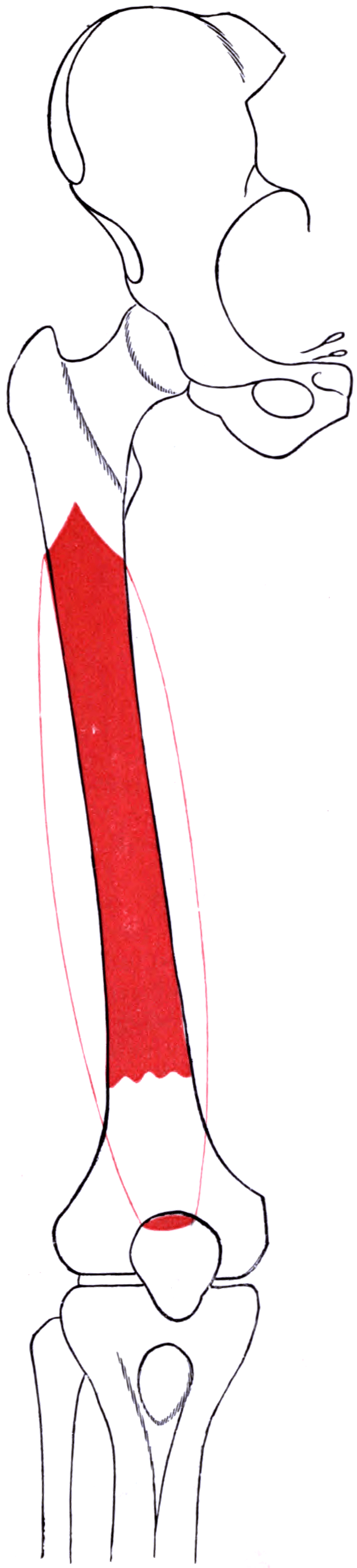
- The vastus intermedius arises from the anterior and lateral surfaces of the femoral shaft (upper two-thirds) and the lower half of the lateral lip of the linea aspera — it is the only quadriceps head arising from the flat anterior surface of the shaft, which is why it scars to a healing femoral fracture.
- The quadriceps tendon is a layered structure: rectus femoris superficially, vastus medialis and vastus lateralis in the intermediate layer, and vastus intermedius forming the deep layer immediately anterior to the suprapatellar pouch.
- Innervated by the femoral nerve, posterior division; root values L2, L3, L4, with branches entering the anterior surface of the muscle; the plane between it and vastus lateralis is often indistinct distally.
- Articularis genu (subcrureus) is the small distal slip of the vastus intermedius that inserts on the apex of the suprapatellar synovial pouch and retracts it during extension, preventing synovial impingement.
- Post-traumatic extension contracture of the knee is characteristically a vastus intermedius problem: the muscle adheres to the anterior femur and to the suprapatellar pouch, abolishing quadriceps excursion — the target of Thompson and Judet quadricepsplasty.
- “Quadriceps tendon autograft is harvested partial-thickness from the superficial rectus femoris layer; going full-thickness breaches the vastus intermedius layer and enters the suprapatellar pouch, converting an extra-articular harvest into an arthrotomy with fluid extravasation during arthroscopy.
- “In V-Y quadriceps turndown the vastus intermedius layer is the depth at which the incision must be full-thickness to gain length — which is exactly why the procedure costs extensor power.
- “The Thompson quadricepsplasty releases the vastus intermedius from the femur and divides the vasti from the rectus femoris, preserving the rectus femoris as the working extensor; the Judet technique is a stepwise proximal-to-distal release preserving more of the extensor mechanism and is generally preferred where power must be retained.
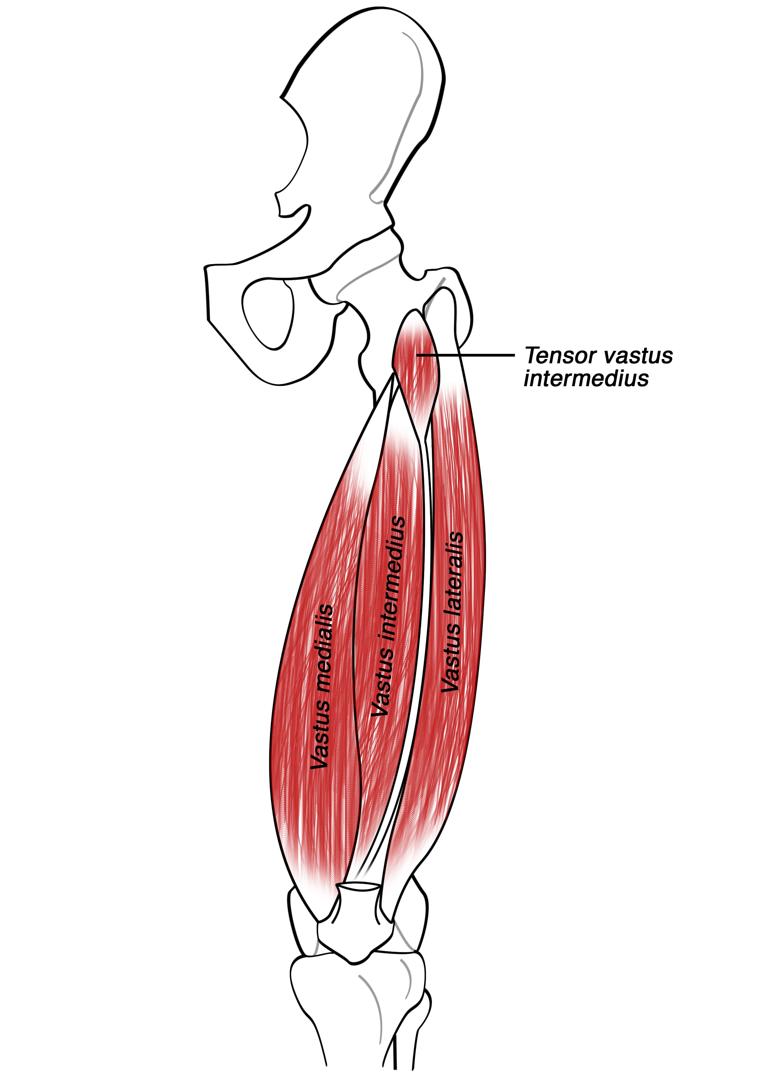
- “A tensor of the vastus intermedius has been described as a distinct fifth component of the quadriceps in most limbs — a genuine anatomical update worth knowing but not a substitute for the classic four-head description.
Overview
The vastus intermedius is the deepest and least accessible head of the quadriceps. It lies directly on the anterior femoral shaft, deep to the rectus femoris and between the vastus medialis and vastus lateralis, with which it is often confluent distally. It is easy to overlook precisely because it is never the muscle you see first — and that is exactly why it causes trouble. Two of its anatomical properties dominate its clinical behaviour:
- It arises from the bone itself. Its origin is the flat anterior and lateral surfaces of the femoral shaft, so any fracture, callus, plate or periosteal insult on the anterior femur is immediately adjacent to the muscle's origin. Healing scars the muscle to the bone, abolishing the excursion the quadriceps needs to flex the knee.
- It is the last layer before the joint. Distally its aponeurosis forms the deep layer of the quadriceps tendon, separated from the suprapatellar synovial pouch by only a thin layer of fat, and it sends the articularis genu slip into the pouch apex.
Together these two facts explain post-traumatic knee stiffness, the depth limits of quadriceps tendon graft harvest, the mechanics of V-Y turndown lengthening and the rationale of quadricepsplasty.
The quadriceps tendon is not one sheet. It is a laminated structure, and knowing which layer you are in determines whether an operation succeeds.
- Layer 1 (superficial, anterior): rectus femoris aponeurosis. This is the layer harvested for quadriceps tendon autograft, and the layer whose fibres continue over the anterior patellar surface into the patellar tendon.
- Layer 2 (intermediate): vastus medialis and vastus lateralis aponeuroses, meeting in the midline. Their expansions continue into the medial and lateral retinacula.
- Layer 3 (deep, posterior): vastus intermedius aponeurosis, lying immediately anterior to the suprapatellar pouch with only a thin fat layer between.
Why it matters, procedure by procedure
- Quadriceps tendon autograft (ACL, multiligament, MPFL): harvest partial-thickness from Layer 1. Typical harvest is a 7-10 mm wide, 6-8 cm long strip taken to about two-thirds to three-quarters of tendon thickness. If you cut through Layer 3 you enter the joint — fluid extravasation during arthroscopy, a suprapatellar arthrotomy to close, and a weakened extensor mechanism.
- V-Y quadriceps turndown for exposure in the stiff knee: the incision must be full-thickness through all three layers to gain length. That is precisely why the procedure buys exposure at the cost of extensor power and extension lag.
- Quadricepsplasty: the pathological tether is in and beneath Layer 3 — vastus intermedius scarred to the femur and to the pouch. Release starts there.
- Total knee arthroplasty arthrotomy: the medial parapatellar incision passes through Layers 1 and 2 medially; recognising the deep layer prevents an unintended and difficult-to-close defect.
R - ML - IQuadriceps Tendon Layers, Superficial to Deep
Hook:Three layers: harvest the first, respect the third.
Attachments, Innervation and Relations
Origin
- Anterior and lateral surfaces of the upper two-thirds of the femoral shaft — the defining feature. No other quadriceps head arises from the flat anterior surface.
- Lower half of the lateral lip of the linea aspera (continuing distally where vastus lateralis leaves off).
- Lateral intermuscular septum distally, in part.
- The muscle is directly applied to the periosteum of the anterior femur over most of its origin. There is no substantial gliding plane; the muscle and the bone are functionally coupled, which is the anatomical root of post-traumatic stiffness.
Insertion
- Fibres converge into a broad, thin aponeurosis on the anterior surface of the muscle which becomes the deep (posterior) layer of the quadriceps tendon, inserting on the base of the patella deep to the rectus femoris and the vasti.
- The aponeurosis is separated from the suprapatellar synovial pouch by only a thin layer of prefemoral fat.
- Medial and lateral expansions blend with the vastus medialis and vastus lateralis aponeuroses, so distally the three are often inseparable — the practical reason the vastus intermedius is described as confluent with the vastus lateralis in its lower part.
Articularis genu (subcrureus)
- A small, variable slip — usually two to five separate fasciculi — arising from the deep surface of the distal vastus intermedius on the anterior femur, above the trochlea.
- It inserts on the apex (proximal reflection) of the suprapatellar synovial pouch.
- Function: it retracts the suprapatellar pouch proximally during knee extension, preventing the redundant synovial fold from being pinched between the patella and the femur.
- Clinical relevance: loss of articularis genu function — by scarring, resection, or adhesion of the pouch — contributes to suprapatellar impingement, pouch obliteration and loss of patellar excursion.
Tensor of the vastus intermedius
- A distinct fifth component of the quadriceps, described by Grob and colleagues as arising from the anterolateral proximal femur between the vastus lateralis and vastus intermedius and inserting through an independent aponeurosis into the medial patellar aspect via the quadriceps tendon.
- Found in all 26 limbs of Grob's series, with its own femoral nerve branch and its own branch of the lateral circumflex femoral artery. Four morphological types were distinguished: Independent 11/26, VI-type 6/26, VL-type 5/26, Common-type 4/26 — so it is consistently present but only about 40 per cent of the time as a fully independent structure. Treat those proportions as indicative: they come from 26 limbs in one laboratory.
- Why it is worth knowing: it accounts for some of the variability in the plane between vastus lateralis and vastus intermedius and for the difficulty in separating them surgically. It does not replace the classical four-head description in an exam answer, but recognising it demonstrates current anatomical awareness.
Named landmarks and measurements
- Relationship
- Direct origin, applied to periosteum
- Practical use
- Site of post-fracture adhesion; released first in quadricepsplasty
- Relationship
- The last layer before the suprapatellar pouch
- Practical use
- Depth limit of a partial-thickness graft harvest
- Relationship
- Articularis genu insertion, roughly 3-5 cm proximal to the patellar base with the knee extended
- Practical use
- Site of adhesions; target of arthroscopic adhesiolysis
- Relationship
- Lies between the deep tendon layer and the pouch
- Practical use
- Its presence confirms you are still extra-articular
- Relationship
- Origin distal to the vastus lateralis origin
- Practical use
- Explains overlap and confluence with vastus lateralis
The graft harvest error that ruins the case.
- The vastus intermedius layer is the last barrier before the suprapatellar pouch.
- Cut through it and you have made a suprapatellar arthrotomy.
- Consequences: fluid extravasation during arthroscopy with a swollen thigh and poor visualisation, a defect that must be closed, and a measurably weaker extensor mechanism.
- Answer: harvest partial-thickness — typically two-thirds to three-quarters of tendon thickness — and use the prefemoral fat appearing in the base as your warning that you are at the limit.
The quadricepsplasty catastrophe.
- The stiff knee after a femoral shaft fracture is stiff because the vastus intermedius is scarred to the healing bone.
- Forceful manipulation under anaesthesia in an incompletely united femur risks refracture through the fracture or the callus, and in an osteopaenic bone a supracondylar femoral fracture or a patellar tendon avulsion.
- Answer: confirm union radiologically, release the tether surgically rather than tearing it, and stop at the point where resistance is soft-tissue rather than bony.
Action and Biomechanics
Actions
- Action
- Knee extension
- Note
- Monoarticular; contributes roughly 15-20 percent of quadriceps cross-sectional area
- Action
- Retracts the suprapatellar pouch proximally during extension
- Note
- Prevents synovial impingement between patella and femur
- Action
- Transmits force to the patellar base; part of the laminated tendon
- Note
- Its integrity is required for a competent extensor mechanism
- Action
- Provides the quadriceps glide over the anterior femur that permits knee flexion
- Note
- Loss of glide, not loss of power, is what makes the post-traumatic knee stiff
The excursion concept — the most important biomechanical point
- To flex the knee to 130 degrees, the quadriceps must glide and lengthen over the anterior femur. Total quadriceps excursion required is substantial, and much of it depends on the vastus intermedius sliding over the femoral shaft.
- If the muscle is adherent to bone, no amount of quadriceps strength produces flexion — the muscle simply cannot lengthen. This is why post-traumatic extension contracture is a length/excursion problem, not a strength problem, and why stretching alone fails.
- Corollary for the exam: in a stiff knee after a femoral fracture, ask "is the block intra-articular (adhesions, arthrofibrosis, pouch obliteration) or extra-articular (vastus intermedius adherent to bone, quadriceps scar)?" The distinction determines whether the answer is arthroscopic adhesiolysis or open quadricepsplasty — and often both are needed.
Length-tension and force
- Being monoarticular, its length depends only on knee angle. It shares the patellar tendon lever with the other heads. As with the other vasti, terminal extension is where the greatest force is required and where deficiency first shows as an extensor lag.
- The articularis genu works isometrically rather than as a mover: it maintains the pouch out of the way.
Synergists and antagonists
- Synergists: rectus femoris, vastus lateralis, vastus medialis.
- Antagonists: hamstrings and gastrocnemius for knee flexion. Note the paradox: in a stiff knee with an adherent vastus intermedius, the hamstrings are the muscles trying to overcome a mechanical block, not a strength imbalance.
What happens when it fails
- Adhesion to the femur: loss of knee flexion, extension contracture, a hard end-point on passive flexion, and a patella with reduced excursion.
- Full-thickness tendon defect: extensor lag, weakness, and in severe cases extensor mechanism dehiscence.
- Denervation or fatty atrophy: modest measurable loss of extension torque; clinically overshadowed by the other three heads.
- Loss of articularis genu function or pouch obliteration: suprapatellar impingement pain, crepitus, and reduced patellar proximal excursion.
The classic scenario is a femoral shaft fracture — treated in traction, in a cast, by plating, or by a nail — followed by a knee that will not flex.
- Vastus intermedius adherent to the anterior femur and to callus — the extra-articular tether, and the dominant lesion. The muscle originates from that bone surface, so healing bridges muscle to bone.
- Loss of the rectus femoris to vastus intermedius plane — the two become a single scarred mass.
- Obliteration of the suprapatellar pouch and loss of articularis genu retraction — an intra-articular component.
- Peripatellar and medial/lateral gutter adhesions — a further intra-articular component.
- Fibrosis and contracture of the quadriceps muscle itself, plus posterior capsular contracture if the knee has been held flexed.
- Patella baja if the patella has been tethered distally, and heterotopic ossification in some.
- Optimise first: confirm union, treat any infection, ensure the extensor mechanism is intact, and give a supervised intensive physiotherapy trial with serial measurement.
- Arthroscopic adhesiolysis for the intra-articular component: release the suprapatellar pouch, the gutters and the peripatellar adhesions.
- Manipulation under anaesthesia only when union is certain, and gently.
- Open quadricepsplasty (Thompson or Judet) for the extra-articular tether when the block persists.
- Continuous postoperative motion — the gains from any of these procedures are lost within weeks without an aggressive, supervised rehabilitation programme starting on day one.
Surface Anatomy and Examination
Palpation and positioning
- The muscle itself cannot be palpated — it lies deep to the other three heads against the femur. This is a legitimate answer and a distinguishing feature.
- What you can assess is its function by proxy:
- Quadriceps glide: with the knee flexed passively, feel whether the anterior thigh soft tissue moves over the femur or is fixed to it. In an adherent vastus intermedius the tissue is tethered and dimples over the fracture site.
- Patellar excursion: with the knee extended and quadriceps relaxed, assess proximal-distal patellar translation. Reduced proximal excursion indicates a tethered extensor mechanism and an obliterated suprapatellar pouch.
- Suprapatellar pouch fullness: a normal pouch admits fluid and can be ballotted. An obliterated pouch feels flat and fibrous.
- Scar assessment: identify previous incisions, note adherence of the skin to underlying tissue, and look for the depression at the fracture or plate site.
Named tests and their interpretation
- How to perform
- Measure maximal passive flexion with the hip flexed to 90 degrees, then with the hip extended (prone)
- Positive finding
- Flexion improves markedly with the hip flexed
- What it means
- The limiting structure crosses the hip — that is the rectus femoris, not the vasti; a purely vastus intermedius tether does not change with hip position
- How to perform
- Passively flex the knee while observing and palpating the anterior thigh over the fracture site
- Positive finding
- Dimpling or fixed tethering of tissue to the femur
- What it means
- Extra-articular adhesion of vastus intermedius to the femur or callus
- False positives
- Adherent skin scar from an open injury or previous surgery
- How to perform
- Knee extended and quadriceps relaxed; translate the patella proximally and distally
- Positive finding
- Reduced proximal excursion
- What it means
- Obliterated suprapatellar pouch and a tethered extensor mechanism
- False positives
- Guarding, patella baja from a separate cause, obesity
- How to perform
- Measure passive then active terminal extension
- Positive finding
- Active extension falls short of passive
- What it means
- Quadriceps power or continuity deficit, including after a V-Y turndown or a full-thickness harvest
- False positives
- Fixed flexion deformity; effusion causing arthrogenic inhibition
- How to perform
- Assess the quality of the end-point on gentle passive flexion under relaxation
- Positive finding
- Hard, abrupt bony end-feel versus a soft, springy soft-tissue end-feel
- What it means
- Bony block (malunion, heterotopic ossification, articular incongruity) versus soft-tissue contracture
- False positives
- Pain-limited guarding preventing a true end-feel assessment
- How to perform
- Standard effusion tests
- Positive finding
- Fluid displacement
- What it means
- An effusion supports intra-articular pathology; an absent effusion with a flat pouch suggests pouch obliteration
- False positives
- Obesity; prepatellar bursitis
- How to perform
- Prone with the hip in neutral extension, briskly flex the knee
- Positive finding
- Ipsilateral pelvic or buttock rise
- What it means
- Implicates the biarticular rectus femoris, NOT the vastus intermedius — useful to exclude the rectus as the limiting structure
- False positives
- Anterior pelvic tilt not controlled; iliopsoas tightness
Pitfalls in examination
- Always distinguish extra-articular from intra-articular block. Passive flexion that changes with hip position implicates the biarticular rectus femoris; passive flexion that is unchanged by hip position and associated with skin dimpling over the femur implicates the vastus intermedius.
- Always assess the end-feel. A hard bony end-point demands imaging for malunion, heterotopic ossification or articular incongruity before any soft-tissue procedure is contemplated.
- Always confirm union radiologically before contemplating manipulation — refracture is a real and avoidable disaster.
- Document the extensor lag and quadriceps power preoperatively. Quadricepsplasty and V-Y turndown both cost extensor power, and a patient who starts with a lag will finish with a worse one.
- Imaging: MRI shows fatty atrophy and fibrosis of the vastus intermedius, adhesion to the femur, loss of the prefemoral fat plane and an obliterated suprapatellar pouch. CT identifies heterotopic ossification and malunion.
Complications
- Mechanism
- Full-thickness cut through the vastus intermedius layer
- Avoidance / management
- Harvest partial-thickness; stop when prefemoral fat appears; close the defect and manage fluid extravasation if it happens
- Mechanism
- Communication between the joint and the harvest site or the extra-articular tissues
- Avoidance / management
- Recognise early, reduce pump pressure, complete the case efficiently, monitor the thigh for compartment signs
- Mechanism
- V-Y turndown, over-deep graft harvest, Thompson quadricepsplasty
- Avoidance / management
- Prefer quadriceps snip; partial-thickness harvest; Judet rather than Thompson where power matters; committed rehabilitation
- Mechanism
- Failure of a repaired full-thickness tendon defect, often in a diabetic, obese or steroid-exposed patient
- Avoidance / management
- Robust layered repair, protected early motion, formal repair or reconstruction if it fails
- Mechanism
- Forceful manipulation with an incompletely united femur; the muscle is adherent to the callus
- Avoidance / management
- Confirm union radiologically first; manipulate gently; release surgically rather than tearing
- Mechanism
- Forceful manipulation in osteopaenic bone or against a dense contracture
- Avoidance / management
- Gentle graded force, abandon manipulation if the end-feel is bony, consider open release instead
- Mechanism
- Inadequate postoperative rehabilitation after manipulation, adhesiolysis or quadricepsplasty
- Avoidance / management
- Analgesia including regional catheters, continuous or supervised motion from day one, daily physiotherapy; do not operate if this cannot be delivered
- Mechanism
- Bleeding, prolonged immobilisation, predisposition, heterotopic ossification
- Avoidance / management
- Meticulous haemostasis, early motion, treat heterotopic ossification only when mature
- Mechanism
- Extensive dissection through scarred, poorly vascularised thigh skin
- Avoidance / management
- Use previous incisions where possible, avoid narrow skin bridges, plastic surgical input for difficult skin
- Mechanism
- Anterior dissection dividing the segmental branches entering the anterior surface
- Avoidance / management
- Preserve the rectus femoris to vastus intermedius plane where possible; stepwise release
- Mechanism
- Resection or scarring of the slip during adhesiolysis
- Avoidance / management
- Preserve the slip; avoid over-resecting the prefemoral fat pad
- Mechanism
- Extensive peripatellar dissection combined with lateral release and fat pad excision
- Avoidance / management
- Avoid unnecessary lateral release; preserve the superior lateral genicular artery
Denervation pattern
- Segmental branches enter the anterior surface of the muscle. Complete denervation is uncommon, but extensive anterior dissection or dense scarring produces patchy denervation and fatty atrophy visible on MRI as high-signal change in the deep muscle against the femur.
- Because the muscle is deep and contributes only roughly 15-20 percent of quadriceps cross-sectional area, isolated denervation is clinically subtle — the functional problem it causes is loss of glide, not loss of power.
Clinical Relevance
Post-traumatic extension contracture of the knee
Definition and presentation
- A knee that will not flex, with the extensor mechanism functionally shortened and tethered. Most commonly follows a femoral shaft or distal femoral fracture, but also prolonged immobilisation, quadriceps contusion with myositis ossificans, repeated intramuscular injections in childhood, burns, and post-arthroplasty arthrofibrosis.
- Functional consequences are considerable: difficulty sitting, climbing and descending stairs, using transport, kneeling and squatting. Rowe measured this directly in 20 elderly normal subjects: gait and slopes need less than 90 degrees, stairs and chairs 90 to 120 degrees, and a bath about 135 degrees — from which the authors set 110 degrees as the rehabilitation goal. A knee stuck at 50-60 degrees is therefore genuinely disabling, and the study did not test squatting or kneeling, which demand more still.
Anatomical basis (in order of contribution)
- Vastus intermedius adherent to the anterior femur and callus — the dominant extra-articular tether.
- Loss of the rectus femoris to vastus intermedius gliding plane.
- Suprapatellar pouch obliteration with loss of articularis genu retraction.
- Peripatellar and gutter adhesions, and patella baja if the patella is tethered distally.
- Quadriceps muscle fibrosis and, if the knee was held flexed, posterior capsular contracture.
- Heterotopic ossification in a subset.
Assessment
- Passive and active range; end-feel; patellar excursion; quadriceps power and extensor lag.
- Confirm union with radiographs (and CT if in doubt).
- MRI for the soft-tissue pattern; CT for heterotopic ossification and malunion.
- Exclude infection (inflammatory markers, aspiration if indicated) and address it first.
Treatment ladder
- Intervention
- Supervised intensive physiotherapy with serial measurement, static progressive or dynamic splinting
- When and caveats
- First-line for at least 3 months; many knees improve substantially without surgery
- Intervention
- Manipulation under anaesthesia
- When and caveats
- Only when union is radiologically certain; gentle, controlled; highest yield in early arthrofibrosis (within about 3 months) and after arthroplasty
- Intervention
- Arthroscopic adhesiolysis
- When and caveats
- Releases the intra-articular component: suprapatellar pouch, gutters, peripatellar adhesions; can be combined with manipulation
- Intervention
- Open quadricepsplasty (Thompson or Judet)
- When and caveats
- For the persistent extra-articular tether; requires committed rehabilitation and costs some extensor power
- Intervention
- Address bony causes
- When and caveats
- Excise mature heterotopic ossification; correct malunion; treat articular incongruity
Every procedure for the stiff knee — manipulation, arthroscopic adhesiolysis, quadricepsplasty — gains motion on the operating table that is lost within weeks unless an aggressive, supervised motion programme starts immediately. Adequate analgesia (including regional catheters where available), continuous passive motion or equivalent, and a physiotherapist who will see the patient daily are not adjuncts; they are the determinants of the result. Do not perform the operation if the rehabilitation cannot be delivered.
Surgical Relevance
Procedure-by-procedure summary
- How vastus intermedius is involved
- Its aponeurosis is the deep layer that must be left intact
- Structures at risk with location
- Suprapatellar pouch immediately deep to the layer; prefemoral fat is the warning sign; extensor mechanism strength
- How vastus intermedius is involved
- The layer must be divided full-thickness to gain length
- Structures at risk with location
- Extensor power and extension lag; patellar vascularity if combined with a lateral release
- How vastus intermedius is involved
- Oblique proximolateral cut; the deep layer is divided only over a short oblique length
- Structures at risk with location
- Minimal; preferred first extensile step
- How vastus intermedius is involved
- Released from the femur; scarred portion excised; vasti separated from the rectus femoris
- Structures at risk with location
- Extensor power; femoral periosteum and nutrient vessels; refracture through an ununited femur
- How vastus intermedius is involved
- Released from the femur in a stepwise proximal-to-distal sequence
- Structures at risk with location
- Nerve branches entering the anterior surface; extensor power (better preserved than Thompson)
- How vastus intermedius is involved
- The suprapatellar pouch deep to its aponeurosis is released
- Structures at risk with location
- Articularis genu (preserve for pouch retraction); prefemoral fat pad bleeding; patellar cartilage
- How vastus intermedius is involved
- The tether it forms with the femur is what resists
- Structures at risk with location
- Refracture through an ununited femur or callus; supracondylar fracture in osteopaenic bone; patellar tendon avulsion
- How vastus intermedius is involved
- The muscle is split or elevated off the anterior cortex
- Structures at risk with location
- Nerve branches entering the anterior surface; extensive denervation; adhesion after healing
- How vastus intermedius is involved
- The muscle is adjacent to the plate or the entry portal and scars to it
- Structures at risk with location
- Quadriceps adhesions with loss of flexion; consider early motion and, in some cases, planned later adhesiolysis
Distances and landmarks worth quoting
- Quadriceps tendon graft: 7-10 mm wide, 6-8 cm long, taken partial-thickness at two-thirds to three-quarters of tendon thickness; the prefemoral fat appearing in the base is the depth warning.
- Where the tendon layers actually fuse — and why a mean is not plannable. Grob traced all six elements in 10 limbs: they fuse over a zone 13 to 90 mm proximal to the patella, with the superficial medial vastus intermedius aponeurosis joining at a mean 56 mm (range 30-90) and the deep at a mean 33 mm (range 13-53). The spread matters more than the average — in some limbs the fusion point is barely a centimetre above the patella, so identify it in the patient rather than predicting it from a textbook figure.
- Suprapatellar pouch apex: approximately 3-5 cm proximal to the patellar base with the knee extended — the articularis genu insertion and the proximal limit of arthroscopic pouch release.
- V-Y turndown apex: the proximal end of the quadriceps tendon, with limbs running distally into the medial and lateral retinacula.
- Quadriceps snip: an oblique cut at approximately 45 degrees proximolaterally from the apex of the medial parapatellar arthrotomy.
- Origin of the muscle: the anterior and lateral surfaces of the upper two-thirds of the femoral shaft — the zone in which fracture callus will tether it.
- Functional flexion targets to quote: less than 90 degrees for level gait and slopes, 90 to 120 degrees for stairs and chairs (Rowe does not separate the two), about 135 degrees for a bath — and 110 degrees as the single rehabilitation goal the authors recommend. Squatting and kneeling need more and were not measured in that study.
Asked how you will expose a stiff knee at revision arthroplasty, give a ladder, not a single answer.
- Optimise the basics: full medial and lateral gutter release, excise the fat pad if it obstructs, release the peripatellar adhesions, and free the suprapatellar pouch — this alone frequently suffices.
- Quadriceps snip — an oblique proximolateral cut through the quadriceps tendon at 45 degrees. Minimal functional cost, no change to rehabilitation. This is the default first extensile step.
- Tibial tubercle osteotomy — when the problem is patellar tendon tension and the priority is protecting extensor power. Adds a bone-healing problem and a tibial fracture risk.
- V-Y turndown — the widest exposure and the only option that deliberately lengthens the extensor mechanism, at the cost of extension lag and quadriceps weakness in a proportion of patients.
State the principle: choose the least costly step that gives adequate exposure, and escalate deliberately rather than tearing tissue. Never allow inadequate exposure to compromise implant position.
Guidelines, Registries & Global Practice
Variation and prevalence
- The degree of fusion between the vastus intermedius, vastus lateralis and vastus medialis aponeuroses varies, as does the thickness of each quadriceps tendon layer. This variability is the practical reason a graft harvest should be guided by what is seen (prefemoral fat appearing) rather than by a fixed millimetre depth, and why measuring tendon thickness on preoperative MRI is helpful.
- The tensor of the vastus intermedius was found in all 26 limbs of Grob's series, though as a fully independent structure in only 11 of 26. It is a genuine anatomical update and explains why the vastus lateralis to vastus intermedius plane can be indistinct, but the classical four-head description remains the standard exam answer.
- Articularis genu varies in the number of fasciculi (commonly two to five) and occasionally is absent. A suprapatellar plica — a remnant of the embryological septum between the suprapatellar bursa and the joint — is present in a substantial minority and ranges from a thin fold to a complete septum.
Differences in described technique
- Emphasis relating to vastus intermedius
- Quadriceps snip is the standard first extensile option in the difficult knee; V-Y turndown is described but discouraged as a default because of extensor lag; tibial tubercle osteotomy widely used in revision.
- Emphasis relating to vastus intermedius
- Strong emphasis on structured non-operative management of post-traumatic stiffness with serial measurement before any surgical release, and on the availability of intensive rehabilitation as a precondition for surgery.
- Emphasis relating to vastus intermedius
- Emphasises early controlled motion after femoral fracture fixation specifically to prevent quadriceps adhesion to the anterior femur, and limits periosteal stripping to the plate footprint.
- Emphasis relating to vastus intermedius
- Quadriceps tendon autograft accepted as a legitimate primary and revision graft, with explicit instruction to harvest partial-thickness and avoid entering the suprapatellar pouch.
- Emphasis relating to vastus intermedius
- Injection-related quadriceps contracture and congenital extension contracture treated by staged release or V-Y quadricepsplasty; intramuscular injections directed to the mid-third of the vastus lateralis with site rotation.
Registry and outcome signals
- Arthroplasty registries do not record the arthrotomy or extensile manoeuvre used, so registry data cannot compare quadriceps snip with V-Y turndown. What they capture is revision for instability, malalignment and infection, which is the argument for prioritising adequate exposure.
- ACL registries (Scandinavian, UK and others) increasingly record graft type; quadriceps tendon autograft use has grown, and reported revision rates are broadly comparable with other autografts in the medium term. Registry data have not identified a distinct complication signal attributable to suprapatellar pouch breach, which is likely under-reported.
- Post-traumatic stiffness is not systematically captured by any registry. Published series of quadricepsplasty are small, single-centre and retrospective, and consistently report substantial flexion gains with a variable rate of extension lag — an honest statement of the evidence base is expected in a viva.
High- versus limited-resource practice
- Well-resourced settings: MRI to characterise the soft-tissue pattern before surgery, CT for heterotopic ossification and malunion, arthroscopic adhesiolysis as a first surgical step, regional analgesia catheters and continuous passive motion devices to protect the gains, and depth-controlled instruments for graft harvest.
- Limited-resource settings: the assessment that matters most is clinical and costs nothing — passive flexion with the hip flexed and extended, end-feel, patellar excursion, quadriceps glide and skin dimpling, and a radiograph to confirm union. Judet-style stepwise release can be performed without special equipment, and the outcome depends far more on the availability of supervised daily physiotherapy than on any device. Where intensive rehabilitation cannot be delivered, the correct decision is to not operate.
- Universal principles: prevent rather than treat — early controlled knee motion after femoral fracture fixation and limited periosteal stripping are the interventions that avoid this problem entirely; confirm union before manipulating; harvest quadriceps graft partial-thickness; and escalate exposure with a quadriceps snip before a turndown.
MCQ Practice Points
Q: What is anatomically unique about the origin of the vastus intermedius? A: It is the only quadriceps head arising from the flat anterior surface of the femoral shaft (upper two-thirds, plus the lower half of the lateral lip of the linea aspera). This is why it scars to a healing femoral fracture.
Q: Name the layers of the quadriceps tendon from superficial to deep. A: Rectus femoris (superficial), vastus medialis and vastus lateralis (intermediate), vastus intermedius (deep). The deep layer lies immediately anterior to the suprapatellar pouch.
Q: How deep should a quadriceps tendon autograft be harvested and what warns you that you are at the limit? A: Partial-thickness, roughly two-thirds to three-quarters of tendon thickness, from the superficial rectus femoris layer. The appearance of prefemoral fat in the base warns you that the deep vastus intermedius layer and the suprapatellar pouch are next.
Q: What is the articularis genu and what does it do? A: A small slip (usually two to five fasciculi) from the deep surface of the distal vastus intermedius, inserting on the apex of the suprapatellar synovial pouch. It retracts the pouch proximally during extension, preventing the synovial fold being pinched between patella and femur.
Q: Why is post-traumatic knee extension contracture predominantly a vastus intermedius problem? A: The muscle arises directly from the anterior femoral shaft with no gliding plane, so callus and scar bind it to the bone, abolishing the quadriceps excursion required for flexion. It is an excursion problem, not a strength problem.
Q: How do the Thompson and Judet quadricepsplasties differ? A: Thompson frees the vastus intermedius from the femur, excises scarred muscle, and divides the vasti from the rectus femoris so that the rectus femoris becomes the extensor — large flexion gains, greater extensor weakness. Judet is a stepwise proximal-to-distal release, stopping when flexion is adequate — better preservation of extensor power.
Q: Why must a V-Y quadriceps turndown be full-thickness, and what is the consequence? A: Length is only gained by dividing all three tendon layers including the deep vastus intermedius layer. The consequence is extensor weakness and extension lag in a proportion of patients — which is why the quadriceps snip is the preferred first extensile step.
Q: Can the vastus intermedius be used as a muscle flap? A: No — it has no single dominant vascular pedicle, being supplied segmentally by perforating branches of the profunda femoris. Quadriceps flaps are raised from the rectus femoris or the vastus lateralis on the descending branch of the lateral circumflex femoral artery.
Q: What must you confirm before manipulating a stiff knee after a femoral shaft fracture? A: Radiological union. The vastus intermedius is adherent to the callus, so forceful manipulation of an incompletely united femur risks refracture, and in osteopaenic bone a supracondylar fracture or patellar tendon avulsion.
Q: How do you tell clinically whether a flexion block is due to the rectus femoris or to the vasti? A: Change the hip position. The rectus femoris is biarticular, so flexion improves when the hip is flexed and a positive Ely test is present. The vasti are monoarticular, so a vastus intermedius tether gives flexion that is unchanged by hip position, with dimpling over the femur on flexion.
Q: What flexion range should a stiff-knee procedure aim to achieve? A: Quote Rowe's figures and his conclusion. Less than 90 degrees for level gait and slopes; 90 to 120 degrees for stairs and chairs; about 135 degrees for a bath — and 110 degrees as the recommended rehabilitation goal. Squatting and kneeling demand more and were not tested in that study. Define the functional target before operating: 110 degrees is what a quadricepsplasty has to achieve to be worth its risk.
Exam Viva Scenarios
Practise clinical reasoning and management decisions out loud
“A 28-year-old man is nine months after plate fixation of a mid-shaft femoral fracture. The femur is united. His knee flexes to 45 degrees actively and passively, with a soft end-feel, and there is dimpling of the anterior thigh over the plate when you flex the knee. Extension is full with no lag. How do you assess and treat him?”
“You are harvesting a quadriceps tendon autograft for a revision ACL reconstruction. On starting the arthroscopy the thigh swells rapidly and your view is poor. What has happened, and how do you proceed?”
“You are revising a total knee arthroplasty. Flexion preoperatively was 70 degrees. After a medial parapatellar arthrotomy and gutter release you cannot translate the patella laterally enough to see the tibia, and the patellar tendon is under alarming tension. What are your options and how do you choose?”
Anatomy
- Origin: anterior and lateral surfaces of upper two-thirds of femoral shaft, lower half of lateral lip of linea aspera
- Only quadriceps head from the flat anterior shaft — hence adhesion after fracture
- Insert: deep layer of quadriceps tendon onto patellar base
- Nerve: femoral, posterior division, L2-L4, segmental branches entering the ANTERIOR surface
- No dominant pedicle — cannot be used as a flap
Tendon Layers
- Layer 1 superficial: rectus femoris — harvest layer
- Layer 2 intermediate: vastus medialis and lateralis
- Layer 3 deep: vastus intermedius — anterior to suprapatellar pouch
- Graft: 7-10 mm wide, 6-8 cm long, partial-thickness
- Prefemoral fat in the base equals depth limit
Articularis Genu
- 2-5 fasciculi from the deep distal muscle
- Inserts on the apex of the suprapatellar pouch
- Retracts the pouch proximally in extension
- Pouch apex 3-5 cm proximal to patellar base
- Preserve during adhesiolysis
Stiff Knee
- Loss of excursion, not loss of strength
- Extra-articular: muscle adherent to femur and callus, skin dimpling on flexion
- Intra-articular: pouch obliteration, gutter and peripatellar adhesions
- Hip position changes flexion equals rectus femoris (biarticular), not vasti
- Confirm union BEFORE manipulation
Operations
- Ladder: physiotherapy, adhesiolysis, manipulation, quadricepsplasty
- Thompson: excise scarred intermedius, rectus femoris becomes extensor — more power loss
- Judet: stepwise proximal to distal, stop when adequate — preserves power
- Exposure ladder: gutters, snip, tubercle osteotomy, V-Y turndown
- Rehabilitation is the operation
Evidence Base
New Insight in the Architecture of the Quadriceps Tendon
- TEN cadaveric lower limbs dissected, tracing every muscle belly of the extensor apparatus distally to its fusion point with the quadriceps tendon
- The tendon is TRI-LAMINAR but formed by SIX elements: the lateral aponeurosis of vastus intermedius, the deep and superficial medial aponeuroses of vastus intermedius, vastus lateralis, tensor vastus intermedius, and rectus femoris
- The LATERAL part of vastus intermedius forms the DEEPEST layer of the tendon, immediately anterior to the suprapatellar pouch; rectus femoris forms the superficial layer
- Fusion of the elements occurs over a wide and variable zone - 13 to 90 mm proximal to the patella. The superficial medial vastus intermedius aponeurosis fused at a mean 56 mm (range 30 to 90) and the deep at a mean 33 mm (range 13 to 53)
- Vastus medialis does not form a discrete layer - it inserts medially into ALL layers
- Fibres of the lateral components run towards the medial femoral condyle and vice versa, so the architecture is crossed rather than parallel
A Newly Discovered Muscle: The Tensor of the Vastus Intermedius
- 26 cadaveric lower limbs - and a tensor vastus intermedius was found in ALL 26, not in a majority. The quadriceps is architecturally a five-headed muscle
- It arises from the anterolateral proximal femur, has its OWN muscular branch of the femoral nerve and its own branch of the lateral circumflex femoral artery, and becomes an independent aponeurosis that merges separately into the quadriceps tendon
- It inserts on the MEDIAL aspect of the patella despite arising laterally - so it crosses the extensor mechanism
- Four morphological types: Independent 11 of 26, VI-type 6 of 26, VL-type 5 of 26, Common-type 4 of 26
- It could not be assigned to either vastus lateralis or vastus intermedius, which is the basis of the claim that it is a separate head
Quadricepsplasty
- Thompson's account of quadricepsplasty, the operation that carries his name
- No abstract or subject headings are indexed for this 1945 paper, so only the title, journal and year are cited here
- The technique attributed to Thompson in the later literature frees the vastus intermedius from the femur, excises irreversibly scarred muscle and separates the vasti from the rectus femoris
- The rectus femoris is preserved as the working extensor, so flexion is gained at the cost of some extensor power
Anterior Cruciate Ligament Reconstruction: A Systematic Review and Meta-analysis of Outcomes for Quadriceps Tendon Autograft Versus Bone-Patellar Tendon-Bone and Hamstring-Tendon Autografts
- 27 clinical studies and 2,856 patients; 581 quadriceps tendon against 514 bone-patellar tendon-bone, and 181 quadriceps tendon against 176 hamstring
- Against BPTB: NO significant difference in instrumented side-to-side laxity, Lachman, pivot shift, Lysholm, subjective IKDC or graft failure - every p value between 0.10 and 0.85
- The one clear advantage: DONOR-SITE PAIN, risk ratio 0.25 (95 per cent CI 0.18 to 0.36, p less than 0.00001) against BPTB - a fourfold reduction
- Against hamstring: no difference in laxity, Lachman, pivot shift, subjective IKDC, donor-site pain or graft failure
- The only difference against hamstring was mean Lysholm, 3.81 points higher (95 per cent CI 0.45 to 7.17, p equals 0.03)
Evolution of the Quadriceps Snip
- SIXTEEN patients (4 women, 12 men), average age 65, operated between May 1990 and December 1992 - 8 for aseptic loosening of a total knee, 2 for septic loosening, and 6 primary arthroplasties in knees with FIBROUS ANKYLOSIS
- Hospital for Special Surgery score at review: 10 excellent and 6 good - no fair or poor results
- Range of motion improved in EVERY knee, by an average of 30 degrees
- THE FINDING THAT DEFENDS THE OPERATION IS THE CYBEX RESULT: extension strength was significantly reduced against the patient's own contralateral NORMAL knee at both 60 and 180 degrees per second - but there was NO DIFFERENCE against a contralateral knee that had itself been replaced
- Postoperative physical therapy did not have to be altered - the extensor mechanism stays in continuity
Knee Joint Kinematics in Gait and Other Functional Activities Measured Using Flexible Electrogoniometry: How Much Knee Motion Is Sufficient for Normal Daily Life?
- TWENTY ELDERLY NORMAL SUBJECTS, mean age 67, measured by flexible electrogoniometry through a circuit of real activities in and around a hospital
- Gait and walking on slopes required LESS than 90 degrees of knee flexion
- Stairs and chairs required 90 to 120 degrees - the study does not separate them, and does not show that rising from a chair demands more than stairs
- Getting into a bath required approximately 135 degrees, the most demanding activity tested
- THE AUTHORS' OWN CONCLUSION IS THE USABLE NUMBER: 110 degrees of flexion is a suitable goal for rehabilitation of knee motion