One Belly, Four Fingers and the Price of Sharing
- One muscle mass with a common belly to the ulnar three digits but a relatively independent index belly - the anatomical basis of the quadriga effect.
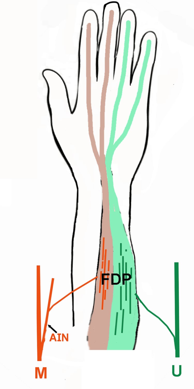
- Dual innervation: the anterior interosseous nerve supplies the index and middle halves and the ulnar nerve supplies the ring and little halves, all C8-T1.
- It is the ONLY flexor of the distal interphalangeal joint, inserting on the volar base of the distal phalanx after passing through Camper chiasm.
- The lumbricals take origin from the FDP tendons in the palm - the only muscles arising from and inserting into tendon.
- Jersey finger is avulsion of the FDP insertion, most commonly in the RING finger, and the Leddy-Packer type determines how urgently it must be repaired.
- “Test FDP by blocking the proximal interphalangeal joint in extension and asking for distal interphalangeal flexion.
- “Leddy-Packer type I retracts to the palm with both vincula torn - Leddy and Packer advise repair within 7 days, and report the first week gives the best results.
- “Leddy and Packer described THREE types. Type IV is Smith 1981 (one case) and type V is Al-Qattan 2001 (four cases) - do not attribute them to the original paper.
- “The 1 cm advancement limit is Malerich 1987 and it is a cadaveric estimate, not a safe allowance: shortening by 10 mm already produces a measurable quadriga, worst from the small and ring fingers.
- “In a high ulnar lesion clawing is LESS marked than in a low one, because the ulnar half of FDP is also denervated - the ulnar paradox.
Overview
Flexor digitorum profundus (FDP) is the deepest and largest of the long digital flexors and the only flexor of the distal interphalangeal joint. It arises as a single sheet from the ulna and interosseous membrane, divides into four tendons, passes deep to superficialis in the forearm and then through Camper chiasm in the digit to insert on the base of the distal phalanx.
Three anatomical peculiarities generate almost all of its clinical and examination content. The tendons to the ulnar three digits share a common muscle belly, so what happens to one tendon happens to all three: the quadriga effect. The lumbricals originate from the FDP tendons in the palm, a mechanical loop between a flexor and the extensor apparatus that produces the lumbrical plus finger when the loop is disturbed. And the muscle has two nerves, the anterior interosseous branch of the median for the radial half and the ulnar nerve for the ulnar half, which makes profundus testing one of the most useful tools in localising a peripheral nerve lesion.
The Roman quadriga was a chariot drawn by four horses abreast on one yoke: shorten one rein and all four horses are held back. The tendons to the middle, ring and little fingers arise from one common muscle belly, so they share one contractile excursion.
Mechanism. If a single profundus tendon is over-advanced at repair, sutured to an amputation stump under tension, tethered by adhesion or held by a stiff joint, that tendon reaches the end of its available excursion early. The common belly cannot shorten further, so the remaining tendons are prevented from completing their arc.
Clinical picture. Incomplete flexion and a weak, "hollow" grip in the uninjured fingers, with a normal-looking hand at rest. Patients describe weakness rather than stiffness and are often disbelieved; a patient with a stiff or over-tightly repaired finger who complains that the other fingers are weak is describing quadriga, not exaggerating.
The experiment, with the asymmetry stated properly. Simulating stiffness in one finger with a thermoplastic wedge significantly reduces the grip strength of the middle, ring and little fingers, in proportion to the degree of stiffness (Horton 2007, ten volunteers). Index finger strength is largely unaffected when other fingers stiffen, the experimental counterpart of its independent belly. But the same paper reports that stiffening the index still weakened the other three, so the independence protects the index as a victim and not as a culprit. Between-subject variation was considerable, so quadriga matters more to some patients than others.
The mirror-image trap. The lumbrical plus finger occurs when the profundus is divided or over-lengthened distal to the lumbrical origin, when a tendon graft is over-long, or after an amputation distal to the origin. The proximal profundus stump now pulls only on the lumbrical, which inserts into the extensor lateral band, so attempted flexion produces paradoxical interphalangeal extension.
Attachments, Innervation and Relations
Origin. The muscle has three origins:
- The proximal three-quarters of the anterior and medial surfaces of the ulna, extending onto the medial side of the coronoid process and the olecranon
- The medial half of the interosseous membrane
- An aponeurosis from the subcutaneous posterior border of the ulna, shared with flexor carpi ulnaris and extensor carpi ulnaris
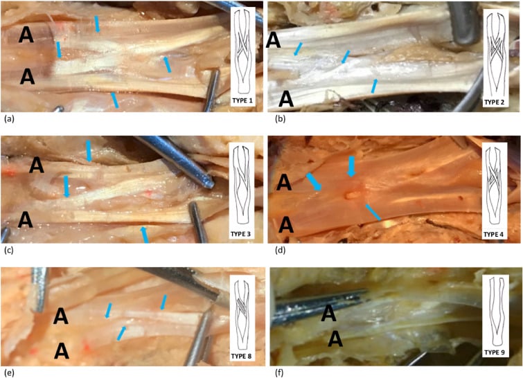
Division into tendons. The muscle divides into four tendons in the distal forearm. The index tendon separates most proximally and has the most distinct belly, which is why the index profundus is functionally the most independent. The middle, ring and little tendons remain fused with a common muscle mass well into the distal forearm and often remain interconnected in the palm.
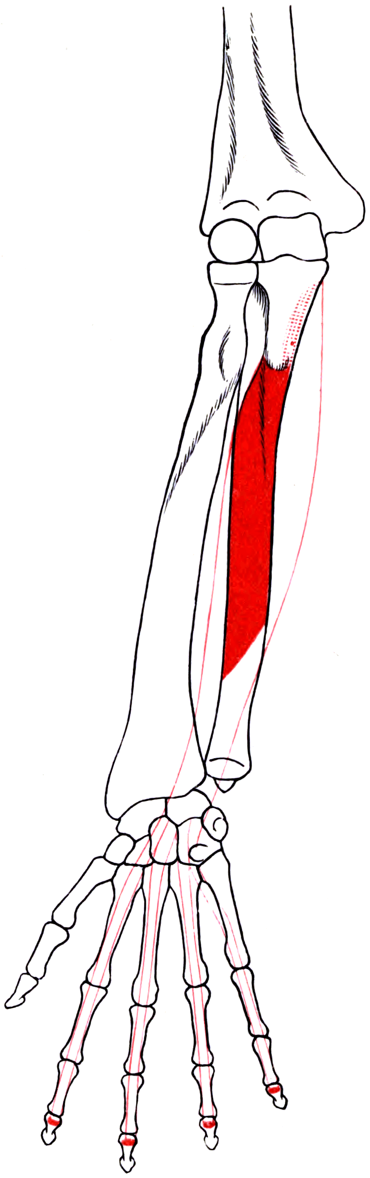
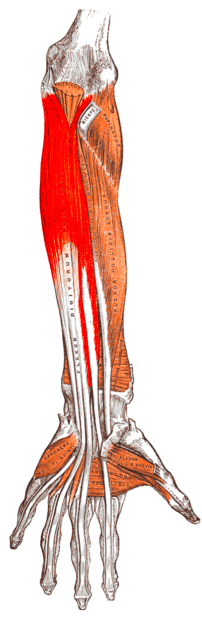
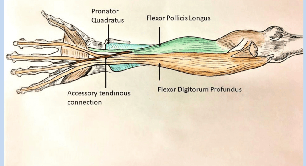
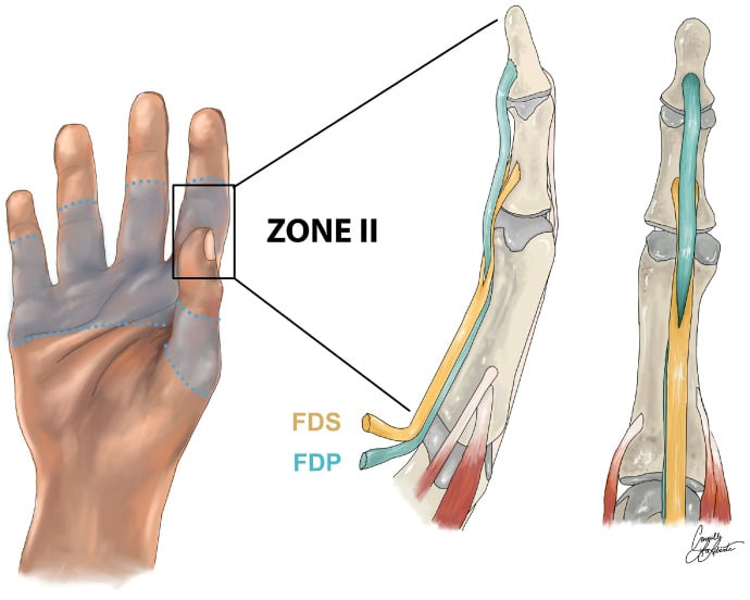
Course and insertion. The tendons pass deep to flexor digitorum superficialis in the forearm and through the carpal tunnel, four of its nine tendons, into the palm. In the digit each lies deep (dorsal) to its superficialis tendon until the proximal phalanx, then passes through the split in FDS at Camper chiasm to become the volar tendon distally. It inserts on the volar base of the distal phalanx, with a broad footprint just distal to the volar plate of the distal interphalangeal joint.
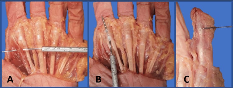
The lumbrical origin. The four lumbricals arise from the FDP tendons in the palm, the only muscles in the body that both originate from and insert into tendon. Each passes volar to the deep transverse metacarpal ligament (the interossei pass dorsal to it) and inserts into the radial lateral band of the extensor expansion, so it converts profundus contraction into extension of the interphalangeal joints and flexion of the metacarpophalangeal joint: the anatomical basis of the lumbrical plus finger.
- First and second lumbricals (index and middle): unipennate, from the radial side of their own profundus tendon
- Third and fourth lumbricals (ring and little): bipennate, from the adjacent sides of two profundus tendons (middle/ring and ring/little respectively)
Action and Biomechanics
Actions. Four:
- Flexion of the distal interphalangeal joint, its unique and defining action; no other tendon does this
- Flexion of the proximal interphalangeal and metacarpophalangeal joints, both of which it crosses and to which it contributes powerfully
- Weak wrist flexion
- Power grip: profundus is the principal power generator of grasp, while superficialis contributes precision and sustained grip
Excursion. The tendon requires approximately 70 mm of excursion, against approximately 64 mm for FDS and 33 mm for the wrist motors, so only a long-excursion donor can replace it.
How it differs from superficialis. FDS has an independent belly for each digit, so quadriga is a profundus phenomenon only. FDS is also wholly median, whereas the two nerves of profundus are what let its testing localise a nerve lesion.
What a single profundus failure costs. Distal interphalangeal flexion is lost and the finger cannot be brought into the palm at the fingertip. Pulp pinch and fine grasp are impaired, and grip strength falls modestly. Many low-demand patients tolerate this well, which is why late untreated jersey fingers that are asymptomatic can legitimately be left alone.
Surface Anatomy and Examination
Palpation. The profundus mass is palpable as the deep bulk on the volar-ulnar forearm along the subcutaneous border of the ulna during resisted finger flexion. The individual tendons are not palpable at the wrist, being deep to the superficialis layer. In the digit the profundus is the tendon that can be felt tightening at the distal phalanx during resisted distal interphalangeal flexion, and the resting cascade of the fingers reflects intact profundus tone.
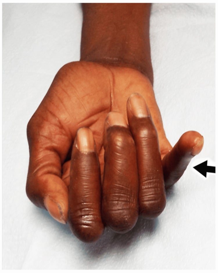
The FDP test. Four steps:
- Hold the proximal interphalangeal joint of the tested finger in full extension, the examiner's finger and thumb stabilising the middle phalanx
- Ask the patient to flex the fingertip
- Any distal interphalangeal flexion must come from FDP, because it is the only tendon crossing that joint
- Grade the power against resistance and compare with the contralateral digit; absent or weak flexion means division, avulsion or denervation of the profundus
Pitfalls that generate wrong answers.
- Tenodesis: if the wrist is allowed to extend the fingers flex passively, so test with the wrist stabilised in neutral
- A partial laceration retains active flexion; the clue is pain and weakness on resisted flexion, sometimes with triggering as the tendon flap catches at a pulley
- Pain inhibition in an acute injury mimics tendon division, as does a fixed distal interphalangeal contracture; a digital block before testing is useful, but assess the resting cascade first because that is unaffected by pain
- A lumbrical plus finger can mislead with apparent distal interphalangeal flexion; watch for the paradoxical interphalangeal extension when the patient tries to make a fist
- A jersey finger often looks normal at rest, the only sign being a slightly extended posture of the distal interphalangeal joint and a palpable, tender lump where the tendon has retracted
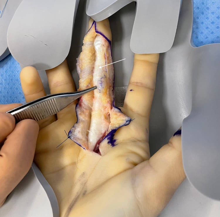
Named tests around FDP. Each has a false positive worth knowing:
- How to perform
- Rest the forearm supinated and observe the finger posture
- Positive finding
- One finger lying more extended than its neighbours
- What it means
- Flexor tendon division in that digit
- False positives
- Old stiffness or a previously fused joint
- How to perform
- Passively extend then flex the wrist
- Positive finding
- Loss of the normal flexion cascade on wrist extension
- What it means
- Flexor tendon discontinuity
- False positives
- Preserved if only FDS is divided
- How to perform
- Ask for a circle of thumb and index
- Positive finding
- Square pinch - thumb IP and index DIP hyperextend
- What it means
- Anterior interosseous palsy - FPL and index FDP
- False positives
- FPL rupture, tendon adhesion or attritional rupture
- How to perform
- Compare individual digit grip strength with and without the stiff or repaired digit blocked
- Positive finding
- Grip in the uninvolved digits improves when the tethered digit is excluded
- What it means
- Quadriga from an over-tight or tethered profundus
- False positives
- Generalised weakness; poor effort
- How to perform
- Ask the patient to make a full fist and watch the interphalangeal joints
- Positive finding
- Paradoxical IP EXTENSION on attempted flexion
- What it means
- Lumbrical plus finger
- False positives
- Extensor adhesion or an intrinsic contracture
- How to perform
- Compare PIP flexion with the MCP joint held extended and then flexed
- Positive finding
- PIP flexion improves with MCP flexion (extrinsic tight) or with MCP extension (intrinsic tight)
- What it means
- Extrinsic flexor or extensor tightness versus intrinsic contracture
- False positives
- Joint contracture limits PIP flexion in both positions equally
Complications
The two iatrogenic syndromes. Quadriga and the lumbrical plus finger are the complications the surgeon makes; their mechanism is in the Overview and their prevention and treatment under Surgical Relevance.
Repair failure.
- Rupture, most often in the first three weeks and usually precipitated by an uncontrolled grasp
- Adhesions, the dominant cause of a poor result in zones II and IV; managed with therapy first, with tenolysis considered at 3 to 6 months once motion has plateaued, the scar is supple and passive range is full
- Bowstringing from excessive pulley loss
- Flexion contracture of the proximal interphalangeal joint from a protected posture and from repair-site scarring
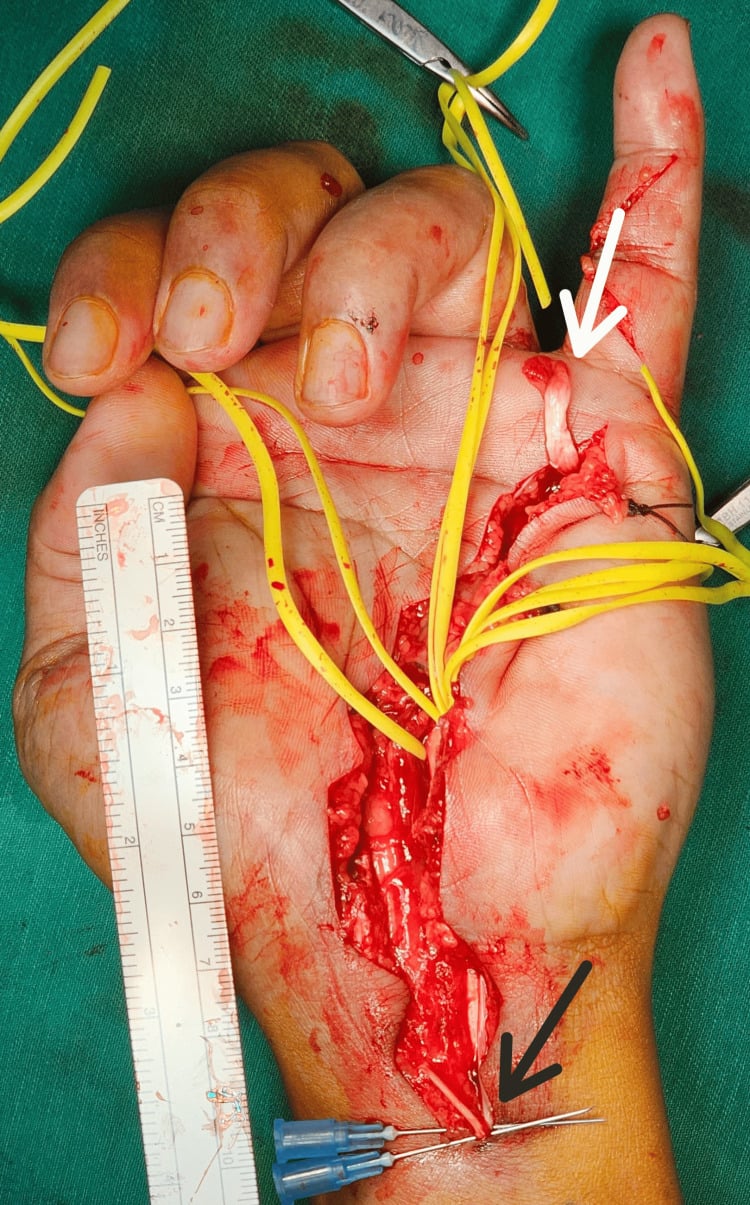
Zone I fixation.
- Nail deformity from a pull-out suture passed through or adjacent to the germinal matrix
- Dorsal button pressure necrosis, infection and pin-site pain
- Anchor problems: dorsal cortical penetration, pull-out from osteoporotic bone, and prominence causing pulp discomfort
- Distal interphalangeal joint stiffness and extension lag
Iatrogenic injury.
- Digital nerve division during volar exposure; keep the bundles in a full-thickness flap
- Anterior interosseous nerve injury during deep forearm dissection
- Missed deep compartment in forearm fasciotomy, a common technical failure that leaves the profundus and flexor pollicis longus to infarct: the direct cause of the classical Volkmann deformity
Clinical Relevance
Mechanism. Forced extension of the distal interphalangeal joint during maximal active profundus contraction, classically a hand grabbing an opponent's jersey in rugby or American football. The ring finger accounts for the large majority of cases in published series, for three reasons:
- In grip the ring fingertip projects furthest, so it is the digit most likely to be caught and forcibly extended
- The ring profundus has the least independent excursion, being most firmly bound to its neighbours in the common mass, so it is least able to yield when suddenly loaded
- Its insertion footprint is comparatively weak, and it is subjected to the greatest differential force at the moment of forced extension against maximal active flexion
Presentation. Pain and swelling on the volar finger, inability to flex the distal interphalangeal joint actively, a tender lump where the tendon has retracted (palm, PIP level, or at A4), and often nothing else. The finger looks deceptively normal, which is why the diagnosis is missed in the emergency department and the patient presents late.
Imaging. Radiographs identify a bony fragment and its level. Ultrasound or MRI localises the retracted tendon end when radiographs are normal and the palpable lump is equivocal, and knowing the level before operating determines the incisions.
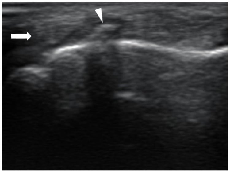
The Leddy-Packer classification, and the two types that are not theirs. Leddy and Packer described three types, from a series of 36 injuries, and they describe a coherent biological gradient of blood supply. Types IV and V were added later, each from a handful of cases: type IV by Smith in 1981 (a single case report) and type V by Al-Qattan in 2001 (four cases). Saying "Leddy-Packer type V" in an exam is a misattribution, and it matters beyond pedantry: IV and V are pattern descriptions resting on five patients between them.
- Level of retraction
- Into the PALM
- Vincula
- Both ruptured
- Bone
- No fragment
- Timing and treatment
- Tendon has lost its vincular blood supply and its nutrition - Leddy and Packer advise repair within 7 days, and report that repair in the first week gave the best results
- Level of retraction
- To the PIP joint
- Vincula
- Long vinculum intact
- Bone
- Small fleck may be present
- Timing and treatment
- Blood supply preserved by the vinculum - repair possible weeks and sometimes a few months later
- Level of retraction
- To the A4 pulley
- Vincula
- Intact
- Bone
- Large bony fragment arrested at A4
- Timing and treatment
- Tendon still attached to bone; fix the fragment - the most forgiving type
- Level of retraction
- Variable - tendon retracts separately
- Vincula
- Variable
- Bone
- Bony fragment PLUS avulsion of tendon FROM the fragment (double avulsion)
- Timing and treatment
- Fix the fragment AND reattach the tendon; the tendon may be in the palm even though a fragment is visible at the distal phalanx
- Level of retraction
- Variable
- Vincula
- Variable
- Bone
- Bony avulsion with a fracture of the distal phalanx itself (5A extra-articular, 5B intra-articular)
- Timing and treatment
- Stabilise the phalanx as well as the tendon - a broken distal phalanx may leave no sound bone for an anchor
The type IV trap. A radiograph showing a bony fragment at the distal phalanx does not prove the tendon is attached to it. If the tendon has avulsed off the fragment it may have retracted to the palm while the fragment stays put, and a surgeon who plans only to fix the fragment will be caught out. Examine for the palmar lump and image if in doubt.
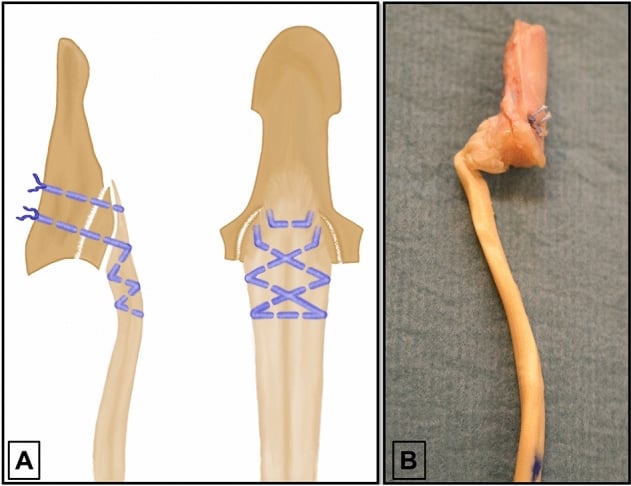
Treatment by type.
- Acute type I: urgent repair. Retrieve the tendon from the palm atraumatically, pass it back through the sheath (a catheter or suture shuttle avoids repeated grasping) and reattach to the distal phalanx with a suture anchor or a pull-out suture over a dorsal button
- Types II and III: repair or fragment fixation, with a more forgiving timescale because the vincular blood supply is intact
- Advancement: as little as the repair allows and never more than 1 cm; if more than a centimetre is needed, choose a graft or a different operation. Where the number comes from, and why it is not an allowance to be spent, is set out under Surgical Relevance
Late presentation. An asymptomatic late case is legitimately left alone. A symptomatic one takes one of the salvage options under Surgical Relevance: distal interphalangeal fusion or tenodesis if the joint is unstable, or two-stage tendon grafting.
Surgical Relevance
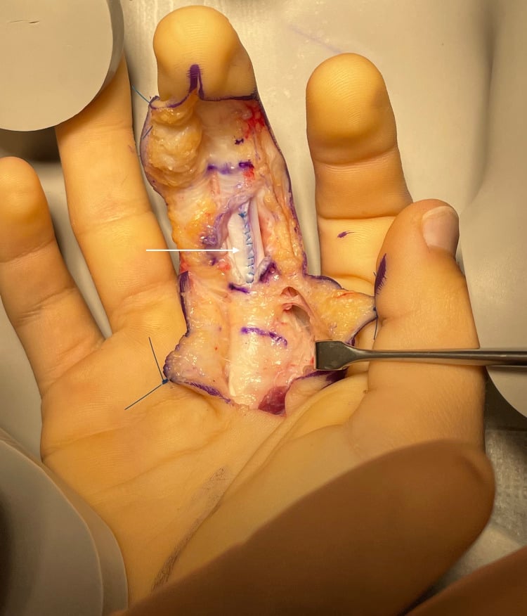
Volar digital exposure. A Bruner zig-zag incision with apices reaching the midlateral line at the flexion creases, raising full-thickness flaps that contain the neurovascular bundles; or a midlateral incision placed at the dorsal limit of the flexion creases, dorsal to the bundle, which gives a less conspicuous scar and more limited volar exposure. Open the sheath through the cruciate segments between annular pulleys to preserve pulley substance.
Tendon retrieval. Plan the incisions for the level of retraction, which the vincula determine. Retrieve by flexing the wrist and fingers and milking the tendon distally; if that fails, open at the A1 pulley and shuttle the tendon distally through the sheath with a small catheter or suture. Do not grasp the tendon repeatedly with toothed forceps, since each bite damages the gliding surface and guarantees adhesion: place a transverse holding suture through the tendon end and handle it by that.
The distal phalanx. Expose the volar base through the distal extension of the Bruner incision, elevating the A4 and A5 region as needed. Identify the footprint just distal to the volar plate and debride to a bleeding bed. When using a dorsal pull-out button, pass the suture through the distal phalanx avoiding the germinal matrix and the nail bed.
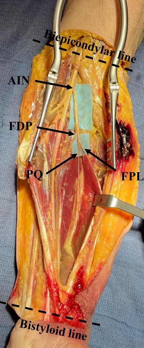
The forearm. The volar (Henry) approach reaches the deep flexor mass; identify the anterior interosseous nerve and artery on the interosseous membrane before deep dissection. The posterior subcutaneous border approach to the ulna exploits the shared aponeurotic origin of FDP, flexor carpi ulnaris and extensor carpi ulnaris. A forearm fasciotomy must decompress the deep compartment, not only the superficial one: after releasing the superficial flexors, retract them and open the fascia over FDP and flexor pollicis longus directly, and release the carpal tunnel.
Where the number comes from. The 1 cm figure is Malerich 1987, a cadaver study written because the limits then quoted ranged from 0.75 to 2.5 cm and were, in the authors' words, based on clinical impressions. Schannen 2017 then shortened cadaveric profundus tendons in 5 mm steps and found a significant quadriga at 10 mm, at the limit itself, not beyond it, and worst from the ulnar side: shortening the small finger profundus by 10 mm left the middle finger 14 mm off the palm.
So treat 1 cm as the point at which a measurable deficit already exists and past which it grows quickly, not as a budget to spend. If the tendon will not reach its footprint comfortably, the answer is a different reconstruction, not more traction.
Guidelines, Registries & Global Practice
Anatomical variation across populations
- Interconnections between profundus tendons in the distal forearm and palm are near-universal but vary in extent, which explains why the severity of quadriga differs so much between individuals - experimental work found considerable inter-subject variability in the strength loss produced by identical simulated stiffness.
- The little finger profundus may be absent, hypoplastic or fused with the ring tendon.
- The middle finger belly frequently receives innervation from both the anterior interosseous and the ulnar nerve, a variant that produces apparently contradictory electrodiagnostic results.
- Lumbrical anomalies, including bellies extending proximally into the carpal tunnel, are described causes of exertional carpal tunnel symptoms in young manual workers.
Differences in described technique between sources
- Position on profundus-related practice
- Multi-strand core repair with epitendinous suture, controlled venting of A4 and distal A2, and early active mobilisation where supervised therapy is available.
- Position on profundus-related practice
- Emphasises prompt recognition of jersey finger, the misleading normal radiograph, and referral within days for suspected type I injuries.
- Position on profundus-related practice
- Recommend that all suspected flexor tendon injuries are referred to a hand service and that early active protocols are used only where formal therapy is available.
- Position on profundus-related practice
- Emphasises complete decompression of the DEEP volar compartment during forearm fasciotomy, since FDP and FPL infarct first and produce the Volkmann deformity.
Evidence positions worth stating
- Suture anchor versus pull-out button in zone I: comparative series show similar functional outcomes, with anchors avoiding the nail-plate and button-site complications and pull-out sutures retaining a role where anchors are unavailable or bone quality is poor. Neither is proven superior in high-level randomised data.
- Timing in jersey finger: the vincular logic of Leddy and Packer remains the accepted framework. Their own recommendation for type I is repair within 7 days, with the best results in the first week; type II has a more forgiving window and type III is the most forgiving of all. Note that this is an expert recommendation from 36 injuries, not a tested threshold - no series has compared early against late repair with functional outcomes.
- Pulley venting: prospective data show venting was required in 64 per cent of repairs - but in a series restricted to injuries distal to the A2 pulley (Tang zones 2A and 2B), and it was A4 that was vented in most (56 per cent), A2 in only 8 per cent. Multi-strand repair with venting produced excellent or good results in 83 per cent of the 28 fingers followed beyond a year (76 per cent of the 25 followed for less), with no ruptures in 54 fingers. No study in this group compared venting against not venting, and none measured bowstringing.
- Doing nothing: the original avulsion series explicitly endorsed leaving asymptomatic late untreated cases alone - a position that remains correct and is frequently the safest answer.
High- versus limited-resource practice
- Well-resourced settings: ultrasound and MRI to localise a retracted tendon before surgery, suture anchors, microsurgical repair, and formal hand therapy with early active protocols and dynamic splinting.
- Limited-resource settings: the diagnosis is entirely clinical - block the proximal interphalangeal joint and test the fingertip - and a pull-out suture over a button remains a reliable, cheap and effective method of distal reattachment. Where supervised therapy is unavailable, a more protective mobilisation regimen and a robust multi-strand repair should be chosen and the patient counselled that adhesion is the likely limiting factor. Distal interphalangeal arthrodesis is an excellent, equipment-light salvage that is often the most appropriate operation in a manual worker presenting late.
MCQ Practice Points
Q: Which nerves supply flexor digitorum profundus? A: The anterior interosseous nerve (median) supplies the index and middle halves; the ulnar nerve supplies the ring and little halves. Roots C8-T1 throughout.
Q: What is the quadriga effect? A: Weakness and incomplete flexion of the other digits because the profundus tendons to the ulnar three fingers share one common muscle belly and one excursion.
Q: How far may an FDP be advanced at repair, and where does that number come from?
A: No more than 1 cm - a figure from Malerich 1987, a cadaver study written because the limits then quoted ranged from 0.75 to 2.5 cm on clinical impression alone. Beyond it you produce quadriga in the neighbours and a lumbrical plus finger in this digit. The refinement worth knowing: Schannen 2017 shortened cadaveric profundus tendons in 5 mm steps and found a significant quadriga at 10 mm, so 1 cm is the point at which a deficit is already measurable rather than a safe allowance - and it is worst when the shortened tendon is the small or ring finger.
Q: Where do the lumbricals arise? A: From the FDP tendons in the palm - the only muscles arising from and inserting into tendon. The radial two are unipennate, the ulnar two bipennate.
Q: What causes paradoxical interphalangeal extension on attempted flexion? A: A lumbrical plus finger - profundus divided or over-lengthened distal to the lumbrical origin, so the pull is diverted through the lumbrical into the extensor lateral band.
Q: Which finger is most commonly affected in jersey finger? A: The ring finger, by a wide margin in published series.
Q: A profundus retracted into the palm - which type and how urgent? A: Type I - both vincula torn, blood supply lost. Leddy and Packer advise repair within 7 days, and report that repair in the first week gave the best results.
Q: What is the relationship of FDP to FDS in the digit? A: FDP lies dorsal to FDS proximally, then passes THROUGH the split in FDS at Camper chiasm to become the volar tendon distally.
Q: Which muscles infarct first in forearm compartment syndrome? A: FDP and flexor pollicis longus - the deep volar compartment. This is why Volkmann contracture produces fixed flexion of the fingers and thumb.
Q: How do you show that a finger flexion contracture is extrinsic? A: The deformity eases with wrist flexion and worsens with wrist extension. An intrinsic contracture changes with metacarpophalangeal position instead.
Q: Define zone I. A: Distal to the FDS insertion on the middle phalanx - profundus only.
Exam Viva Scenarios
Practise clinical reasoning and management decisions out loud
“A 24-year-old rugby player felt a sudden pain in his ring finger while tackling three weeks ago. He was told it was a sprain. He cannot bend the fingertip. Radiographs are reported as normal. Assessment and plan?”
“Eight months after a traumatic amputation through the middle phalanx of the middle finger, a 52-year-old joiner complains that his whole hand is weak. He cannot fully close his ring and little fingers, but they are not stiff and they extend fully. What has happened and what would you do?”
“A 33-year-old cannot flex the tip of her index finger or the tip of her thumb after an insidious onset over two weeks preceded by severe shoulder and arm pain. Sensation is entirely normal. Where is the lesion and what else do you check?”
Anatomy
- Origin: proximal 3/4 anteromedial ulna, coronoid, interosseous membrane
- Insert: volar base of the distal phalanx - the only DIP flexor
- Passes THROUGH Camper chiasm to become volar to FDS
- Nerve: AIN (index and middle) plus ulnar (ring and little), C8-T1
- Excursion approximately 70 mm
The Shared Belly
- One common belly to the ulnar three; index relatively independent
- Quadriga: tether one, weaken three
- Advance as little as possible - 1 cm is the limit (Malerich), and 10 mm already causes quadriga
- Never suture the profundus tightly to an amputation stump
Lumbricals
- Arise from the FDP tendons in the palm
- Radial two unipennate (median), ulnar two bipennate (ulnar)
- Volar to the deep transverse metacarpal ligament
- Lumbrical plus: paradoxical IP extension - divide the lumbrical
Jersey Finger
- Ring finger commonest; forced DIP extension against active flexion
- I - palm, both vincula gone, repair within 7 days
- II - PIP, long vinculum intact, more forgiving
- III - bony fragment at A4; IV - double avulsion; V - comminuted phalanx
Pitfalls
- Normal radiograph does not exclude the injury
- A fragment on X-ray does not prove the tendon is attached to it
- Deep compartment infarcts first - Volkmann contracture
- Extrinsic tightness: worse with wrist extension, better with wrist flexion
Evidence Base
Avulsion of the Profundus Tendon Insertion in Athletes
- Thirty-six profundus insertion avulsions in athletes reviewed over five years; the injury was most common in the RING finger
- Classified by the presence of a bony fragment, the level of tendon retraction and the status of the tendon blood supply
- If the tendon retracts into the palm it should be repaired within 7 days
- If it retracts only to the proximal interphalangeal joint it can often be repaired a few months after injury
- Late untreated patients who were relatively asymptomatic were left alone; unstable distal interphalangeal joints were treated by fusion or tenodesis
- A flexor tendon graft through an intact functioning superficialis in the ring finger was seldom indicated
The Effect of Flexor Digitorum Profundus Quadriga on Grip Strength
- Thermoplastic wedges were used to simulate mild, moderate and severe stiffness of each finger in turn, in ten healthy adults, and the individual strength of each finger during power grip was measured on a digit-grip dynamometer
- Middle, ring and little finger strength fell significantly (p less than 0.05) when EACH of the other fingers was stiffened - and the authors state explicitly that this INCLUDED the index
- Index finger strength was largely unaffected by simulated stiffness of the other fingers - so the index is protected as a victim of quadriga, but stiffening it still weakens the other three
- The degree of stiffness influenced the strength lost, but there was considerable variation between individuals, so the significance of quadriga differs from person to person
A Simultaneous Distal Phalanx Avulsion Fracture with Profundus Tendon Avulsion
- Case report and literature review of the double avulsion pattern - a bony fragment with the tendon avulsed from that fragment
- The injury was classified using the Leddy and Packer system and its distinct management requirements described
- Emphasis on pathomechanism, physical findings and the surgical repair method
- Stabilisation of the distal interphalangeal joint was necessary even at the expense of early motion
Anatomy of the Finger Flexor Tendon Sheath and Pulley System
- Sixty-one fresh cadaver fingers dissected under the operating microscope
- Synovial sheath extends from the metacarpal neck to the distal interphalangeal joint
- Five annular pulleys, three cruciform pulleys and the palmar aponeurosis pulley described
- Substantial morphological variation in the A1 and cruciform pulleys
Venting or Partial Lateral Release of the A2 and A4 Pulleys After Repair of Zone 2 Flexor Tendon Injuries
- 126 consecutive zone 2 flexor tendon injuries studied prospectively, in each of which at least one flexor tendon had been completely divided
- Venting of one or other pulley was required in 81 repairs (64%) either to allow the repair to be performed or to let it glide freely
- A4 was vented between 10 and 100 per cent of its length in 71 fingers (56%)
- The distal edge of A2 was vented by only 4 to 10 mm, and in just 10 fingers (8%) - so the pulley that is usually vented is A4, not A2
Outcomes of the Six-Strand M-Tang Repair for Zone 2 Primary Flexor Tendon Repair in 54 Fingers
- Fifty-four fingers with complete zone 2 divisions repaired with a six-strand core M-Tang technique, followed by early passive motion for 3 to 4 weeks and then full active range
- Of the 28 fingers followed for MORE than one year, 24 (83%) were excellent or good, four fair and one poor; of the 25 followed for LESS than a year, 19 (76%) were excellent or good
- There were NO repair ruptures in the series
- Outcomes were significantly better in patients younger than 37, in male patients, and where the A2 pulley had been vented
- Fingers followed beyond a year had significantly SMALLER extension deficits than those followed for less - the deficit continues to improve well past the point most series stop looking
Avulsion of a Profundus Tendon with Simultaneous Intraarticular Fracture of the Distal Phalanx
- The original report of what is now universally called the TYPE IV injury - a profundus avulsion occurring together with an intra-articular fracture of the distal phalanx
- Published four years after Leddy and Packer, and indexed by PubMed under the subheading 'classification', which is how it entered the scheme
- The point of the report is that the fragment and the tendon are two separate problems: the bone can be where the radiograph shows it while the tendon has gone elsewhere
- Treatment therefore has to address the joint surface and the tendon insertion as distinct steps
Type 5 Avulsion of the Insertion of the Flexor Digitorum Profundus Tendon
- Four cases of profundus avulsion with an osseous fragment in which there was ANOTHER significant fracture of the distal phalanx
- The existing classification was reviewed and an extended classification proposed to accommodate this pattern - the origin of type V
- The distinction that matters is that the distal phalanx itself is broken, so there may be no sound bone to which the tendon can be reattached
- Type V is conventionally subdivided into 5A, where the distal phalanx fracture is extra-articular, and 5B, where it is intra-articular
Permissible Limits of Flexor Digitorum Profundus Tendon Advancement - an Anatomic Study
- This is the source of the '1 cm' rule, and it opens by pointing out that the limits then cited in the English-language literature varied from 0.75 to 2.5 cm and 'appear to be based on CLINICAL IMPRESSIONS'
- In their cadaver model the degree of advancement tolerable was 1 cm - which is where the familiar figure comes from
- The authors' own framing is that a DELICATE BALANCE exists in the profundus system, and that this balance should be considered whenever advancement is contemplated
- The context is a laceration distal to the superficialis insertion, where the proximal cut end can be advanced to the insertion rather than grafted
A Novel Cadaveric Model of the Quadriga Effect
- Five cadaveric specimens; the profundus tendons to the middle, ring and small fingers were shortened in 5 mm increments and the tip-to-palm distance of the ADJACENT fingers was recorded
- Shortening by as little as 10 mm produced a significant quadriga effect - so the commonly taught 1 cm 'safe' advancement is already at the level where a deficit appears
- The effect is DIGIT-DEPENDENT and worst from the ulnar side: shortening the SMALL finger profundus by 10 mm left the middle finger 14 mm and the ring finger 10 mm from the palm
- Shortening the RING finger by 10 mm cost the middle finger 11 mm and the small finger 9 mm, whereas shortening the MIDDLE finger cost the ring only 6 mm and the small only 5 mm