A radiographic pattern, not a diagnosis — the differential runs from abuse to brittle bone disease to normal healing variants
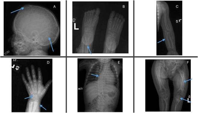
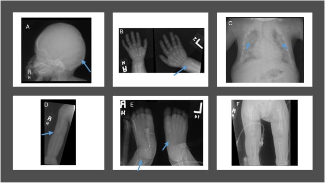
- Multiple fractures of DIFFERENT AGES in a non-ambulant infant is non-accidental injury until proven otherwise — the burden is on you to exclude it, not to prove it.
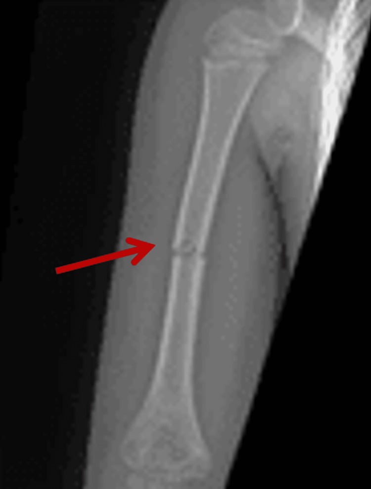
- Fractures in a child who is not yet cruising or walking are inherently suspicious: a 4-month-old cannot generate the force to fracture a femur.
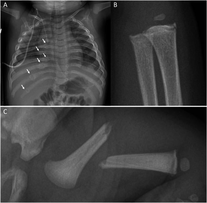
- Classic metaphyseal lesion (bucket-handle / corner fracture), posterior rib fractures, scapular, spinous process and sternal fractures are strongly associated with inflicted injury. Phrase that carefully: Kemp's systematic review could compute a probability of abuse for ribs, humerus, femur and skull only — for the CML the comparative studies do not exist, so 'most specific sign' is received teaching rather than a measured quantity.
- Osteogenesis imperfecta is the principal mimic and must be actively considered — but OI type I rarely produces posterior rib fractures or metaphyseal corner lesions.
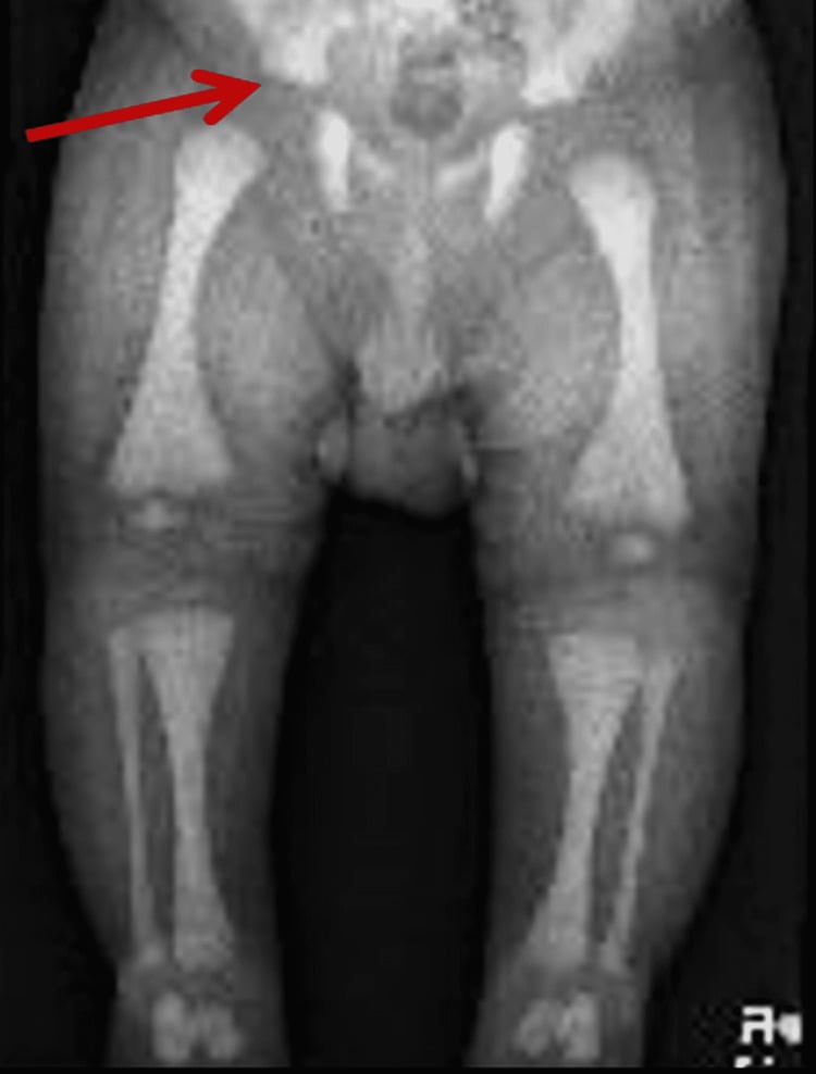
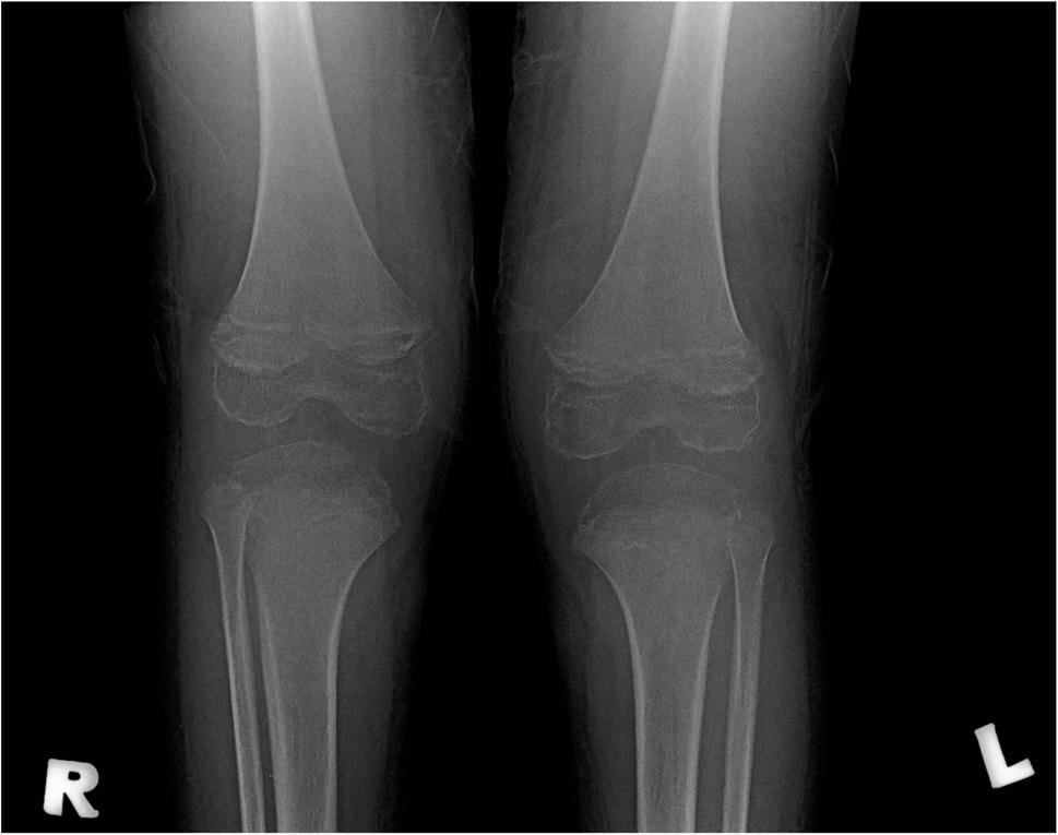
- Rickets, osteopenia of prematurity, and copper deficiency produce metaphyseal fraying that mimics CML — look at the whole metaphysis, not just its corner.
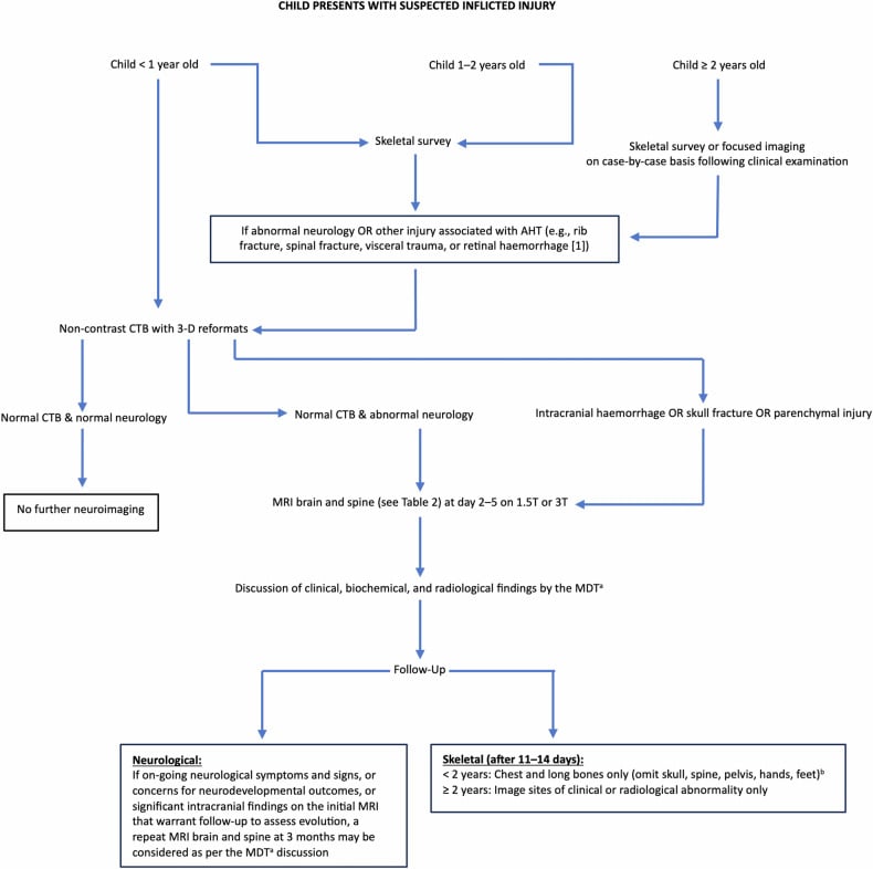
- A skeletal survey is a legal document as much as a clinical one: full series, named radiographer, repeat imaging at 11–14 days to unmask healing occult fractures.
- “Say 'fractures of differing ages' out loud — but keep the claim COMPARATIVE. Prosser's systematic review found radiological dating to be 'an inexact science' resting mostly on personal experience, so 'clearly different ages' is defensible and 'this fracture is 10 to 14 days old' is not.
- “Posterior rib fractures near the costovertebral junction arise from antero-posterior chest compression (squeezing) and are almost never caused by CPR in children.
- “Absence of blue sclerae does not exclude OI: types III and IV frequently have white or greyish sclerae.
- “Spiral fracture of the tibia in a toddler who has just started walking (toddler's fracture) is a benign accidental pattern — do not overcall it.
- “Always examine and document the retina, skin, frenulum and neurology: the skeleton is one part of the case.
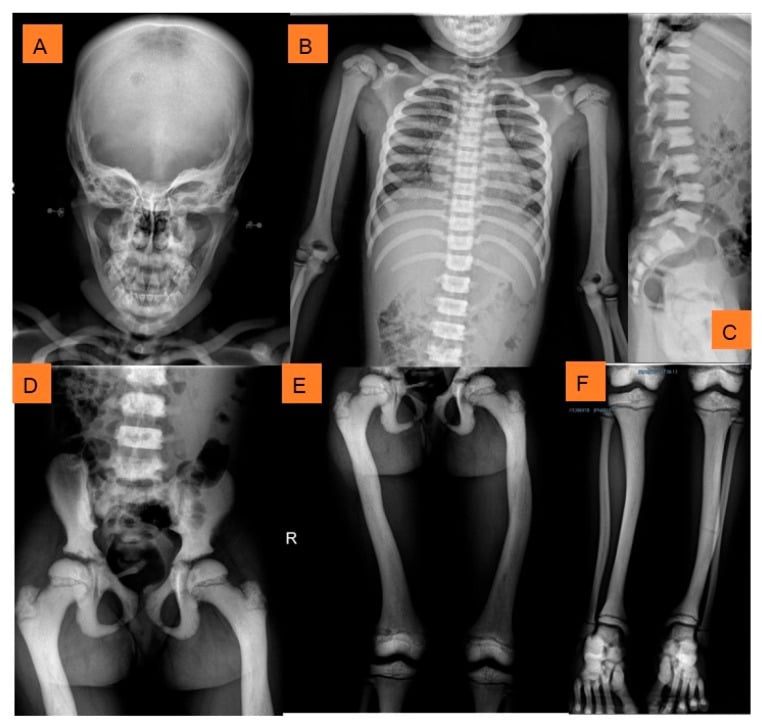
Reaching for osteogenesis imperfecta as a diplomatic answer without evidence is a fail. OI type I bones are typically gracile and osteopenic with wormian bones and a positive family history. Normal bone density with normal cortical thickness and a normal family history makes OI very unlikely.
Developmental stage is the most powerful single discriminator and it is free. Any fracture in a child who cannot yet roll, crawl or cruise demands explanation. Candidates who never ask "is the child walking?" lose the case.
Paediatric CPR uses antero-posterior compression and produces, if anything, anterior or lateral rib injury. Posterior rib fractures at the costovertebral articulation are a squeezing injury and remain highly specific for inflicted trauma. This is not merely taught, it is observed: in Weber's series of 546 infant post-mortems, every rib fracture attributed to resuscitation was anterolateral, whereas inflicted fractures were anterolateral and/or posterior, and anterior costochondral junction fractures appeared in some inflicted cases and in none of the resuscitation cases. Say so when the defence is raised.
Physiological periosteal new bone is symmetrical, smooth, less than 2 mm thick, spares the metaphysis, appears between roughly 1 and 6 months of age and involves the diaphyses of femora, tibiae and humeri. Asymmetry or metaphyseal involvement makes it pathological.
Recognising the Pattern
The finding. Two or more fractures in a single child, either simultaneously or of demonstrably different healing stages, in the absence of a mechanism that plausibly accounts for them.
Confirming the pattern is genuine.
- Are they truly fractures? Nutrient foramina, vascular grooves, unfused ossification centres (bipartite patella, os odontoideum, secondary ossification of the acromion), and the physeal plate itself are all mistaken for fracture lines. Fracture lines cross cortex and are not corticated.
- Are they truly of different ages? Dating rests on soft tissue swelling (0–10 days), periosteal new bone (typically appearing from around 7–14 days), soft callus, hard callus and finally remodelling over months. A fracture with crisp non-corticated margins and no periosteal reaction is recent; one with bridging callus is weeks old. If both appear on the same film, that is a fracture of differing ages.
- Know how far this framework is entitled to take you. Prosser's systematic review, written for exactly this context, concluded that radiological dating is "an inexact science", that most radiologists date fractures from personal clinical experience, and that the literature provides "little consistent data to act as a resource" — with an urgent call to validate the criteria in children under 5. The ranges above are a teaching framework, not a validated timetable. So make the comparative claim, which the evidence supports — these fractures are of clearly different ages — and refuse the absolute one. Attaching a narrow window to a single fracture is not defensible, and in this topic the opinion may be tested in court.
- Is the bone itself normal? Assess cortical thickness, trabecular pattern, metaphyseal morphology and the skull vault for wormian bones. Abnormal underlying bone reframes the entire differential.
Saying it out loud. "This is a frontal radiograph of the left femur of a skeletally immature child. There is a transverse mid-diaphyseal fracture with no periosteal reaction, so it is recent. On the chest film there are healing fractures of the posterior fourth, fifth and sixth ribs with bridging callus, indicating an injury several weeks old. The bone density and cortical thickness appear normal. These are fractures of differing ages in what I suspect is a non-ambulant infant — my primary concern is inflicted injury and I would escalate to the safeguarding team while completing a full skeletal survey."
Mimics — the false positives.
- Why it fools you
- Symmetrical diaphyseal periosteum in a 2–5 month old resembles healing fractures
- How to unmask it
- Symmetrical, smooth, thin, spares metaphysis, no cortical break
- Why it fools you
- Normal variant of the distal femoral and proximal tibial metaphyses
- How to unmask it
- Symmetrical, no adjacent lucent zone, no subperiosteal bone
- Why it fools you
- Metaphyseal fraying mimics classic metaphyseal lesion
- How to unmask it
- Whole metaphysis is cupped, splayed and frayed with widened physis, not just the corner
- Why it fools you
- Fragile bones, incidental healing rib fractures
- How to unmask it
- Extreme prematurity, prolonged parenteral nutrition, generalised osteopenia
- Why it fools you
- Corticated fragment near a joint mistaken for avulsion
- How to unmask it
- Smooth corticated margins, contralateral comparison
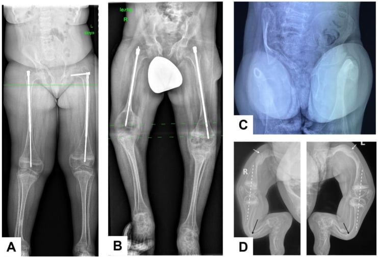
Next Investigation
Bone scintigraphy is a supplementary tool, not a replacement for skeletal survey. It is sensitive for rib and diaphyseal injury but poor at the metaphyses because of physiological physeal uptake — exactly where classic metaphyseal lesions occur.
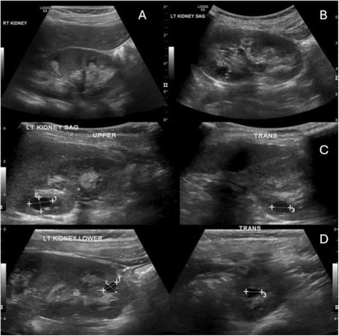
The Differential
- Typical age / setting
- Under 2 years, peak under 12 months, non-ambulant infant
- Discriminating feature
- Fractures of DIFFERING ages with normal bone density; posterior rib and classic metaphyseal lesions; history that changes or does not fit the injury
- What confirms it
- Full skeletal survey plus repeat at 11–14 days, CT head, ophthalmology for retinal haemorrhage, multidisciplinary safeguarding assessment
- Typical age / setting
- Any age; type III fractures in utero or at birth
- Discriminating feature
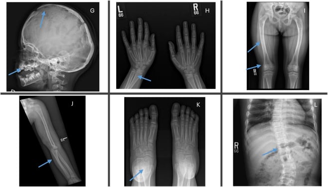
- Generalised osteopenia with THIN cortices, wormian bones in the lambdoid suture, bowing deformity, blue sclerae, dentinogenesis imperfecta, positive family history
- What confirms it
- COL1A1 / COL1A2 sequencing (plus other genes for recessive types); skin fibroblast collagen analysis if genetics negative
- Typical age / setting
- 6 months to 2 years; dark skin, exclusive breastfeeding without supplementation, malabsorption
- Discriminating feature
- Cupped, splayed, frayed WHOLE metaphysis with widened lucent physis; rachitic rosary; genu varum in the walking child
- What confirms it
- Low 25-hydroxyvitamin D, low or normal calcium, low phosphate, high alkaline phosphatase, high parathyroid hormone; wrist and knee radiographs
- Typical age / setting
- Ex-premature infant, birthweight under 1500 g, prolonged parenteral nutrition
- Discriminating feature
- Diffuse osteopenia with rib and long bone fractures occurring during routine handling in the neonatal unit; clear documented context
- What confirms it
- Alkaline phosphatase markedly raised, low phosphate; nursery records; fractures cease with mineral supplementation
- Typical age / setting
- Any age; road traffic collision, fall from significant height
- Discriminating feature
- All fractures of the SAME age with a single high-energy mechanism corroborated by independent witnesses and paramedic record
- What confirms it
- Consistent history, appropriate injury pattern and energy, trauma series imaging
- Typical age / setting
- Neonate, first 2 weeks of life
- Discriminating feature
- Clavicle, humerus or femur fracture presenting within days of a difficult delivery, shoulder dystocia or breech extraction; typically solitary
- What confirms it
- Obstetric record; healing appropriate to age of the infant; no other fractures on survey
- Typical age / setting
- Any age; unwell, febrile child
- Discriminating feature
- Periosteal reaction with underlying bone destruction and a permeative pattern, focal to one bone, with systemic sepsis
- What confirms it
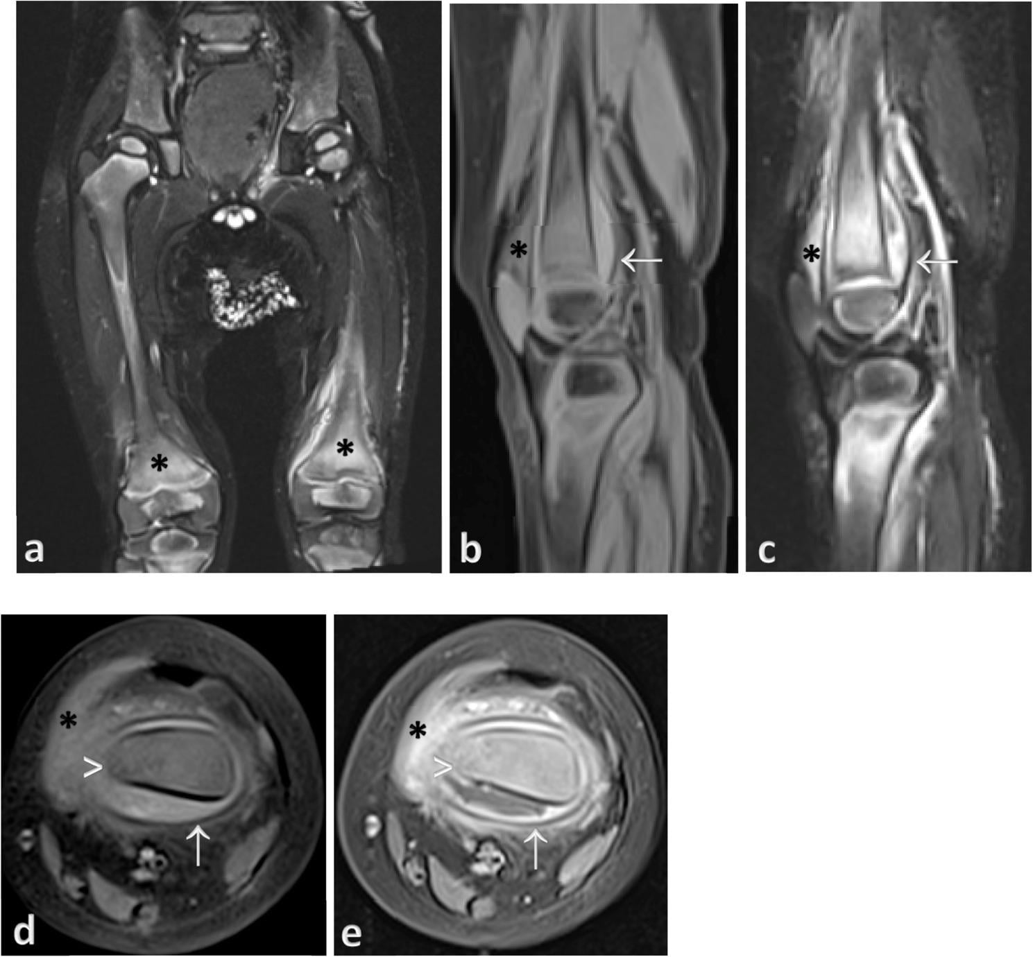
- Raised C-reactive protein and white cell count, blood cultures, MRI showing marrow oedema and abscess
- Typical age / setting
- 2–5 years for leukaemia; under 2 years for neuroblastoma
- Discriminating feature
- Metaphyseal lucent bands, permeative diaphyseal lysis and diffuse osteopenia with pallor, hepatosplenomegaly, bruising and bone pain
- What confirms it
- Full blood count with film, bone marrow aspirate, urinary catecholamines, MIBG scan
- Typical age / setting
- Non-ambulant child, often over 4 years
- Discriminating feature
- Disuse osteopenia with slender diaphyses; supracondylar femoral fractures after physiotherapy or transfers; joint contractures
- What confirms it
- Established neurological diagnosis, DXA, low-trauma fracture during documented care activity
- Typical age / setting
- Toddler onset once mobile
- Discriminating feature
- Painless fracture, self-mutilation of tongue and fingertips, repeated Charcot-like joint destruction, anhidrosis with unexplained fevers
- What confirms it
- NTRK1 / SCN9A genetics, absent nociception on formal sensory testing, normal bone mineral density
- Typical age / setting
- Under 1 year; parenteral nutrition or intestinal failure
- Discriminating feature
- Metaphyseal spurring and cupping WITH sideburn periosteal reaction plus hypopigmented kinky hair, anaemia and neutropenia
- What confirms it
- Low serum copper and caeruloplasmin; ATP7A mutation in Menkes
- Typical age / setting
- Infancy (malignant recessive form) or later (benign dominant)
- Discriminating feature
- DENSE bone — the opposite of OI — with bone-within-bone appearance, obliterated marrow cavity, transverse brittle diaphyseal fractures
- What confirms it
- Radiographs diagnostic; anaemia and cranial nerve palsies from marrow and foraminal encroachment; TCIRG1 genetics
- Typical age / setting
- Perinatal to childhood presentation
- Discriminating feature
- Fractures with rickets-like metaphyses but LOW alkaline phosphatase; premature loss of deciduous teeth with roots intact
- What confirms it
- Low serum alkaline phosphatase, raised urinary phosphoethanolamine, ALPL mutation
- Typical age / setting
- Older child, 5–15 years
- Discriminating feature
- Ground-glass expansile lesions with shepherd's crook proximal femoral deformity; café-au-lait macules with irregular coast-of-Maine borders
- What confirms it
- Radiographic appearance, bone scan for polyostotic disease, GNAS mutation on lesional tissue
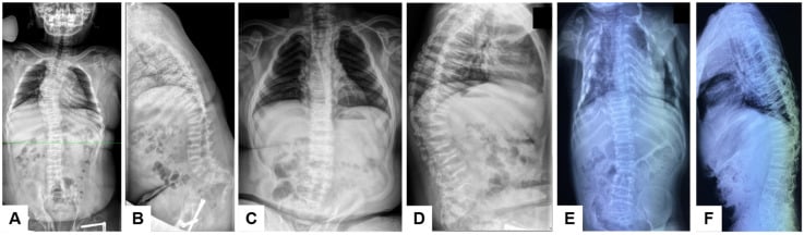
Narrowing It Down
- 1Step 1 — What is the developmental stage, not just the age?
Ask, in these words, whether the child rolls, crawls, cruises or walks - and record the answer before looking at another film.
A fracture in a NON-AMBULANT infant carries a very high probability of inflicted injury regardless of the history offered. An ambulant toddler generates real force and accidental patterns become plausible. Kemp found the developmental stage to be an important discriminator specifically for FEMORAL fractures, where the pooled probability of abuse spans 0.28 to 0.43. This single question reorders the entire differential.
- 2Step 2 — Are the fractures of the same age or different ages?
Compare fractures against each other for soft tissue swelling, periosteal reaction, soft and hard callus. Make the COMPARATIVE judgement; do not assign a date to any single fracture.
Same-age fractures with a credible high-energy mechanism support accidental polytrauma. Different ages in a normally mineralised skeleton is the signature of repeated inflicted trauma or of an intrinsic bone fragility disorder. Prosser's systematic review found radiological dating to be 'an inexact science' resting largely on personal experience, so 'clearly of different ages' is defensible and a narrow window for one fracture is not.
- 3Step 3 — Is the intervening bone normal?
Assess cortical thickness, trabecular density, metaphyseal morphology and the skull vault for wormian bones on every available film.
Normal cortex and trabeculae push you towards inflicted or accidental injury. Generalised osteopenia with thin cortices suggests osteogenesis imperfecta, prematurity, disuse or malignancy. Abnormally DENSE bone suggests osteopetrosis. Metaphyseal fraying with physeal widening suggests rickets or hypophosphatasia.
- 4Step 4 — Where in the bone, and which bones?
Map every fracture by site, then weigh each against what the comparative literature can and cannot quantify.
RIBS carry the highest measured probability of abuse - 0.71 once major trauma is excluded. Metaphyseal corner and bucket-handle lesions, scapula, spinous processes and sternum are strongly associated but were NOT quantifiable in Kemp's review for want of comparative studies. Two findings point the other way: a SUPRACONDYLAR humeral fracture is less likely to be inflicted, and a linear skull fracture is equally common in abuse and non-abuse. Mid-diaphyseal transverse fractures in bowed osteopenic bone suggest OI; a solitary spiral distal tibial fracture in a new walker is a toddler's fracture.
- 5Step 5 — Are there extraskeletal features?
Examine and document the skin, frenulum, retina and neurology in every case - the skeleton is one part of the assessment.
Blue sclerae, dentinogenesis imperfecta and joint hypermobility favour OI. Cafe-au-lait macules favour fibrous dysplasia or neurofibromatosis. Bruising in a non-mobile infant, torn frenulum, retinal haemorrhage and burns favour abuse. Pallor, hepatosplenomegaly and lymphadenopathy favour leukaemia. Kinky hypopigmented hair favours Menkes.
- 6Step 6 — What does the biochemistry say?
Send calcium, phosphate, alkaline phosphatase, parathyroid hormone, 25-hydroxyvitamin D, full blood count, clotting screen, and renal and liver profile.
High alkaline phosphatase with low phosphate points to rickets; LOW alkaline phosphatase points to hypophosphatasia. A normal panel with normal bone density leaves inflicted injury as the leading diagnosis - but a normal panel is not by itself a diagnosis of abuse.
- 7Step 7 — Does the history explain the injuries?
Take the history from each carer separately and record it VERBATIM, including the exact mechanism, the timing and who was present.
Congruence is the decisive discriminator, and Kemp's central conclusion is why: no fracture, on its own, can distinguish an abusive from a non-abusive cause. A mechanism that is absent, vague, changing between carers, developmentally impossible for the child, or disproportionate to the injury severity - combined with delayed presentation - is as diagnostically powerful as any radiograph.
MCQ Practice Points
Q: Which fracture carries the highest probability of non-accidental injury in an infant?
A: Ribs — probability of abuse 0.71 (95% CI 0.42 to 0.91) in Kemp's systematic review of 32 comparative studies, once major trauma is excluded. Then humerus (0.48 to 0.54) and femur (0.28 to 0.43), with skull lowest at 0.30. Answer the second half before the examiner asks it. The classic metaphyseal lesion, scapular, spinous process and sternal fractures are conventionally taught as the most specific signs and are strongly associated with inflicted injury — but Kemp found insufficient comparative studies to compute a probability for any of them, so that specificity is received teaching rather than a measured quantity. Two findings run the other way: supracondylar humeral fractures are less likely to be inflicted, and linear skull fractures are equally common in abuse and non-abuse. And the review's overarching conclusion: no fracture, on its own, distinguishes an abusive from a non-abusive cause.
Q: A 4-month-old has symmetrical smooth periosteal new bone along both femora and both tibiae, 1 mm thick, sparing the metaphyses. What is it?
A: Physiological periosteal reaction of infancy — a normal variant between roughly 1 and 6 months. Asymmetry, thickness greater than 2 mm, or metaphyseal involvement makes it pathological.
Q: Which biochemical result distinguishes hypophosphatasia from nutritional rickets?
A: Alkaline phosphatase. Markedly raised in rickets and low in hypophosphatasia, despite both producing rickets-like metaphyseal changes and fractures. It is the one result on the panel where a low value is the abnormal one, which is why it is missed.
Q: What is the correct timing and content of the repeat skeletal survey, and what is the evidence for it?
A: A limited repeat at 11 to 14 days including chest, long bones and any equivocal region, to detect periosteal new bone and callus from fractures that were radiographically occult at presentation. Kemp's imaging review found that a repeat survey at two weeks gave significant additional information on tentative findings and on the number and age of fractures — and that the skeletal survey commonly misses rib fractures while bone scintigraphy misses skull, metaphyseal and epiphyseal ones. Hence oblique rib views on every survey, and hence neither modality alone being adequate. The yield is significant specifically under 2 years, which is where imaging becomes mandatory rather than discretionary.
Q: Which gene defects underlie the majority of osteogenesis imperfecta cases?
A: COL1A1 and COL1A2, encoding the pro-alpha-1 and pro-alpha-2 chains of type I collagen; most cases are autosomal dominant, with rarer recessive forms involving genes such as CRTAP and P3H1. Remember that OI and inflicted injury are not mutually exclusive, and that a negative genetic panel does not exclude every fragility phenotype.
BRITTLECauses of Multiple Fractures in a Child
Hook:BRITTLE runs the differential, but it deliberately opens on B. Work through all seven - a mimic missed is a family destroyed - and hold two rules while you do it: no fracture on its own proves abuse (Kemp), and no negative test on its own excludes it, because a normal first survey misses most fresh rib fractures and normal biochemistry does not exclude inflicted injury. The discriminator that costs nothing and outperforms every letter here is whether the child can yet move independently.
Exam Viva Scenarios
Practise clinical reasoning and management decisions out loud
“You are shown this radiograph of a 5-month-old infant brought to the emergency department with a swollen thigh. There is a transverse mid-diaphyseal femoral fracture. The parents report the child rolled off a sofa. How do you proceed?”
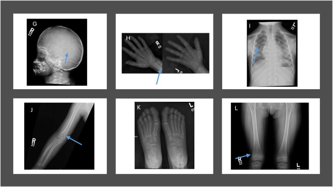
“You are shown a chest radiograph of a 9-month-old showing healing fractures of the left posterior fourth to sixth ribs with callus, and a separate wrist film showing a corner fracture of the distal radial metaphysis with no periosteal reaction. What is your interpretation?”
“You are shown radiographs of a 3-year-old with bowed, gracile femora, two healing diaphyseal fractures at different stages, and a lateral skull film with multiple small intrasutural bones. What is your diagnosis and how do you manage it?”
Red flags for inflicted injury
- Any fracture in a non-ambulant infant
- Fractures of differing ages with normal bone density
- Classic metaphyseal lesion, posterior rib, scapular, spinous process, sternal fracture
- History absent, changing, developmentally implausible or disproportionate
- Delayed presentation; bruising in a child who is not yet mobile
Osteogenesis imperfecta on plain film
- Generalised osteopenia with thin gracile cortices
- Wormian bones in the lambdoid suture
- Bowing and shepherd's crook or anterolateral tibial deformity
- Vertebral compression and codfish vertebrae
- Clinical: blue sclerae, dentinogenesis imperfecta, hearing loss, hypermobility
Rickets on plain film
- Cupped, splayed, frayed metaphyses at the wrist and knee
- Widened lucent physis
- Rachitic rosary at the costochondral junctions
- Genu varum or valgum once weight bearing
- High alkaline phosphatase, low phosphate, low vitamin D, high parathyroid hormone
Investigation bundle
- Full skeletal survey if under 2 years, plus repeat limited survey at 11–14 days
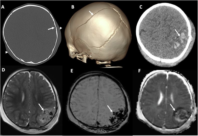
- CT head if under 1 year or any neurological sign; MRI brain and spine if positive
- Ophthalmology for retinal haemorrhage
- Bone profile, alkaline phosphatase, parathyroid hormone, 25-hydroxyvitamin D
- Full blood count with film and clotting screen
- COL1A1/COL1A2 genetics where OI is suspected
Things that are NOT abuse
- Toddler's fracture — spiral distal tibia in a new walker
- Physiological periosteal new bone at 1–6 months
- Clavicle fracture after documented shoulder dystocia
- Same-age fractures after a witnessed high-energy road traffic collision
- Metaphyseal fraying of established, biochemically confirmed rickets
Non-negotiables in the viva
- Ask whether the child is walking
- Date every fracture out loud
- Comment on the density of the intervening bone
- Escalate to safeguarding the same day and admit for protection
- Assess siblings under 2 years with skeletal survey
- Remember OI, rickets and abuse can coexist
Evidence Base
Patterns of Skeletal Fractures in Child Abuse: Systematic Review
- 32 comparative studies, all languages, each independently reviewed by paediatricians, paediatric radiologists, orthopaedic surgeons and named child-protection nurses; random-effects meta-analysis
- RIB FRACTURES carry the highest probability of abuse once major trauma is excluded: 0.71 (95% CI 0.42 to 0.91)
- HUMERUS 0.48 (0.06 to 0.94) to 0.54 (0.20 to 0.88) depending on the definition of abuse used - but SUPRACONDYLAR fractures specifically were LESS likely to be inflicted
- FEMUR 0.28 (0.15 to 0.44) to 0.43 (0.32 to 0.54), with the child's DEVELOPMENTAL STAGE an important discriminator
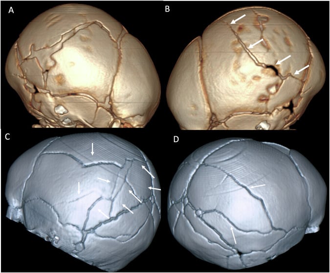
- SKULL 0.30 (0.19 to 0.46); linear fractures were the commonest pattern in abuse AND in non-abuse
- For every other fracture type - INCLUDING THE CLASSIC METAPHYSEAL LESION - there were insufficient comparative studies to calculate a probability at all
- The authors' conclusion: no fracture, on its own, can distinguish an abusive from a non-abusive cause
How Old Is This Fracture? Radiologic Dating of Fractures in Children: A Systematic Review
- Systematic review of the evidence for radiological dating of fractures in children, conducted specifically for the child-protection context
- The conclusion is unambiguous: radiologic dating of fractures 'is an inexact science'
- Most radiologists date fractures on the basis of their PERSONAL CLINICAL EXPERIENCE, and the literature 'provides little consistent data to act as a resource'
- The authors identify an URGENT need for research to validate the criteria used to date fractures in children under 5
- No validated timetable of soft tissue swelling, periosteal reaction, soft callus and hard callus emerged from the literature