A slip in the wrong patient is a metabolic diagnosis until proven otherwise
- Idiopathic SUFE occurs in a tall-for-age or obese child aged 10-16 (girls 10-14, boys 12-16) around the pubertal growth spurt. Any slip outside this window demands an endocrine or renal workup.
- Short stature with a slip is the single most powerful red flag — idiopathic slippers are of normal or above-average height for age.
- Hypothyroidism, growth hormone therapy, hypogonadism, panhypopituitarism, renal osteodystrophy and prior pelvic irradiation are the classic secondary causes.
- Renal osteodystrophy slips are usually bilateral (9 of 11 patients) in a young child (mean 10.6 years), and in about a third of hips the displacement is METAPHYSEAL rather than physeal — which changes where fixation must sit. Medical control of hyperparathyroidism is part of the treatment, not an aside.
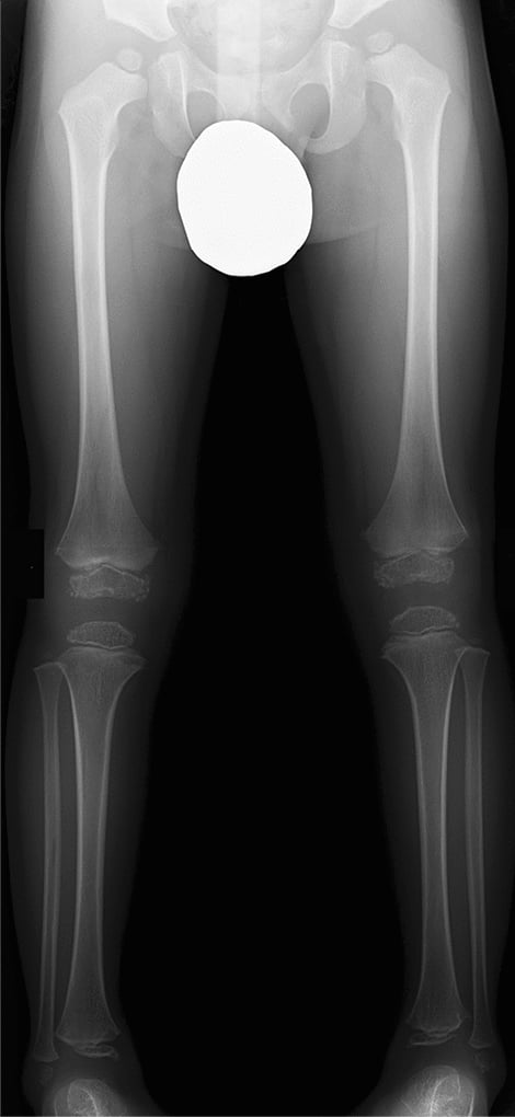
- The commonest secondary slip in a modern tertiary practice may not be endocrine at all: 19 of 22 atypical children in Chung's series were childhood cancer survivors, chemotherapy or irradiation. Atypical slips are significantly more often VALGUS with genu valgum, which Klein's line will not detect.
- Growth hormone therapy is a genuine risk factor — 92 per cent of Loder's growth-hormone-deficient patients slipped during or after supplementation — but the risk is NOT confined to the first year: Hwang found a median 3.6 years from endocrine diagnosis to slip, with 27 per cent occurring beyond five years. Monitor throughout childhood.
- Bilateral simultaneous slip, or a metachronous slip in a child under 10, is a secondary cause until the bloods return normal.
- Screening bloods do not delay surgery: fix the slip, investigate in parallel.
- “The examiner's opening gambit is 'this 9-year-old has a slip' — the answer is never 'obesity'.
- “Trethowan's sign, Klein's line and metaphyseal blanch of Steel are how you prove the slip; the patient's phenotype is how you explain it.
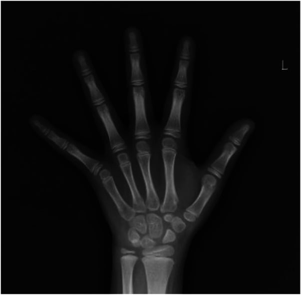
- “Bone age lagging behind chronological age by 2 years or more points to hypothyroidism or panhypopituitarism.
- “Radiation-induced slip follows treatment fields for Wilms tumour, neuroblastoma or pelvic sarcoma, typically 2-5 years later.
- “Prophylactic contralateral pinning is far more readily justified in secondary SUFE: bilaterality was 61 per cent in Loder's endocrine series against roughly a fifth to a quarter in idiopathic disease, so the default flips and the conversation with the family starts from pinning both.
Idiopathic SUFE children are at or above the 50th centile for height and heavy. A child below the 10th centile with a slip has an endocrinopathy until the thyroid function and pituitary axis are cleared. Plot the height on a growth chart in front of the examiner.
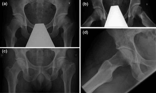
Never accept a single AP. A frog-lateral of both hips is mandatory. In endocrine and renal slips the contralateral hip is abnormal in a majority of cases, and a subtle physeal widening with no displacement is a pre-slip that must be pinned.
Referred distal thigh and knee pain via the obturator nerve is the classic delay. In secondary SUFE the onset is insidious and atraumatic, so the delay is even longer and slips present as chronic, severe deformity rather than acute unstable slips.
In renal disease the femoral neck can show generalised physeal widening, subperiosteal resorption and a rachitic frayed metaphysis without true epiphyseal displacement. Check Klein's line on both sides and compare — the metabolic change is symmetrical, a slip is not.
Recognising the Pattern
The pattern is a slipped upper femoral epiphysis occurring in a patient whose age, stature, sex, laterality or comorbidity does not fit the idiopathic mechanical model. It is defined by context rather than by a distinct radiographic appearance — the slip itself looks the same.
Confirming the slip is genuinely present
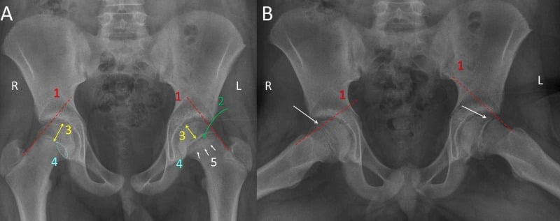
- Klein's line: a line along the superior femoral neck on the AP should intersect the lateral epiphysis. If it fails to do so, or intersects less epiphysis than the opposite side (the modified Klein or Southwick comparison), the epiphysis has slipped posteroinferiorly.
- Trethowan's sign: the same finding named — Klein's line lying entirely superior to the epiphysis.
- Metaphyseal blanch of Steel: a crescent of increased density in the proximal metaphysis on the AP, produced by the overlapped posteriorly displaced epiphysis. Present in early slips before Klein's line breaks.
- Physeal widening and irregularity with loss of the normal epiphyseal height — the earliest change, and the only change in a pre-slip.
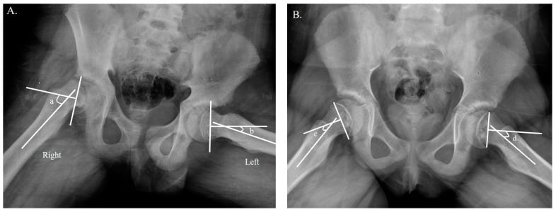
- Frog-lateral quantifies it: the Southwick head-shaft angle difference between sides gives mild (less than 30 degrees), moderate (30-50 degrees) and severe (greater than 50 degrees).
Confirming the cause is secondary
- Age less than 10 years or greater than 16 years.
- Height below the 10th centile, or a body habitus that is short and stocky rather than tall and obese.
- Bilateral simultaneous slip at presentation.
- Known chronic kidney disease, growth hormone therapy, thyroid disease, prior craniospinal or pelvic irradiation, or a recognised syndrome.
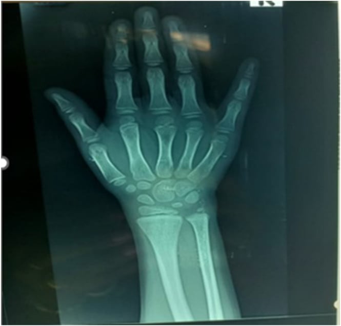
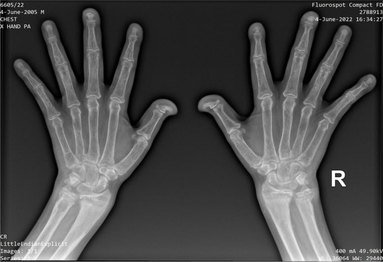
- Bone age discrepancy of 2 years or more on a left-hand-and-wrist film.
How to say it out loud
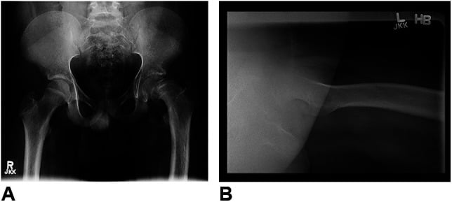
"This is an AP pelvis of a skeletally immature child. Klein's line drawn along the superior femoral neck fails to intersect the lateral epiphysis on the left, with a metaphyseal blanch sign and physeal widening — the appearances of a slipped upper femoral epiphysis. What concerns me is the context: the epiphyses look immature for the stated age and the physes elsewhere on this film are widened, so I would not accept this as idiopathic. I would plot height and weight, obtain a frog-lateral of both hips, and send thyroid function, urea and electrolytes, calcium, phosphate, alkaline phosphatase and parathyroid hormone."
Mimics — what is not a slip
- Femoral neck stress fracture / femoral neck fatigue line — the lucency crosses the metaphysis, not the physis, and Klein's line is intact.
- Perthes disease, fragmentation stage — epiphyseal sclerosis, collapse and subchondral fracture, but the physis is not displaced; typical age 4-8, younger than SUFE.
- Septic arthritis with epiphyseal destruction — systemic sepsis, raised CRP, joint space change; the epiphysis is eroded not displaced.
- Renal osteodystrophy without slip — symmetrical physeal widening and rachitic metaphyseal fraying; Klein's line intact bilaterally.
- Rotated pelvis / poor positioning — an externally rotated hip mimics posterior displacement. Always confirm on the frog-lateral before committing.
- Chondrolysis or Klein's line false positive — Klein's line has poor sensitivity for mild slips; the modified version comparing both sides is far more reliable.
Next Investigation
The Differential
- Typical age / setting
- Any age, often 8-12; short, stocky, bradycardic, constipated, poor school performance
- Discriminating feature
- Bone age markedly delayed (2 years or more); epiphyses fragmented and stippled; often bilateral atraumatic slip with no obesity
- What confirms it
- TSH markedly raised with low free T4; bone age film
- Typical age / setting
- Child aged 8-14 with known CKD, dialysis or transplant
- Discriminating feature
- Bilateral slips plus rickets-like metaphyseal fraying, subperiosteal resorption of radial phalangeal borders, rugger-jersey spine
- What confirms it
- Raised urea, creatinine, phosphate and PTH with low calcium; skeletal survey
- Typical age / setting
- Any age; profound short stature, delayed puberty, visual field defect, headaches
- Discriminating feature
- Absent pubertal growth spurt yet a slip — a growth plate that never closed; multiple axis deficiencies
- What confirms it
- Pituitary hormone profile (IGF-1, cortisol, TSH, LH/FSH) and MRI pituitary
- Typical age / setting
- 2-5 years after treatment for Wilms tumour, neuroblastoma, rhabdomyosarcoma or leukaemia craniospinal fields
- Discriminating feature
- Slip confined to the irradiated side; adjacent bone shows growth arrest, coxa valga or vara, and asymmetric pelvic hypoplasia
- What confirms it
- Radiotherapy field records; asymmetric skeletal maturation on radiograph
- Typical age / setting
- Girls 10-14, boys 12-16; BMI above 95th centile; height at or above 50th centile
- Discriminating feature
- Normal bone age, normal endocrine screen, unilateral in about half at presentation; femoral retroversion
- What confirms it
- Diagnosis of exclusion — normal TFT, U&E and growth parameters
- Typical age / setting
- Turner syndrome, Prader-Willi, GH deficiency; risk highest in first 12 months of treatment
- Discriminating feature
- Rapid recent height velocity acceleration in a previously short child; slip often bilateral
- What confirms it
- Treatment history; documented growth velocity jump on the chart
- Typical age / setting
- Boys aged 14-17 with delayed secondary sexual characteristics
- Discriminating feature
- Open physes persisting well beyond expected closure — prolonged window of vulnerability; eunuchoid proportions
- What confirms it
- Low testosterone with low or inappropriately normal LH/FSH; karyotype if Klinefelter suspected
- Typical age / setting
- Any age; global ligamentous laxity
- Discriminating feature
- Slip often mild and chronic, may coexist with atlantoaxial instability and hip dysplasia or habitual dislocation; hypothyroidism frequently coexists
- What confirms it
- Clinical phenotype plus mandatory thyroid function screen
- Typical age / setting
- Post-transplant adolescent on maintenance steroids
- Discriminating feature
- Steroid effect plus pre-existing renal bone disease; may be complicated by concurrent avascular necrosis of the femoral head
- What confirms it
- Transplant history; MRI if AVN suspected
- Typical age / setting
- Adolescent; bone pain, renal stones, abdominal symptoms
- Discriminating feature
- Brown tumours, subperiosteal resorption, physeal widening from osteoclastic resorption at the metaphyseal side
- What confirms it
- Raised PTH with raised calcium; sestamibi scan
- Typical age / setting
- Toddler to adolescent; bow legs, wide wrists, rachitic rosary
- Discriminating feature
- Symmetrical cupped and frayed metaphyses with widened physes at knee and wrist as well as hip
- What confirms it
- Low phosphate with raised alkaline phosphatase; vitamin D and FGF-23 profile
- Typical age / setting
- Short-trunk or short-limb disproportion, waddling gait
- Discriminating feature
- Symmetrical epiphyseal irregularity at multiple joints and platyspondyly; family history
- What confirms it
- Skeletal survey with lateral spine; genetic testing (COMP, COL2A1)
- Typical age / setting
- Survivor of ALL on or after high-dose steroids and methotrexate
- Discriminating feature
- Osteopenia with metaphyseal lucent bands; concurrent osteonecrosis at multiple sites
- What confirms it
- Oncology history; MRI showing multifocal osteonecrosis
- Typical age / setting
- Any age; high-energy mechanism, normal physis beforehand
- Discriminating feature
- Single violent event with immediate inability to weight-bear; no antecedent limp or thigh pain
- What confirms it
- Mechanism history; contralateral hip completely normal
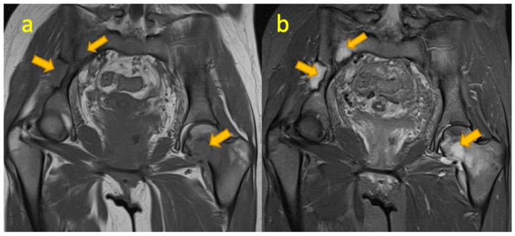
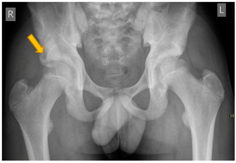
Narrowing It Down
- 11. Is a slip actually present, and in which direction?
Apply the modified Klein's line to BOTH hips, look for the metaphyseal blanch of Steel and physeal widening, and confirm on a frog-lateral. Then ask explicitly whether the epiphysis has gone posteroinferior or superolateral.
If the physes are widened symmetrically with intact Klein's lines bilaterally you are looking at metabolic bone disease rather than a slip — but that same disease is why a slip will follow. Do not stop at Klein's line: atypical slips are significantly more often VALGUS, in which the epiphysis displaces superolaterally and Klein's line stays intact. Rotation on the AP is the commonest false positive, so never diagnose a slip on a single AP.
- 22. How old is the child, how tall, and what sex?
Plot height and weight on the growth chart before ordering anything, and note the sex as a datum rather than an assumption.
Age 10-16 with height at or above the 50th centile and obesity supports idiopathic. Under 10, over 16, or height below the 10th centile mandates an endocrine and renal workup — atypical children in Chung's series averaged 132 cm and 36 kg against 149 cm and 57 kg. Loder's split is sharper still: only hypothyroid and growth-hormone-deficient patients slipped under 10, while every other endocrinopathy at an atypical age presented over 16. And the sex ratio inverts: 73 per cent of non-endocrine slips are male but 53 per cent of endocrine-associated slips are female.
- 33. Is it unilateral or bilateral, simultaneous or sequential?
Look at the contralateral hip on every film and record whether the slips were simultaneous or metachronous.
Bilateral simultaneous slip strongly favours a secondary cause — hypothyroidism, renal osteodystrophy or growth hormone therapy. Sequential slip occurs in idiopathic disease too, but a metachronous slip in a child under 10 remains secondary until proven otherwise. Bilaterality was 61 per cent in Loder's endocrine series and 9 of 11 in Oppenheim's renal series, against roughly a fifth to a quarter in idiopathic disease.
- 44. What has this child been treated with?
Ask directly about growth hormone, thyroid replacement, chemotherapy, and radiotherapy fields — and about dialysis.
In Chung's tertiary series 19 of 22 atypical children were childhood cancer survivors: 16 chemotherapy-associated and 9 radiotherapy-associated, against only 4 primary endocrine. Radiation slips follow fields for Wilms tumour, neuroblastoma or pelvic sarcoma, typically 2 to 5 years later. Growth hormone deficiency carries an SCFE incidence of 583.8 per 100,000, and 92 per cent of Loder's growth-hormone-deficient patients slipped during or after supplementation.
- 55. What do the rest of the bones look like, and what is the bone age?
Scan the whole pelvis and any incidentally included bone, then request a left hand and wrist radiograph.
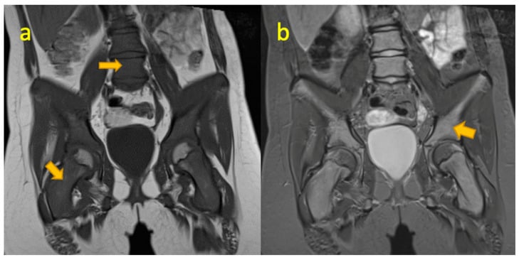
Subperiosteal resorption and a rugger-jersey spine point to renal osteodystrophy. Frayed cupped metaphyses point to rickets. Fragmented stippled epiphyses point to hypothyroidism. Asymmetric hypoplasia with growth arrest lines points to radiotherapy. Symmetrical multi-joint epiphyseal irregularity points to a dysplasia. On bone age, a delay of two years or more supports hypothyroidism, panhypopituitarism or chronic disease — Loder's endocrine patients averaged a bone age of 11.6 against a chronological age of 16.5. Bone age also grades remaining growth and therefore contralateral slip risk.
- 66. What do the bloods say, and who follows this child up?
Send TSH and free T4, urea and electrolytes with creatinine, calcium, phosphate, alkaline phosphatase, parathyroid hormone and vitamin D; add IGF-1, cortisol and gonadotrophins if pituitary disease is suspected. Refer to paediatric endocrinology or nephrology in parallel with fixation, and agree who will keep asking about the other hip.
Hypothyroidism is usually diagnosed AT the slip — the orthopaedic team finds it — whereas growth hormone deficiency is usually already known. Risk rises with the number of deficient hormones. In renal osteodystrophy the metabolic control is part of the treatment: the single fixation failure in Oppenheim's series occurred with inadequately controlled renal disease and was salvaged by subtotal parathyroidectomy, not by revising the metalwork. And surveillance must be long: Hwang found a median 3.6 years from endocrine diagnosis to slip, with 27 per cent occurring beyond five years.
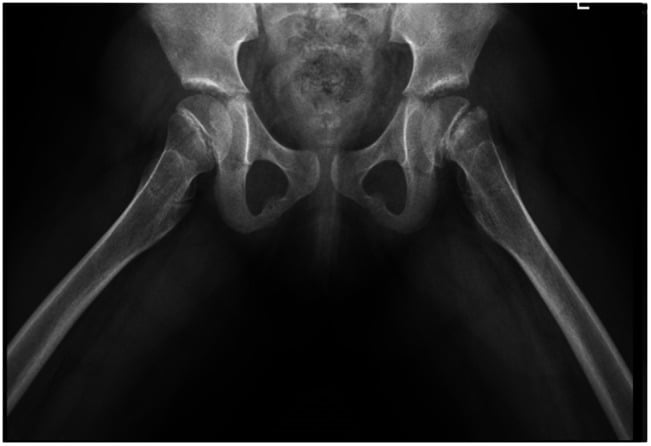
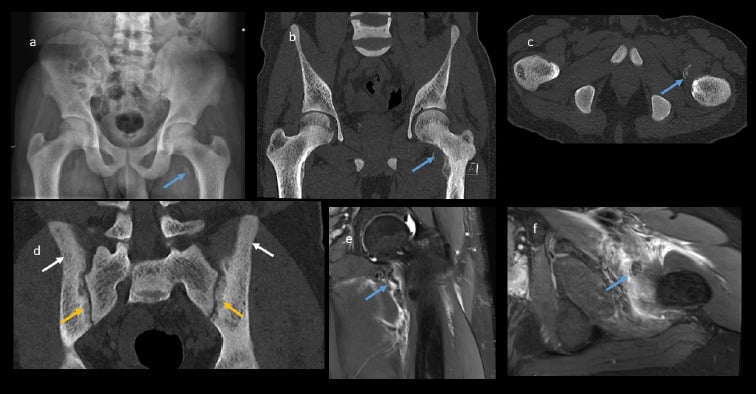
MCQ Practice Points
Q: Which single clinical parameter most strongly suggests a secondary cause of SUFE?
A: Height below the 10th centile. Idiopathic SUFE children are obese but of normal or above-average stature. Short stature with a slip points to hypothyroidism, panhypopituitarism, chronic renal disease or a skeletal dysplasia, and mandates an endocrine and renal screen. Answer the question as asked, but carry the caveat into clinic: the cut-off is a convention drawn from group means with wide overlap, so a child of normal height with an atypical age, a bilateral simultaneous slip, a valgus slip or an oncological history is still atypical.
Q: A child on growth hormone for Turner syndrome develops a slip. When is the risk highest?
A: In the first year of treatment, during the phase of accelerated growth velocity. Growth hormone widens and destabilises the hypertrophic zone of the physis. The therapy is usually continued with endocrine input after fixation rather than stopped outright, and both hips are commonly fixed.
Q: What radiographic sign distinguishes renal osteodystrophy physeal widening from a true slip?
A: Symmetry. Metabolic physeal widening is bilateral and symmetrical with intact Klein's lines and no epiphyseal displacement, and is accompanied by subperiosteal resorption and rachitic metaphyseal fraying. A slip produces asymmetric posteroinferior epiphyseal displacement with a positive Trethowan's sign and a metaphyseal blanch.
Q: What is the metaphyseal blanch sign of Steel and why does it matter?
A: A crescentic band of increased density in the proximal femoral metaphysis on the AP, produced by superimposition of the posteriorly displaced epiphysis over the metaphysis. It is visible in early slips before Klein's line becomes clearly abnormal, so it is a key early sign in the pre-slip or minimally displaced slip.
Q: How does a secondary cause change the decision on prophylactic contralateral fixation?
A: It lowers the threshold substantially. In idiopathic SUFE, contralateral fixation is selectively considered using bone age, the posterior sloping angle and modified Oxford bone age score. In endocrine, renal and syndromic slips the systemic physeal weakness affects both hips and contralateral slip rates are high, so most units fix both sides prophylactically at the index procedure.
Q: Which slip presentation carries the highest risk of avascular necrosis?
A: The acute unstable slip — one in which the child cannot weight-bear even with crutches. The retinacular vessels are at risk from the displacement itself. Secondary causes more commonly present as chronic stable slips because the onset is insidious and atraumatic, but where they present acutely and unstably the AVN risk is the same or worse given the abnormal bone.
Exam Viva Scenarios
Practise clinical reasoning and management decisions out loud
“You are shown this AP pelvis of a 9-year-old girl who has limped for six months. Klein's line fails on both sides and the epiphyses appear fragmented. She is on the 3rd centile for height.”
“You are shown this radiograph of a 12-year-old boy, three years after nephrectomy and flank radiotherapy for a Wilms tumour, who presents with left thigh pain. There is a left-sided slip and the left hemipelvis looks smaller than the right.”
“You are shown this pelvis of a 13-year-old on haemodialysis for congenital renal dysplasia. Both femoral necks show widened irregular physes and there is subperiosteal resorption along the medial femoral necks.”
Red flags for a secondary cause
- Age less than 10 or greater than 16 years
- Height below the 10th centile
- Bilateral simultaneous slip at presentation
- Known CKD, dialysis or transplant
- Current growth hormone therapy
- Prior pelvic or craniospinal radiotherapy
- Delayed puberty or eunuchoid proportions
- Bone age delayed by 2 years or more
The core secondary causes
- Hypothyroidism — short, stocky, delayed bone age, fragmented epiphyses
- Renal osteodystrophy — subperiosteal resorption, rugger-jersey spine, raised PTH
- Panhypopituitarism — no pubertal spurt, absent axes, MRI pituitary
- Growth hormone therapy — risk peaks in first 12 months
- Hypogonadism — physes stay open beyond normal closure
- Radiotherapy — 2-5 year latency, asymmetric hypoplasia in the field
- Down syndrome — laxity plus frequent coexistent hypothyroidism
Radiographic signs of the slip
- Klein's line fails to intersect the lateral epiphysis (Trethowan's sign)
- Modified Klein's line — compare epiphyseal width intersected on both sides
- Metaphyseal blanch sign of Steel — early, before Klein's line breaks
- Physeal widening and irregularity — the pre-slip
- Southwick head-shaft angle: mild less than 30, moderate 30-50, severe greater than 50 degrees
Workup
- AP pelvis plus frog-lateral of both hips (cross-table lateral if unstable)
- Growth chart — height, weight, BMI centiles
- TSH, free T4
- U&E, creatinine, calcium, phosphate, ALP, PTH, vitamin D
- Bone age — left hand and wrist
- IGF-1, cortisol, LH/FSH and MRI pituitary if pituitary disease suspected
Management principles
- Bloods never delay theatre — fix the slip, investigate in parallel
- In-situ fixation with a cannulated screw perpendicular to the physis, central in the epiphysis
- Low threshold for prophylactic contralateral fixation in all secondary slips
- Joint care with paediatric endocrinology or nephrology is mandatory
- Anticipate slower healing, fixation failure and premature physeal closure in abnormal bone
- Counsel on AVN and chondrolysis risk, higher in unstable slips
Evidence Base
Incidence and Clinical Characteristics of Slipped Capital Femoral Epiphysis in Patients with Endocrinopathy: A Population-Based Cohort Study
- Nationwide South Korean database, every new diagnosis of endocrinopathy or slipped capital femoral epiphysis between 2002 and 2019 in the 2002-2005 birth cohorts
- SCFE incidence was 37.1 per 100,000 children WITH endocrinopathy against 9.0 per 100,000 WITHOUT - relative risk 4.1 (95 per cent CI 2.8 to 6.1)
- Growth hormone deficiency carried by far the highest incidence at 583.8 PER 100,000 - roughly 65 times the background rate
- Risk rose linearly with the NUMBER of deficient hormones (Cochran-Armitage, p less than 0.001)
- The sex ratio INVERTS: 73 per cent male in non-endocrine slips but 53 per cent FEMALE in endocrine-associated slips (p equals 0.009); 22 of 30 endocrine slips came after the endocrine diagnosis at a median of 3.6 years, and 6 of those 22 (27 per cent) occurred more than FIVE YEARS later
Slipped Capital Femoral Epiphysis Associated with Endocrine Disorders
- 85 patients with an endocrine disorder and slipped capital femoral epiphysis: hypothyroidism 40 per cent, growth hormone deficiency 25 per cent, other 35 per cent
- Bone age lagged chronological age markedly - 11.6 years against 16.5 years in the 53 hips where both were recorded (p less than 0.01)
- Age at first slip ranged from 7 to 35 years, and the split is diagnostic: ONLY hypothyroid and growth-hormone-deficient patients presented under 10, while every other endocrinopathy presenting at an atypical age presented OVER 16
- Hypothyroid children usually had the endocrine diagnosis made AT the slip; growth-hormone-deficient children usually had it made BEFORE, and 92 per cent slipped during or after supplementation
- BILATERALITY WAS 61 PER CENT (p less than 0.01), on which the authors base their recommendation to consider prophylactic fixation of the opposite hip
Clinical and Radiographic Characteristics of Atypical Slipped Capital Femoral Epiphysis
- 53 patients (66 hips) treated 1995-2016 at one centre; 29 slips were atypical and 38 typical, compared directly
- THE CAUSES WERE NOT MAINLY ENDOCRINE: of 22 children with atypical slips, 19 had a NEOPLASM (8 neuroblastoma, 3 medulloblastoma, 3 retinoblastoma, 2 ependymoma, 1 craniopharyngioma, 1 Wilms tumour, 1 immature teratoma) and only 4 had a primary endocrine disorder
- By mechanism: 16 chemotherapy-associated, 9 radiotherapy-associated, 4 endocrine-associated
- Atypical children were lighter (36.2 versus 56.9 kg, p less than 0.001), shorter (132.1 versus 148.9 cm, p less than 0.001) and had a much lower body mass index (19.4 versus 25.8, p less than 0.001)
- Atypical slips were significantly more often VALGUS, and significantly more often accompanied by GENU VALGUM (both p less than 0.001)
Valgus SUFE - Why Klein's Line Cannot Find It, and Why the Screw Is Dangerous
- Ordinary SUFE is a POSTEROMEDIAL displacement of the epiphysis on the metaphysis; the valgus slip is SUPEROLATERAL - the opposite direction
- 4 children with 7 stable valgus slips found among 105 children with 141 idiopathic SUFEs at one institution
- Pooling the authors' cases with the literature gave 22 children and 30 valgus slips, mean age 12.4 years, weight 69.3 kg, height 155.3 cm, BMI 27.1
- Demographics matched routine SUFE except for sex - 76% of valgus slips occurred in GIRLS
- Valgus slips tended to be LESS severe, and differed significantly from varus slips in symptom duration and body mass index
- The authors state plainly that in a valgus SCFE Klein's line WILL ALWAYS BE NORMAL, which is why lateral radiographs are needed in every child assessed for a slip
- Single central screw fixation must be approached with caution: the more medial entry point places the screw path in immediate proximity to the femoral neurovascular bundle
Outcome of Slipped Capital Femoral Epiphysis in Renal Osteodystrophy
- 11 consecutive patients with renal osteodystrophy-associated slipped capital femoral epiphysis, 20 hips, mean age at presentation 10.6 years
- NINE OF ELEVEN were bilateral
- The slip was PHYSEAL in 13 hips but METAPHYSEAL in 7 - the displacement is not always through the growth plate in this group
- All had prompt medical treatment of the bone disease and 9 underwent surgical stabilisation with multiple custom Steinmann pins, threaded proximally and smooth distally to allow continued growth
- 14 of 16 operated hips (88 per cent) stabilised at a mean radiographic follow-up of 5.7 years; the single progression occurred in a patient with INADEQUATELY CONTROLLED renal disease and needed subtotal parathyroidectomy plus revision fixation