Loss of the crisp zone of provisional calcification at the growing end of a long bone — a pattern, not a diagnosis
- Fraying is loss of the sharp metaphyseal margin; cupping is concavity; splaying is transverse widening. They travel together in rickets but can occur alone.
- Image the fastest-growing physes: distal femur, proximal tibia, distal radius/ulna, anterior rib ends. A normal-looking ankle does not exclude rickets.
- A widened, lucent physis with fraying equals rickets until proven otherwise. Normal physeal width with metaphyseal irregularity points to dysplasia, infection or infiltration.
- Alkaline phosphatase is the single most useful screening blood test: markedly raised in rickets, normal in metaphyseal chondrodysplasia, low in hypophosphatasia.
- Classic metaphyseal lesions (corner/bucket-handle) of non-accidental injury are focal and unilateral or asymmetric — not a symmetrical generalised fraying.
- Healing rickets after treatment produces a dense band of provisional calcification; do not call this a new pathology.
- Metaphyseal lucent bands in a miserable toddler with limp and no rickets biochemistry means leukaemia until the blood film says otherwise - but bands appear in only about 10 per cent of children with acute leukaemia, so a normal metaphysis excludes nothing. The full blood count and film do the ruling out, not the radiograph.
- Vitamin D deficiency does NOT explain away inflicted injury. The Global Consensus is explicit that biochemical deficiency ALONE carries no increased fracture risk - the risk attaches to radiographically confirmed rickets. A low 25OHD with a normal metaphysis is not a metabolic cause of fractures, and finding one does not discharge the safeguarding duty.
- “Describe before diagnosing: 'symmetrical fraying, cupping and splaying of the distal radial and ulnar metaphyses with widening of the physis'.
- “Rachitic rosary, Harrison sulcus and craniotabes are the clinical partners of the radiographic sign.
- “Normal physiological metaphyseal irregularity at the distal femur (medial cortical spur/notch) is a common false positive.
- “Hypophosphataemic (X-linked) rickets: normal calcium, low phosphate, raised ALP, normal PTH, and a child who is bow-legged but not tetanic.
- “Scurvy gives a dense white line (Frankel) with a lucent Trümmerfeld zone beneath it and corner Pelkan spurs — not true fraying.
Recognising the Pattern
Definition. The metaphysis normally ends in a thin, dense, sharply defined line, the zone of provisional calcification, where the columns of hypertrophic chondrocytes mineralise before ossification. Loss of that line produces three related appearances, which travel together in rickets but can occur alone:
- Fraying - the metaphyseal margin becomes ragged, brush-like, indistinct
- Cupping - the metaphyseal end becomes concave, saucered, embracing the physis
- Splaying - the metaphysis flares transversely, wider than the diaphysis
The widened physis. Unmineralised hypertrophic cartilage accumulates and is radiolucent, so alongside the frayed margin the physis appears widened. That widening is the single most discriminating feature of a mineralisation defect, and genuine widening is obvious, not subtle.
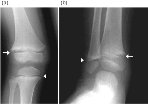
Where to look. Check at least two fast-growing sites: the distal radius and ulna, the distal femur, the proximal tibia and the anterior rib ends. Ankles and hips grow slowly, and a negative ankle film has misled many candidates and some clinicians; if you want to see rickets, film the wrist and the knee.
The company it keeps. Rickets softens and coarsens the trabeculae, whereas a dysplasia leaves density normal, so assess density across the whole film rather than the margin alone. Look also for bowing, cortical thinning, Looser zones and, in older children, slipped epiphyses, the companions of osteomalacic bone.
Saying it out loud. "This is an anteroposterior radiograph of the wrist in a young child. There is symmetrical fraying, cupping and splaying of the distal radial and ulnar metaphyses, with widening and lucency of the physes and coarsened trabeculae. The epiphyseal ossification centres are indistinct. The appearances are those of a metabolic mineralisation defect, most likely rickets. I would complete this with a knee film, a bone profile including alkaline phosphatase, phosphate, calcium, PTH and 25-hydroxyvitamin D, and a renal profile."
Mimics. Several appearances are called fraying and are not, and the false positives are worth knowing by name:
- Physiological metaphyseal irregularity at the medial distal femur and proximal tibia in toddlers: focal, cortical, no physeal widening
- Normal distal femoral cortical irregularity, the cortical desmoid, in older children
- Healing rickets: a dense sclerotic band forming beneath a still-irregular margin is a treatment response, not disease progression, and not a new pathology
- Overlying bowel gas or soft-tissue shadow on poorly collimated films
- Scurvy: the margin is dense and sharply white (the Frankel line) and the lucency lies beneath it in the metaphysis; that is not fraying
- Chondroblastoma or infection adjacent to the physis, which produce focal rather than generalised irregularity
Next Investigation
The list runs from the tests every child with the sign needs to the ones a particular pattern earns, and it ends with the child who needs nothing further.
When a metaphyseal lesion raises the question of inflicted injury, a low 25-hydroxyvitamin D is often produced as the innocent explanation. It is not one, and you should be able to say why in three steps.
- The Global Consensus is explicit that biochemical vitamin D deficiency alone does not increase fracture risk. The increased risk attaches to radiographically confirmed rickets, not to a number on a blood form. Deficiency is common; rickets is not.
- The lesions are different objects. Rickets is a generalised failure to mineralise, and its fraying is symmetrical, along the whole metaphyseal margin, with a widened lucent physis. The classic metaphyseal lesion is a focal planar fracture through the primary spongiosa separating a discrete fragment, with a normal physeal width. One is a mineralisation defect, the other is a fracture.
- So the two can coexist without one excusing the other. A vitamin D deficient infant can also be assaulted. Finding a low 25OHD does not close the safeguarding question; only the radiographic pattern, the fracture inventory and the history do that.
Treat the deficiency, and follow the safeguarding pathway on its own evidence. The two run in parallel.
The Differential
Each row gives the setting in which the cause presents, the feature that separates it from the rest, and the test that confirms it. Two causes have no row of their own: cytomegalovirus sits with syphilis and rubella among the congenital infections, and neuroblastoma metastases sit with leukaemia among the malignancies.
- Typical age / setting
- 2–5 years, unwell, limp, pallor, bone pain at night
- Discriminating feature
- Transverse lucent metaphyseal bands plus periosteal reaction and diffuse osteopenia; child is systemically ill; physis is NOT widened
- What confirms it
- Full blood count and blood film, then bone marrow aspirate
- Typical age / setting
- Less than 18 months, non-ambulant infant with fracture
- Discriminating feature
- Focal PLANAR fracture through the metaphysis (not an avulsion) - projects as a corner or bucket-handle depending on the beam; asymmetric, often multiple bones of differing ages, rib fractures
- What confirms it
- Skeletal survey with follow-up films at 11–14 days; safeguarding referral
- Typical age / setting
- Neonate, positive maternal serology, hepatosplenomegaly
- Discriminating feature
- Wimberger sign — symmetrical erosion of the medial proximal tibial metaphysis; diaphyseal periostitis
- What confirms it
- Treponemal serology, PCR; long-bone series
- Typical age / setting
- Perinatal to childhood; premature tooth loss, fractures
- Discriminating feature
- Fraying with tongue-like lucencies extending into the metaphysis and a LOW alkaline phosphatase — the reverse of rickets
- What confirms it
- Serum ALP low, raised urinary phosphoethanolamine, ALPL gene testing
- Typical age / setting
- 6–24 months; breastfed without supplements, dark skin, limited sunlight
- Discriminating feature
- Symmetrical fraying, cupping, splaying with WIDE lucent physis; rachitic rosary; genu varum once walking
- What confirms it
- Low 25-OH vitamin D, raised ALP and PTH, low or normal calcium, low phosphate
- Typical age / setting
- Presents at walking age, 1–2 years; family history, short stature
- Discriminating feature
- Rickets with NORMAL calcium and NORMAL PTH but persistently low phosphate and phosphaturia; lower limbs affected out of proportion to wrists
- What confirms it
- Serum phosphate low, tubular reabsorption of phosphate reduced, raised FGF23, PHEX mutation
- Typical age / setting
- Older child, chronic kidney disease, growth failure
- Discriminating feature
- Rickets PLUS sclerosis, subperiosteal resorption of radial phalangeal margins and rugger-jersey spine; slipped epiphyses
- What confirms it
- Urea and creatinine, raised phosphate, very high PTH, low 1,25-dihydroxyvitamin D
- Typical age / setting
- Walking age onwards; short stature, waddling gait, well child
- Discriminating feature
- Metaphyseal irregularity with NORMAL physeal width and NORMAL biochemistry; coxa vara and bowing; spine spared
- What confirms it
- Normal bone profile; COL10A1 mutation; skeletal survey
- Typical age / setting
- Infancy onwards; fine sparse light hair, Amish/Finnish ancestry
- Discriminating feature
- Metaphyseal changes with hair abnormality, immunodeficiency and ligamentous laxity; fibular overgrowth at ankle
- What confirms it
- RMRP gene testing; lymphocyte subsets
- Typical age / setting
- Infancy; severe short-limbed dwarfism
- Discriminating feature
- Grossly expanded, bulbous, fragmented metaphyses with HYPERcalcaemia despite normal PTH
- What confirms it
- Serum calcium raised, PTH suppressed; PTH1R activating mutation
- Typical age / setting
- 6 months–2 years; restricted diet, autism spectrum, refusal to bear weight
- Discriminating feature
- Dense white Frankel line with lucent Trümmerfeld zone beneath and Pelkan corner spurs; subperiosteal haemorrhage
- What confirms it
- Serum ascorbate low; dramatic response to vitamin C within days
- Typical age / setting
- School age, insidious multifocal pain, often clavicle and metaphyses
- Discriminating feature
- Multifocal symmetrical metaphyseal lysis with sclerosis, no organism, culture negative, relapsing course
- What confirms it
- Whole-body MRI showing multifocal marrow oedema; biopsy to exclude malignancy
- Typical age / setting
- Any age; fever, focal tenderness, raised CRP
- Discriminating feature
- SINGLE metaphysis, focal lysis with periosteal reaction, soft-tissue swelling and refusal to move that limb
- What confirms it
- MRI with contrast, blood cultures, aspiration
- Typical age / setting
- Preterm infant, prolonged parenteral nutrition, cholestasis
- Discriminating feature
- Metaphyseal cupping with spurs plus osteopenia and sideroblastic anaemia/neutropenia in copper deficiency
- What confirms it
- Serum copper and caeruloplasmin; nutritional review; ALP for osteopenia of prematurity
Narrowing It Down
Seven questions, asked in order, take the film to a diagnosis. The first two are answered on the radiograph, the next two from the history and examination, the fifth and sixth from the blood form, and the last by looking beyond the metaphysis.
- 1Step 1 - Is the physis widened?
Compare the height of the physeal lucency against a normal physis on the same film - distal ulna against distal radius, or the contralateral side.
Widening PLUS fraying is a mineralisation defect: rickets of any cause, or hypophosphatasia. Normal physeal width redirects you to dysplasia, infiltration, infection or trauma. This single question halves the differential.
- 2Step 2 - Is it symmetrical and generalised, or focal?
Check every fast-growing metaphysis you can see, and check both sides on the same exposure.
Symmetrical involvement of all fast-growing metaphyses is metabolic or dysplastic. A single metaphysis is infection, trauma or tumour. Asymmetric focal corner lesions in a non-ambulant infant are the classic metaphyseal lesion of inflicted injury until proven otherwise.
- 3Step 3 - How old is the child, and are they walking?
Place the child on the developmental timeline before interpreting the film.
Under 6 months suggests congenital infection, hypophosphatasia or severe maternal vitamin D deficiency. 6 to 24 months is the peak for nutritional rickets and scurvy. Progressive bowing at walking age with normal calcium suggests X-linked hypophosphataemic rickets. School age with multifocal pain suggests CRMO.
- 4Step 4 - Is the child systemically well?
Examine the child, not only the radiograph: pallor, fever, night pain, hepatosplenomegaly, growth and hydration.
A thriving child with short stature and normal bloods is metaphyseal chondrodysplasia. A pale, febrile, night-waking child with bone pain is leukaemia or infection. Failure to thrive with polyuria and growth failure points to renal osteodystrophy.
- 5Step 5 - What does the alkaline phosphatase do?
Read ALP first and interpret it against an age-specific reference range, because ALP is physiologically high in growing children.
Markedly raised ALP with low phosphate confirms active rickets. Normal ALP with metaphyseal irregularity effectively excludes rickets and supports dysplasia. A LOW ALP is hypophosphatasia - the result candidates forget, and the one that contraindicates bisphosphonates.
- 6Step 6 - Split the rickets by calcium, phosphate and PTH
Take the bone profile as a pattern rather than reading each value alone.
Low vitamin D with raised PTH is nutritional. Low phosphate with normal calcium and normal PTH is X-linked hypophosphataemic. Raised phosphate with very high PTH and raised creatinine is renal osteodystrophy. Normal phosphate with hypercalciuria and low PTH suggests a tubular or hereditary variant needing specialist review.
- 7Step 7 - Look beyond the metaphysis before committing
Review the spine, hands, ribs, skull, hair and teeth before you name the diagnosis.
Rugger-jersey spine in renal disease, platyspondyly in spondylometaphyseal dysplasia, subperiosteal resorption in the hands, rosary or fractures in the ribs, craniotabes and wormian bones in the skull, and the hair and dental changes of cartilage-hair hypoplasia and hypophosphatasia.
MCQ Practice Points
Q: Which radiographic feature best distinguishes rickets from metaphyseal chondrodysplasia?
A: Widening of the physis. Both give an irregular metaphyseal margin, but only the mineralisation defect of rickets accumulates unmineralised hypertrophic cartilage and widens the growth plate. Bone density is also reduced in rickets and normal in dysplasia.
Q: A child has metaphyseal fraying, fractures and early loss of deciduous teeth. Alkaline phosphatase is below the reference range. Diagnosis?
A: Hypophosphatasia, from a loss-of-function ALPL mutation. A low ALP is the discriminating result in this context; urinary phosphoethanolamine and plasma pyridoxal-5-phosphate are elevated. Never give bisphosphonates.
Q: Which biochemical profile fits X-linked hypophosphataemic rickets?
A: Low serum phosphate, normal calcium, raised alkaline phosphatase, normal or near-normal PTH and normal 25-hydroxyvitamin D, with renal phosphate wasting driven by excess FGF23. This distinguishes it from nutritional rickets, in which PTH is raised and vitamin D is low.
Q: Which metaphyseal finding is most specific for inflicted injury in an infant?
A: The classic metaphyseal lesion - a corner fracture or bucket-handle appearance, representing a planar fracture through the primary spongiosa. It is focal, often bilateral but asymmetric, and typically found at the distal femur, proximal tibia and proximal humerus in a non-ambulant infant.
Q: Which sign in a neonate suggests congenital syphilis rather than rickets?
A: The Wimberger sign - symmetrical destruction of the medial aspect of the proximal tibial metaphyses - accompanied by diaphyseal periostitis and metaphyseal lucent bands, with normal physeal width and positive treponemal serology.
Exam Viva Scenarios
Practise clinical reasoning and management decisions out loud
“You are shown this AP radiograph of both knees in an 18-month-old, exclusively breastfed, of South Asian background, who has been walking for three months with progressive bowing. Describe what you see.”
“You are shown this AP knee radiograph of a 4-year-old who refuses to weight-bear and wakes at night with pain. There are transverse lucent bands at the distal femoral and proximal tibial metaphyses with faint periosteal reaction. Bloods are pending.”
“You are shown radiographs of a 5-year-old with short stature and a waddling gait. There is irregularity and flaring of the metaphyses of the femora and tibiae with coxa vara, but the physeal width is normal, bone density is normal and the spine is unremarkable. Bone profile including ALP is entirely normal.”
Describe in this order
- Fraying (ragged margin), cupping (concavity), splaying (transverse flare)
- Physeal width — widened or normal
- Bone density and trabecular pattern
- Symmetry and number of sites involved
- Bowing, fractures, periosteal reaction, soft tissues
Wide physis (mineralisation defect)
- Nutritional vitamin D deficiency rickets — low vitamin D, high PTH, high ALP
- X-linked hypophosphataemic rickets — low phosphate, normal PTH
- Renal osteodystrophy — high phosphate, very high PTH, raised creatinine
- Hypophosphatasia — LOW alkaline phosphatase
Normal physis (not rickets)
- Leukaemia — lucent bands, unwell child, periosteal reaction
- Non-accidental injury — focal corner / bucket-handle lesions
- Metaphyseal chondrodysplasia (Schmid, McKusick, Jansen) — well child, normal bloods
- Osteomyelitis / CRMO — focal or multifocal, raised inflammatory markers
- Scurvy — dense Frankel line, Trümmerfeld zone, Pelkan spurs
First-line workup
- AP wrist and AP knee radiographs
- Calcium, phosphate, ALP, PTH, 25-hydroxyvitamin D, urea and creatinine
- Full blood count and film if systemically unwell
- Skeletal survey and safeguarding if under 2 years with focal metaphyseal lesions
Red flags
- Night pain, refusal to weight-bear, pallor or hepatosplenomegaly
- Fracture in a non-ambulant infant
- Low alkaline phosphatase
- Failure of radiographic healing after 3 months of adequate vitamin D therapy
Evidence Base
Radiographic Scoring Method for the Assessment of the Severity of Nutritional Rickets
- The origin of the 10-point Thacher score, which grades wrist and knee radiographs on the degree of metaphyseal FRAYING and CUPPING and the proportion of the growth plate affected, in half-point increments from 0 (normal) to 10 (severe)
- Four trained physicians scored radiographs from 67 children with active rickets on two separate occasions
- Reliability was good: interobserver correlation 0.84 or greater for every observer pair, and intraobserver correlation 0.89 or greater for each observer
- Correlation with alkaline phosphatase was only MODERATE at r = 0.58 - the radiograph and the biochemistry measure related but not interchangeable things
Global Consensus Recommendations on Prevention and Management of Nutritional Rickets
- An evidence-based consensus from 33 nominated experts representing 11 international scientific organisations, developed with a systematic literature search and graded using GRADE
- Defines vitamin D status by serum 25-hydroxyvitamin D: DEFICIENCY under 30 nmol/L, INSUFFICIENCY 30-50 nmol/L, SUFFICIENCY above 50 nmol/L
- Rickets is diagnosed on history, examination and biochemistry and must be CONFIRMED BY RADIOGRAPHS - a low 25OHD alone does not make the diagnosis
- Biochemical vitamin D deficiency ALONE does not increase fracture risk; the raised risk attaches to radiographically confirmed rickets
- Ranks risk factors in mothers and infants, and makes prevention recommendations for both the clinical and the public health setting, including supplementation and food fortification
- Does NOT recommend routine 25OHD screening in healthy children, and frames rickets as a preventable global public health problem
High-Resolution CT with Histopathological Correlates of the Classic Metaphyseal Lesion of Infant Abuse
- Long bone specimens from five fatally abused infants whose skeletal surveys showed definite or suspected classic metaphyseal lesions, imaged post-mortem by micro-CT at 45 micrometre resolution and correlated with histopathology
- Confirms that the CML is a FRACTURE extending in a PLANAR fashion through the metaphysis, separating a disk-like mineralised fragment - it is not an avulsion and not a metabolic lesion
- The separated fragment has two components: a thick PERIPHERAL part encompassing the subperiosteal bone collar, and a thin CENTRAL part made up mostly of the radiographic zone of provisional calcification
- That geometry is why the same lesion projects as a CORNER fracture when viewed obliquely and as a BUCKET-HANDLE when viewed en face - one lesion, two appearances, depending on the beam
Musculoskeletal Manifestations in Pediatric Acute Leukemia
- 122 children with acute leukaemia reviewed retrospectively; 83.6 per cent had acute lymphoblastic and 16.4 per cent acute myeloid leukaemia, mean age 6.6 years
- Musculoskeletal complaints were present at presentation in 38.3 per cent - pain in 34.4 per cent, functional impairment 22.9 per cent, limping 12.3 per cent, swelling 10.6 per cent
- 40.2 per cent had at least one radiographic abnormality at presentation, but METAPHYSEAL BANDS were present in only 9.8 per cent - the sign is far less common than its teaching prominence suggests
- Correlation between symptoms and radiographic lesions was POOR, and radiographic abnormality was not a prognostic factor (p = 0.400)