Paediatric ACL Equivalent | Meyers-McKeever Classification | Arthroscopic Fixation
- Paediatric ACL equivalent - tibial spine fracture in children = ACL injury in adults
- Meyers-McKeever classification guides treatment: Type I-II non-operative, Type III-IV surgical
- Intermeniscal ligament may block reduction in Type II and III - requires arthroscopic debridement
- Arthroscopic suture fixation is gold standard for Type III-IV (avoids hardware removal)
- Excellent outcomes with proper treatment: 85-95% good results, low complication rate
- “Tibial spine fracture = paediatric ACL injury - mechanism is hyperextension with valgus
- “Type II may be reducible with extension - if blocked by intermeniscal ligament, needs surgery
- “Arthroscopic suture fixation avoids hardware removal and allows early ROM
- “Complications: arthrofibrosis (~7-10% pooled, the dominant problem), residual laxity (usually mild and asymptomatic), growth disturbance (rare)
Overview and Epidemiology
Tibial spine fractures, also called tibial eminence fractures, are avulsion fractures of the anterior tibial spine where the ACL inserts. They are the paediatric equivalent of the adult ACL injury, occurring in skeletally immature patients before the ACL is strong enough to tear midsubstance.
Mechanism. The classic mechanism is hyperextension with a valgus force, which arises in several ways:
- Non-contact: jumping or landing with the knee hyperextended, as in soccer, basketball and gymnastics
- Contact: a direct blow to the flexed knee that drives it into hyperextension
- Bicycle accidents, common in children, when the knee strikes the handlebar
Who gets it. The peak age is 8-14 years, in the skeletally immature, with a male predominance of about 2:1. The injury accounts for 3-5% of paediatric knee injuries and is usually unilateral; bilateral fractures are rare.
Anatomy and Pathophysiology
The tibial spine. The anterior tibial spine, or tibial eminence, is the bony prominence in the anterior intercondylar area of the tibia, lying in the intercondylar notch. Both the anteromedial and posterolateral bundles of the ACL insert here, and the spine shares the ACL's blood supply from the middle genicular artery. It is cartilaginous in children and ossifies with age.
The physis. The proximal tibial physis lies 2-3 cm distal to the tibial spine, and fixation must avoid crossing it (see Growth Plate Protection under Surgical Technique).
Why the bone fails first. In a child the ACL is relatively strong, its collagen already mature, while the tibial spine is cartilaginous, immature and weak. A hyperextension force therefore makes the ACL avulse the tibial spine rather than tear the ligament. How far the fragment displaces depends on the magnitude and direction of that force.
The shift to ligament tears. As the child grows, the tibial spine ossifies and strengthens while the ACL becomes relatively weaker, and the injury pattern moves from avulsion fracture to ligament tear. The transition comes at 14-16 years, with skeletal maturity.
In children, bone is weaker than ligament. This is why ACL injuries in children under 14 are almost always tibial spine fractures, not ligament tears.
Classification
The Meyers-McKeever classification (1959, modified by Zaricznyj in 1977) grades the fracture by displacement on the lateral radiograph, and the grade, together with whether a Type II will reduce, sets the treatment.
- Description
- Minimal displacement, less than 3mm elevation
- Radiographic finding
- Anterior edge slightly elevated; posterior hinge intact
- Reducible?
- N/A - minimal
- Treatment
- Extension cast 4-6 weeks
- Description
- Posterior hinge intact; anterior 1/3-1/2 elevated
- Radiographic finding
- Anterior elevation with the posterior hinge visible and in contact
- Reducible?
- May be reducible
- Treatment
- Attempt closed reduction with extension; cast if reducible
- Description
- Complete displacement
- Radiographic finding
- Fragment completely separated, no contact with the tibial bed, may be rotated
- Reducible?
- No - blocked
- Treatment
- Surgical fixation (arthroscopic preferred)
- Description
- Comminuted
- Radiographic finding
- Multiple fragments, may be rotated or displaced
- Reducible?
- No
- Treatment
- Surgical fixation (arthroscopic preferred)
Type II is where the decision lies. Because its posterior hinge is intact, a Type II may reduce with knee extension. If the intermeniscal ligament blocks the reduction, the fracture needs arthroscopic debridement and fixation instead.
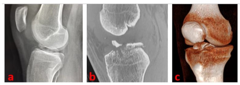
Clinical Assessment
History. The child reports the hyperextension injury described above, followed by immediate pain and swelling and an inability to bear weight. The knee may give way, and it locks if the fragment blocks extension.
Look and feel. There is an effusion, which is a haemarthrosis, with an antalgic gait and the knee held in slight flexion. The anterior tibial spine is tender; joint line tenderness suggests a meniscal injury, and MCL tenderness an associated MCL injury.
Movement. Flexion is limited by pain and effusion. Extension is limited if the fragment blocks it, and an extensor lag appears if the extensor mechanism is affected.
Ligament testing. The avulsed tibial spine allows anterior tibial translation, so the tests are those of an ACL injury:
- Lachman test: positive (anterior translation)
- Anterior drawer: positive (anterior translation)
- Pivot shift: may be positive (rotational instability)
- Valgus stress: may be positive if the MCL is injured
Lachman test is positive in tibial spine fractures, as in the adult ACL injury. That shared instability pattern, anterior translation and pivot shift, is why the fracture is called the paediatric ACL equivalent.
Associated injuries. These are the injuries to look for alongside the fracture:
- Meniscal tears in 10-20%, the lateral meniscus more commonly
- MCL injuries in 5-10%
- Bone bruises of the posterolateral tibia and lateral femoral condyle, the kissing contusion
- Extensor mechanism injury (patellar tendon avulsion), which is rare
Investigations
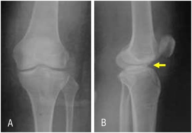
Radiographs. AP and lateral views of the knee, with oblique views if needed. The lateral view is the one that diagnoses and classifies: it shows the anterior elevation of the fragment from its bed, the displacement (under 3mm in Type I, complete in Type III), the fragment's size and comminution, and its rotation.
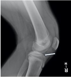
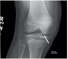
The AP view may show the fragment in the intercondylar notch and screens for an associated plateau injury, but the lateral is diagnostic. When displacement or soft-tissue entrapment remains uncertain, obtain a true lateral and an MRI.
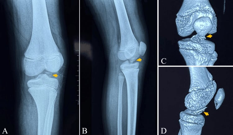
CT. CT is not routine but is helpful in complex cases and for surgical planning, defining fragment size and comminution, articular involvement and any associated fracture, and so the fixation strategy. It is most useful when plain films underestimate rotation or when the choice of fixation depends on fragment size and comminution. Three-dimensional reconstruction helps with complex comminution (Type IV), with fragment rotation and with preoperative planning.
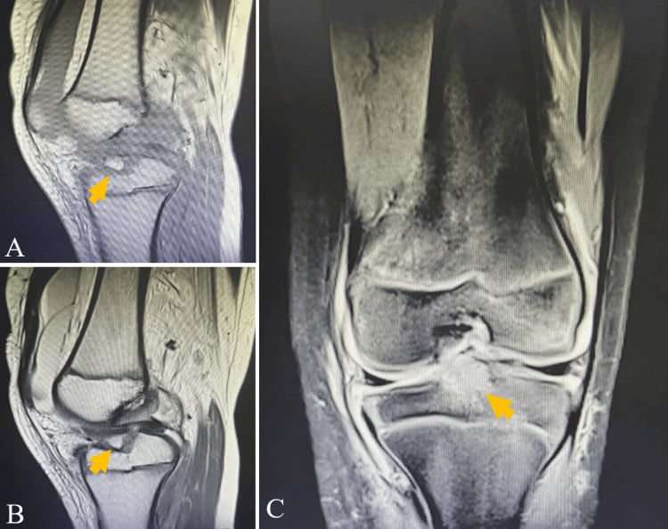
MRI. MRI is not routine but is indicated when associated injuries are suspected: meniscal tears, MCL injuries, bone bruises in the kissing contusion pattern and cartilage injuries. It also shows the ACL fibres, which are usually intact, and their relationship to the fragment, and it defines intermeniscal-ligament entrapment that can block reduction.
Differential Diagnosis
The acutely swollen, haemarthrosis knee in a child has a focused differential. The tibial spine fracture is distinguished by the avulsed eminence on the lateral radiograph.
- Mechanism / clue
- Hyperextension/valgus; bicycle, sport
- Imaging
- Lateral X-ray: avulsed eminence
- Distinguishing feature
- Bony fragment at ACL footprint; Lachman positive
- Mechanism / clue
- Pivot/deceleration in older adolescent
- Imaging
- MRI: discontinuous ACL, no bony fragment
- Distinguishing feature
- No avulsion fragment; usually skeletally mature
- Mechanism / clue
- Twisting with quadriceps contraction
- Imaging
- Skyline / MRI: MPFL injury, medial patella bruise
- Distinguishing feature
- Lateral patellar apprehension; medial tenderness
- Mechanism / clue
- Shear during dislocation/twist
- Imaging
- X-ray/MRI: loose body, donor defect
- Distinguishing feature
- Mechanical locking, intra-articular fragment
- Mechanism / clue
- Direct/indirect force across physis
- Imaging
- X-ray: physeal widening, metaphyseal/epiphyseal line
- Distinguishing feature
- Tenderness over physis, not joint line
- Mechanism / clue
- Twist on planted foot
- Imaging
- MRI: meniscal signal
- Distinguishing feature
- Joint-line tenderness, no avulsion fragment
Loss of Reduction During Non-Operative Treatment
Non-operative care of a Type I or a reduced Type II fracture is an active, monitored process, not "cast and forget" — this answers the viva follow-up "What if it displaces to Type II in the cast?"
- Interval radiographs are mandatory. Films at roughly one to two weeks confirm the fragment has held its position; a marginally reduced Type II can re-elevate within the cast as swelling settles.
- Immobilisation angle is debated and matters. Near-full extension helps seat the elevated anterior fragment of a Type II (the femoral notch presses the fragment down), but frank hyperextension can tension the ACL and lift the fragment, so a position of near-extension (commonly about 10 to 20 degrees of flexion) is often chosen to balance these effects.
- Loss of reduction changes the plan. If interval imaging shows progression to a displaced Type II or III, or a fragment now blocked by the interposed intermeniscal (transverse) ligament, abandon closed treatment and convert to arthroscopic reduction and fixation. Accepting a malunited, elevated fragment risks a mechanical extension block (the prominent fragment impinges in the intercondylar notch), residual laxity, and secondary arthrofibrosis.
Q: A Type I or reduced Type II tibial spine fracture is found to have displaced further on the two-week radiograph — what do you do? A: Do not accept malunion. A fragment that has lost reduction (progressed to a displaced Type II/III or become blocked by the intermeniscal ligament) is an indication to convert to arthroscopic reduction and fixation. This is precisely why interval radiographs during cast treatment are essential rather than optional.
Management
Before choosing, classify the fracture, assess a Type II for reducibility and check for associated meniscal and MCL injuries.
Non-operative treatment. Type I fractures, and Type II fractures that reduce with extension and are stable, are treated in an extension cast for 4-6 weeks. The child is non-weight-bearing initially, for 2-3 weeks, then progressively weight-bearing (3-4 weeks), and range of motion starts when the cast comes off. Outcomes are excellent for Type I and good for a reducible Type II. The exact immobilisation angle is debated; the section on Loss of Reduction During Non-Operative Treatment sets out the argument.
Follow-up in the cast. A radiograph at 2 weeks checks the position and one at 4-6 weeks checks healing. The cast is removed when the fracture has healed, at 4-6 weeks.
Surgical indications. Surgery is indicated as follows.
Absolute
- Type III (complete displacement)
- Type IV (comminuted)
- Type II that will not reduce, blocked by the intermeniscal ligament
Relative
- Type II with persistent instability after reduction
- An associated meniscal tear requiring repair
- The high-demand athlete
Timing. Operate within 1-2 weeks. That allows the swelling to resolve, but comes before the fragment becomes fixed.
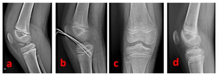
The operation. Type III and IV fractures are reduced and fixed arthroscopically, with suture or screw, and opened if arthroscopic reduction fails. Range of motion starts early after surgery.
Surgical Technique
Arthroscopic reduction with suture fixation through transosseous tunnels is the gold standard for Type III-IV fractures. Screw fixation is acceptable but suture fixation is preferred, and open reduction is reserved for complex cases.
Why it is preferred. Suture fixation needs no hardware removal and allows early range of motion; the arthroscope gives excellent visualisation, and the complication rate is low.
Set-up. The patient lies supine on a standard table with a thigh tourniquet and a leg holder or lateral post, in a standard arthroscopy set-up. The anterolateral portal is for viewing, the anteromedial portal for working, and a superomedial portal is an optional outflow.
Steps.
- Diagnostic arthroscopy: assess the fragment, meniscus and cartilage
- Debride the intermeniscal ligament if it blocks reduction
- Reduce the fragment with a probe or shaver
- Prepare the fragment bed, debriding to bleeding bone
- Pass 2-3 sutures through the fragment
- Create 2-3 transosseous tunnels, avoiding the physis
- Pass the sutures through the tunnels
- Tie the sutures over the bone bridge with the knee in extension
- Confirm reduction and stability
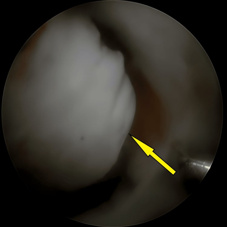
Look before you reduce. Probe the fragment's integrity and mobility, then inspect both menisci and the intermeniscal ligament before choosing screw or suture fixation. The posterior horn of a meniscus can be torn or trapped beneath the avulsed fragment, and forcing the fragment down may incarcerate or damage it, so remove interposed tissue before accepting the reduction.
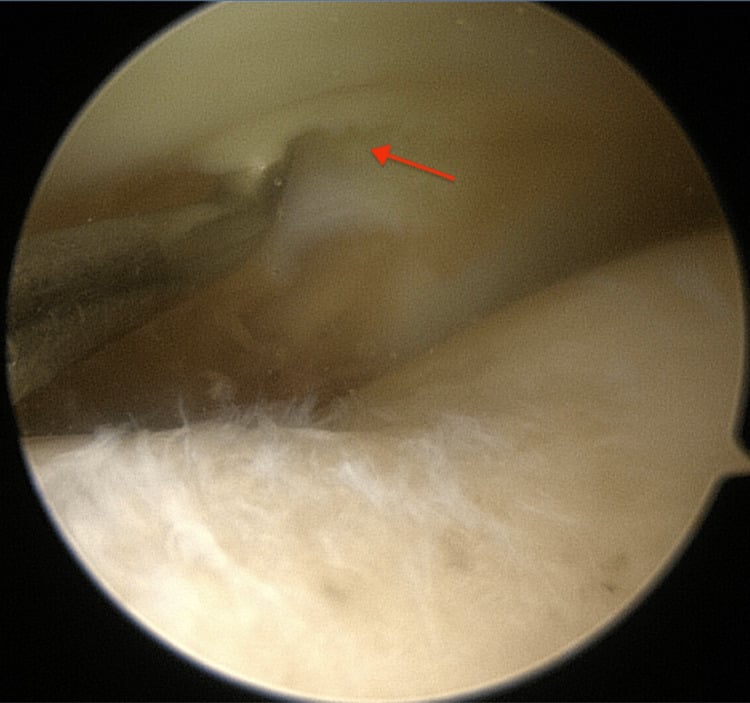
The bed. Haematoma and fibrous tissue are cleared to expose bleeding cancellous bone, preserving the fragment and its ACL attachment. Reduction cannot be anatomic while the bed, the anterior horn or the intermeniscal ligament remains interposed.
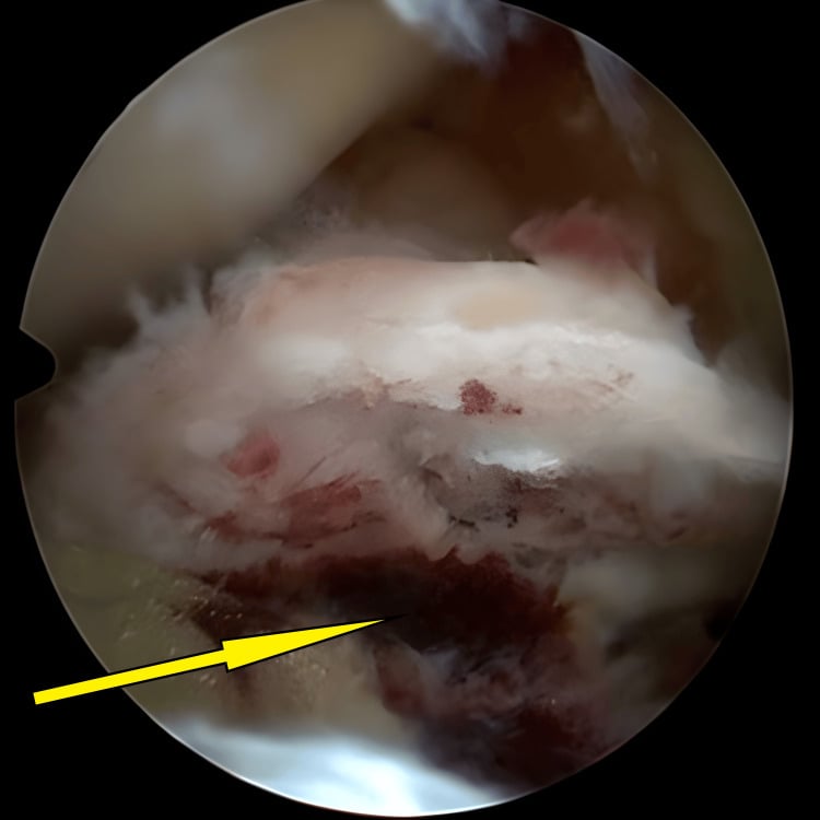
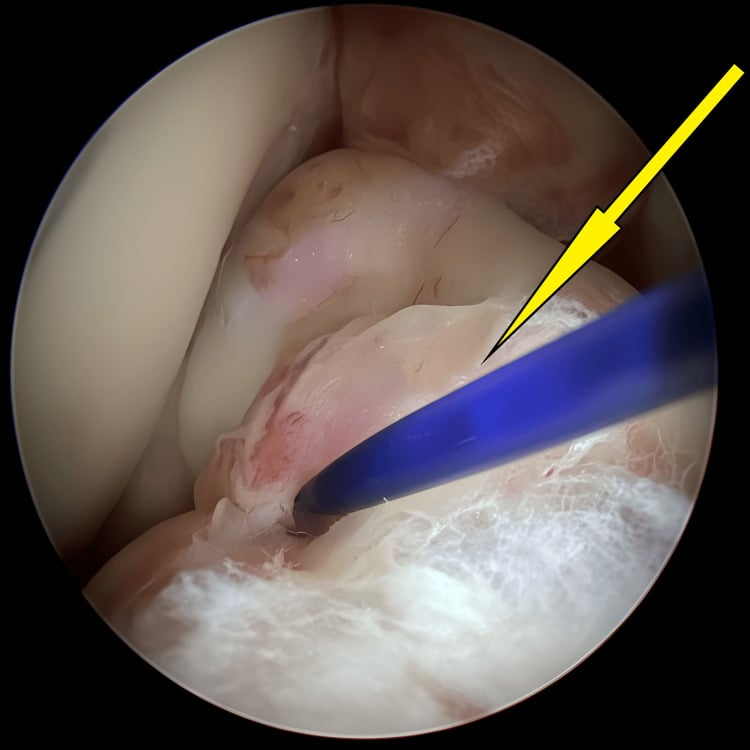
Sutures and tunnels. Use No. 2 non-absorbable suture (Ethibond or Fiberwire), passed through the ACL insertion on the fragment and then through the transosseous tunnels. The tunnels stay proximal to the physis (see Growth Plate Protection), and 2-3 of them give stability. Before the fixation is final, cycle the knee and inspect for anterior notch impingement or loss of terminal extension.
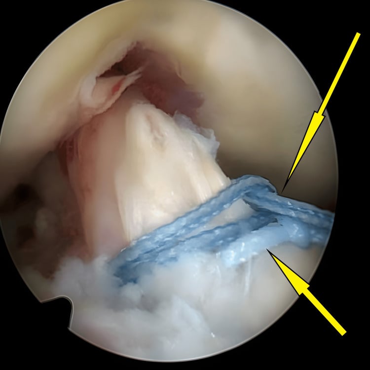
Anchors instead of tunnels. Sutures passed around the ACL and fragment can instead be tensioned into a metaphyseal knotless anchor, which avoids transphyseal tunnels. Confirm compression through flexion and extension, and make sure the anchor does not violate the physis. Suture-bridge constructs combine anchors, crossing sutures and cortical fixation to compress the fragment broadly.
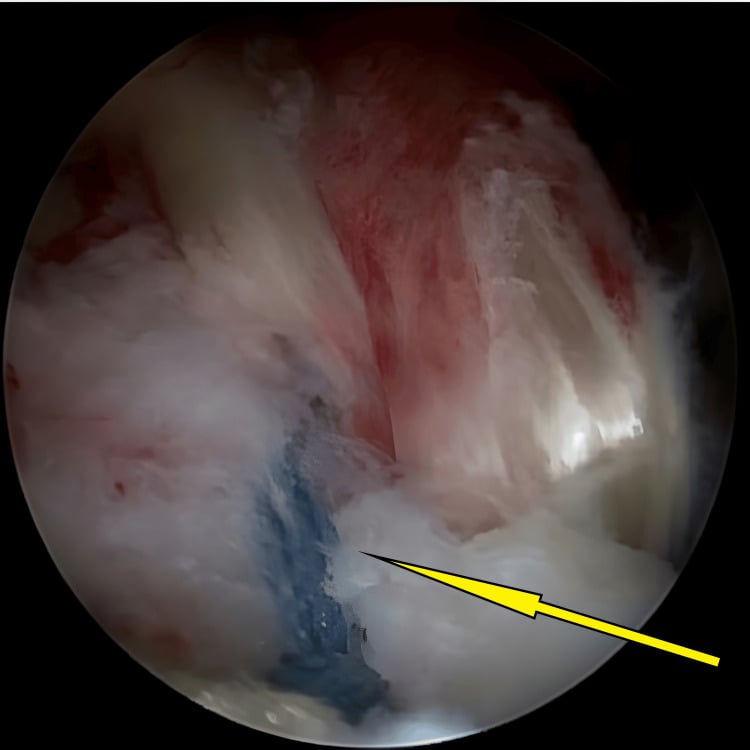
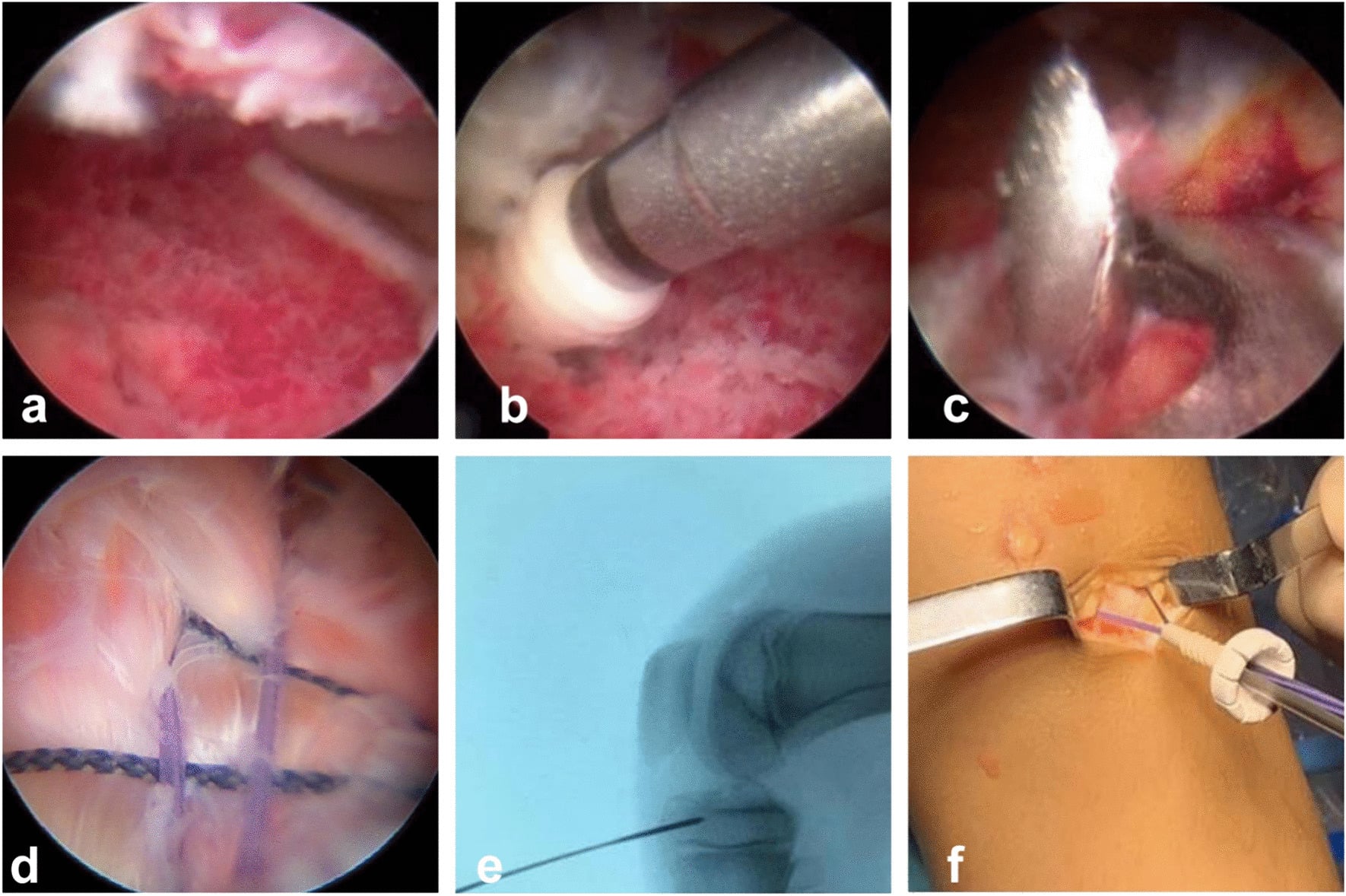
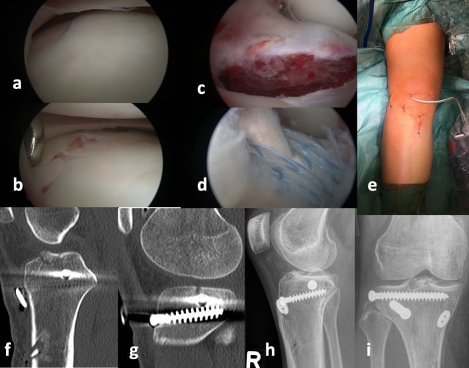
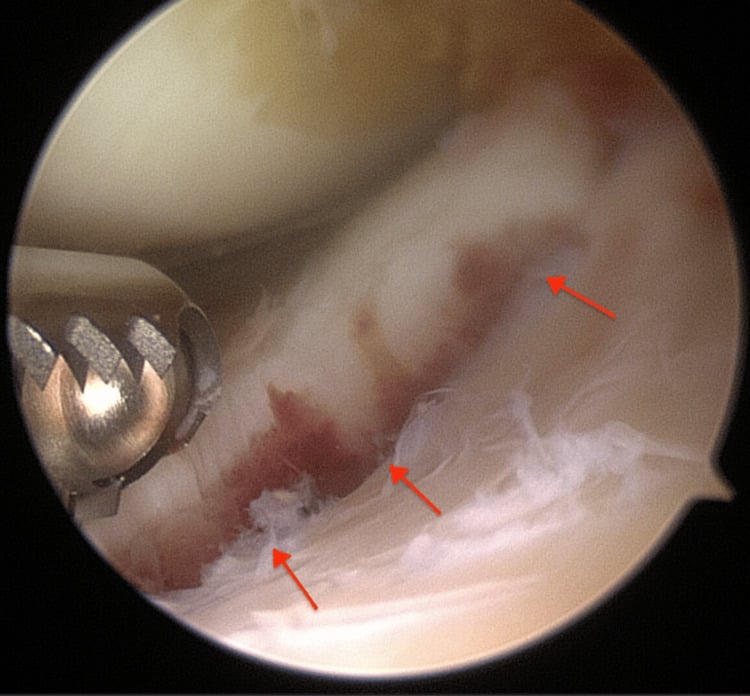
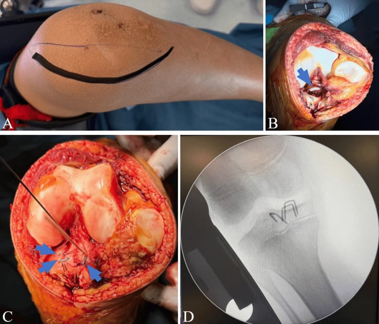
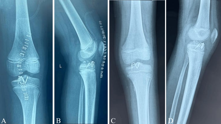
Avoid crossing the proximal tibial physis with fixation. The physis is 2-3 cm distal to the joint line. Use transosseous tunnels that stay proximal to the physis, or suture anchors in the tibial spine itself. Crossing the physis can cause growth disturbance, which is rare but devastating.
Complications
- Incidence
- ~7-10% (pooled)
- Risk Factors
- Delayed ROM, prolonged immobilisation
- Prevention/Management
- Early ROM (2-4 weeks), aggressive physiotherapy
- Incidence
- ~3-4% reported as translation over 3mm
- Risk Factors
- Malreduction, inadequate fixation, fragment resorption
- Prevention/Management
- Anatomic reduction, secure fixation
- Incidence
- Less than 5%
- Risk Factors
- Inadequate fixation, poor reduction, fragment devascularisation
- Prevention/Management
- Secure fixation, bone bed preparation
- Incidence
- Less than 1% with proper technique
- Risk Factors
- Crossing physis with fixation
- Prevention/Management
- Avoid physis, use proximal tunnels
- Incidence
- 5-10% (screw)
- Risk Factors
- Prominent hardware, need for removal
- Prevention/Management
- Use suture fixation to avoid removal
- Incidence
- Long-term risk; less than 5% at 10 years with proper treatment
- Risk Factors
- Malreduction, persistent instability
- Prevention/Management
- Anatomic reduction, restore stability
Arthrofibrosis. Stiffness is the most common complication. The Farinelli meta-analysis (49 studies, 2,017 patients) pooled an incidence of 7.4% after ARIF and 10.3% after ORIF, with no significant difference between them. Other reviews report 16-21%, so treat it as roughly one knee in ten, not a precise figure.
Treating the stiff knee. Established arthrofibrosis is managed with manipulation under anaesthesia and arthroscopic lysis of adhesions. The two go together: Vander Have's series in the Evidence Base is the reason manipulation is not performed alone.
Residual laxity. Farinelli recorded translation over 3mm in roughly 3-4% of pooled cases, though instrumented testing finds mild laxity far more often, and that figure counts the laxity a surgeon chose to report. Mild laxity is usually asymptomatic and does not affect function; symptomatic instability is rarer still. If it is symptomatic, the options are revision fixation or, if needed, ACL reconstruction.
Nonunion. When it occurs, it is treated by revision fixation, with bone graft if needed.
Growth disturbance. Where there is concern, monitor the child until skeletal maturity.
Postoperative Care
The first weeks. A hinged knee brace is locked in extension for 2-4 weeks and the child is non-weight-bearing for the first 2-3 weeks. Quadriceps sets and straight leg raises start immediately, and the brace is unlocked for passive range of motion at 2-4 weeks.
- Brace and ROM
- Brace locked in extension
- Weight bearing
- Non-weight bearing
- Exercise
- Quadriceps sets, straight leg raises, ice and elevation
- Brace and ROM
- Brace unlocked, ROM 0-90 degrees
- Weight bearing
- Progressive, partial to full
- Exercise
- Stationary bike when ROM allows; continue quadriceps strengthening
- Brace and ROM
- Full ROM
- Weight bearing
- Full weight bearing
- Exercise
- Progressive strengthening, balance and proprioception
- Brace and ROM
- -
- Weight bearing
- -
- Exercise
- Sport-specific training; physiotherapy continues for 3-6 months
Return to sport. Return comes when strength and range of motion are normal and the criteria below are met, usually at 3-6 months after surgery depending on the sport and level. 80-90% return to their pre-injury level, the outcome depending on age, sport level and compliance with rehabilitation.
- Full range of motion, equal to the contralateral side
- Quadriceps strength greater than 90% of the contralateral side
- No effusion
- No instability: negative Lachman and pivot shift
- Single-leg hop test greater than 90% of the contralateral side
- Agility testing passed and sport-specific drills completed
Prevention. Primary prevention rests on proper landing technique, with the knee flexed rather than hyperextended, together with quadriceps and hamstring strength training, balance and proprioception training, and sport-specific conditioning. After an injury, complete rehabilitation before returning to sport and continue strength and conditioning work; bracing is controversial and may not prevent reinjury.
Outcomes and Prognosis
Outcomes are excellent with proper treatment. Non-operatively treated Type I-II fractures give 90-95% good or excellent results, and surgically treated Type III-IV fractures 85-95%. At 5-10 years, 85-90% maintain good results.
Guidelines, Registries & Global Practice
Global Epidemiology
- Incidence: ~3 per 100,000 per year; peak age 8-14 years; male predominance (~2:1).
- Represents roughly 2-5% of paediatric knee injuries with haemarthrosis; rising with youth sport participation.
- Bony ACL avulsion equivalent in the skeletally immature knee; adults more often tear the ACL mid-substance.
Society Guidance (Side by Side)
There is no formal single-society guideline dedicated to tibial spine fractures; the table summarises principles endorsed across major bodies and the Tibial Spine Research Interest Group consensus.
- Classification used
- Meyers-McKeever (+ MRI)
- Operative threshold
- Displaced (type III-IV); irreducible type II
- Emphasis
- Arthroscopic fixation, early ROM
- Classification used
- Meyers-McKeever
- Operative threshold
- Displaced / blocked reduction
- Emphasis
- Refer to paediatric centre, MRI for associated injury
- Classification used
- Meyers-McKeever / OTA
- Operative threshold
- Articular incongruity or instability
- Emphasis
- Stable fixation that respects the physis
- Classification used
- Meyers-McKeever
- Operative threshold
- Displaced; meniscal entrapment
- Emphasis
- Suture over screw; meniscal preservation
Registries and Practice Variation
- Joint registries (NJR, AJRR, AOANJRR, SHAR) do not track tibial spine fixation; they are relevant only for the rare late ACL reconstruction or arthroplasty after malreduction and post-traumatic arthritis.
- High-resource settings: arthroscopic reduction with suture fixation is the prevailing standard, enabling early motion.
- Limited-resource settings: open reduction and internal fixation (screw or suture over bone bridge) remains common and effective where arthroscopy or implants are unavailable; outcomes are good when reduction is anatomic and rehabilitation is supervised.
Related pages: Paediatric ACL Injury is the sibling diagnosis and the one this fracture is the childhood equivalent of — the same mechanism, the same haemarthrosis, and the decision between them is made on the radiograph before the MRI; ACL Injuries for the adult mid-substance tear and for the reconstruction that a small minority of these knees eventually need — and note the distinction this page turns on, that here the ligament is in continuity but plastically elongated, so anatomic bony reduction cannot shorten it; Knee Arthrofibrosis is the dominant complication and carries the staged management in full, including why manipulation must be combined with lysis of adhesions rather than performed alone; Meniscus Tears and Discoid Meniscus matter here for a specific reason — entrapment of the anterior horn or the intermeniscal ligament under the fragment is the commonest block to closed reduction, and the Najdi series found it in half of its patients; MCL Injuries for the concomitant ligamentous injury that turns mild residual laxity into symptomatic instability; Physeal Injuries: Salter-Harris and Proximal Tibial Physeal Injuries for the growth plate that constrains every implant choice, and Distal Femoral Physeal Injuries because the iatrogenic distal femoral fracture caused by manipulation is a physeal injury with the highest arrest risk in the body; Tibial Plateau Fractures is the adult intra-articular differential in the same region; Osteochondral Defects of the Knee for the other cause of a paediatric haemarthrosis with a fragment on the film; and Return to Sport Criteria for the functional testing that should govern clearance rather than time alone.
Controversies and Areas of Uncertainty
Suture (pull-through or suture-bridge) fixation avoids a second operation for implant removal and shows higher patient-reported scores in a randomised trial and systematic review, but screws give immediate compression and excellent results in large series. Choice often reflects fragment size, comminution and surgeon experience rather than proven superiority for every case.
The classic "3 mm" cut-off and the reducibility of type II fractures are debated. Many type II fractures are blocked by the intermeniscal (transverse) ligament or entrapped meniscus, so post-reduction radiographs and a low threshold for arthroscopy are increasingly favoured over a fixed millimetre rule.
MRI is not mandatory but detects entrapped soft tissue, chondral injury and meniscal tears (reported in a notable minority) and refines classification. Routine versus selective MRI remains practice-dependent.
Mild residual anterior laxity (Lachman/KT-1000) is common after healing but is usually asymptomatic and rarely requires later ACL reconstruction. Whether this laxity predisposes to long-term degenerative change is not yet settled.
Residual Anterior Laxity: Why It Persists After Anatomic Union
- Instrumented testing detects it more than patients do: side-to-side differences on Lachman and KT-1000 arthrometry are found more often than the child reports any symptom. Most such laxity is mild, functionally silent, and does not require later ACL reconstruction — consistent with the excellent Lysholm scores and absence of clinical laxity reported in the operative series in the Evidence Base.
- Clinically important laxity is the minority and clusters with malreduction, fragment resorption, inadequate fixation, or a missed concomitant ligamentous injury rather than with the plastic-elongation mechanism alone. Pooled series record translation over 3 mm as a reported complication in only about 3-4 percent, which almost certainly understates objective laxity because it counts what surgeons chose to report rather than what arthrometry would find in everyone.
- Do not conflate with a mid-substance ACL tear — the primary treatment of a torn ACL and its reconstruction are covered on the dedicated ACL topics; here the ligament is in continuity but lengthened.
Q: Why can a patient have a positive Lachman test after a tibial spine fracture that has healed in anatomic position? A: The ACL is plastically elongated (interstitial stretch) at the moment of avulsion, so anatomic bony reduction cannot shorten the already-lengthened ligament. The resulting laxity is usually mild and asymptomatic; symptomatic instability instead points to malreduction, fragment resorption, or a missed associated ligament injury.
MCQ Practice Points
Q: What is the most common complication following tibial spine fracture fixation? A: Arthrofibrosis (pooled ~7-10%, and the dominant complication) - stiffness is more common than instability or nonunion. Prevention requires early ROM.
Q: Why do children sustain tibial spine fractures instead of ACL tears? A: Bone is weaker than ligament - The cartilaginous tibial spine avulses before the collagen of the ACL fails midsubstance.
Q: What is the absolute indication for surgery in Meyers-McKeever classification? A: Type III (Complete displacement) - Also Type II if reducible but blocked by meniscus (intermeniscal ligament).
Q: How does ACL function compare between anatomically healed tibial spine fractures and native ACL? A: Normal function expected - Studies show 95% of patients achieve normal Lachman and KT-1000 testing when healed in anatomic position.
Q: What structure may block reduction of a displaced tibial spine fracture? A: Intermeniscal ligament (transverse ligament) - It can become interposed between the fragment and its bed, preventing closed reduction.
Exam Viva Scenarios
Practise clinical reasoning and management decisions out loud
“A 10-year-old boy presents after falling off a bicycle. Knee is swollen. X-ray shows Meyers-McKeever Type I tibial spine fracture.”
“A 12-year-old female gymnast lands awkwardly. X-ray shows a Type III tibial spine fracture (completely displaced).”
“A patient returns 3 months after fixation of a tibial spine fracture with a 15-degree extension deficit.”
Key Concepts
- Pediatric ACL Equivalent
- Meyers-McKeever Classification
- Hyperextension + Valgus mechanism
- Lachman positive (anterior laxity)
Classification (Meyers-McKeever)
- Type I: Non-displaced → Extension Cast
- Type II: Hinge intact → Reduce/Cast or Surgery
- Type III: Complete → Surgery
- Type IV: Comminuted → Surgery
Surgical Goals
- Anatomic reduction
- Stable fixation (suture preferred over screw)
- Avoid Physis (Growth plate protection)
- Early ROM (Prevent arthrofibrosis)
Complications
- Arthrofibrosis (Most common)
- Residual Laxity (Usually asymptomatic)
- Growth Disturbance (Rare)
- Nonunion (Rare)
Pearl
- Intermeniscal ligament often blocks reduction
- Lateral meniscus tear is most common associated injury
- Lateral X-ray is diagnostic view
- Arthroscopic suture fixation is gold standard
Evidence Base
Meyers-McKeever Classification
- Original radiographic classification (Type I-III) based on displacement of the fragment
- Type I-II reduce with extension and do well non-operatively
- Type III (complete separation) requires reduction and fixation
State of the Art Review (Incidence and Principles)
- Incidence approximately 3 per 100,000 per year, peak age 8-14 years
- Tibial spine fracture is the bony ACL avulsion equivalent in skeletally immature knees
- Most common complications: arthrofibrosis, residual laxity, nonunion/malunion, physeal arrest
Surgical Treatment - Systematic Review (ARIF vs ORIF, Suture vs Screw)
- 12 studies pooled for Meyers-McKeever type II-IV operative outcomes
- Arthroscopic suture fixation gave higher Tegner/IKDC/Lysholm scores than arthroscopic screw fixation (p less than 0.001)
- Screw fixation had higher implant-removal rates; suture fixation had higher arthrofibrosis rates
ARIF vs ORIF - Meta-analysis of 49 Studies (arthrofibrosis and laxity rates)
- PRISMA meta-analysis of 49 studies (2004-2025), 2,017 patients - 1,785 ARIF (88%) and 232 ORIF (12%)
- Arthrofibrosis 7.4% (95% CI 6.2-8.7) after ARIF and 10.3% (95% CI 6.1-13.8) after ORIF; comparative studies showed NO difference (OR 0.98, 95% CI 0.41-2.37, p=0.97)
- Residual anterior tibial translation over 3 mm in only 3.7% (ARIF) and 2.8% (ORIF)
- Functional scores near-identical: IKDC 92.1 vs 92.4, Lysholm 94.5 vs 96.7
- Conclusion: ARIF and ORIF are comparable; neither is superior
Arthroscopic Suture vs Screw Fixation - Randomised Trial
- Randomised trial of 90 patients (45 suture vs 45 screw) with tibial ACL avulsion
- Suture group superior subjective IKDC (91.4 vs 85.7) and Lysholm (92.0 vs 86.0), both p=0.001
- 7 screw-fixation patients required reoperation for implant removal; none in the suture group
Arthroscopic Screw Fixation (Type II-III)
- 24 children (mean age 11y), Meyers-McKeever type II (n=15) and III (n=9), intra-epiphyseal ASNIS screws
- Mean Lysholm 99.3 (type II) and 98.6 (type III); no anterior laxity at 12 weeks
- Meniscal entrapment present in 12 cases without affecting functional result
Arthrofibrosis After Surgical Fixation
- 32 children/adolescents who developed arthrofibrosis after fixation of displaced tibial eminence fractures
- 24 required reoperation for loss of motion; manipulation under anaesthesia caused distal femoral fracture and growth arrest in 3
- Stable fixation enabling early rehabilitation is key; manipulation should be combined with lysis of adhesions
Complications - Prevention and Treatment
- Tibial eminence fixation may be complicated by failed fixation, knee stiffness and arthrofibrosis
- Arthrofibrosis is rare in children overall but occurs most frequently after tibial eminence injuries
- Emphasises secure fixation and early controlled motion to limit stiffness