Failure of metaphyseal tubulation — a wide, under-modelled metaphysis with a narrow diaphysis, classically at the distal femur
- The sign is a modelling (tubulation) defect, not a destructive lesion — the cortex is intact and the outline is smooth.
- It reflects failure of osteoclastic funnelisation of the metaphysis during growth, from marrow packing, osteoclast failure or a primary dysplasia.
- Bone density is the single most useful discriminator: dense in osteopetrosis and pyknodysostosis, osteopenic in Gaucher and thalassaemia, normal in Pyle disease.
- Bilateral and symmetrical is the rule; a unilateral flask-shaped metaphysis is far more likely a local process (fibrous dysplasia, enchondromatosis, healed osteomyelitis).
- Gaucher is confirmed biochemically (leucocyte beta-glucocerebrosidase), not radiologically.
- A wide metaphysis on an infant radiograph is often normal physiology — do not over-call in the first two years.
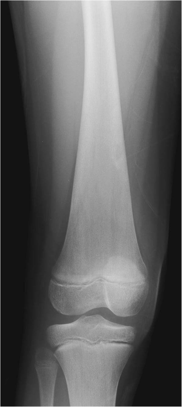
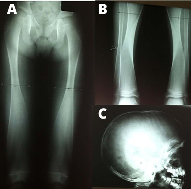
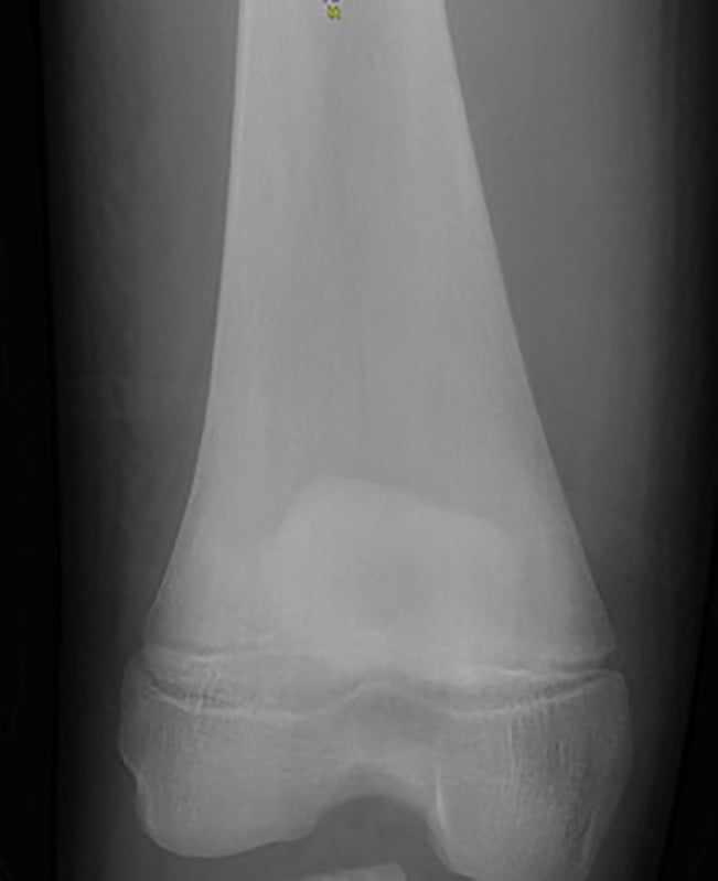
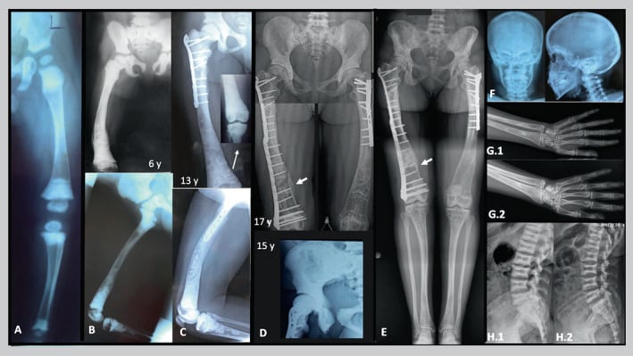
- “Say the words: 'There is loss of the normal metaphyseal tubulation of the distal femora with widening and an abrupt transition to a narrow diaphysis — an Erlenmeyer flask deformity.'
- “Then immediately: 'I would assess bone density, look for a marrow-packing process, and check whether it is bilateral.'
- “Gaucher = flask plus osteopenia plus avascular necrosis of the femoral head plus hepatosplenomegaly.
- “Thalassaemia = flask plus hair-on-end skull plus coarsened trabeculae plus a transfusion history.
- “Pyle metaphyseal dysplasia is the purest form — massive metaphyseal flaring with a strikingly normal skull, in contrast to craniometaphyseal dysplasia.
Metaphyses are relatively broad and under-modelled in infancy. The deformity is only meaningful when the metaphyseal width clearly exceeds the expected flare and the diaphysis is disproportionately narrow. Compare with the contralateral limb and the age-matched norm before committing.
Candidates list ten causes and never state whether the bone is dense or lucent. Density splits the list in half in one sentence: sclerotic points to osteopetrosis or pyknodysostosis; lucent points to Gaucher, thalassaemia or Niemann-Pick.
In Gaucher the flask deformity is a bystander finding — the clinical problem is avascular necrosis, bone crisis and pathological fracture. Always comment on the hips and on any bone infarcts before moving on.
A modelling defect has a smooth, intact cortex and no matrix. An expansile lesion has a lesion — ground glass, endosteal scalloping, chondroid rings. Fibrous dysplasia and enchondromatosis can produce a flask-like contour but the bone is abnormal internally.
Recognising the Pattern
Definition. The Erlenmeyer flask deformity is a failure of metaphyseal remodelling (tubulation, or funnelisation) in which the metaphysis and distal diaphysis remain abnormally wide and the shaft is comparatively narrow, so the bone takes the profile of a conical laboratory flask. It is best appreciated at the distal femur, then the proximal tibia, proximal humerus and the ribs (anterior rib ends).
Mechanism. During growth, osteoclasts on the outer periosteal surface of the metaphyseal cut-back zone narrow the wide metaphysis into the tubular diaphysis. Anything that (a) packs the marrow and mechanically resists cut-back, (b) impairs osteoclast function, or (c) is a primary defect of bone modelling will produce the sign.
How to confirm the sign is genuinely present.
- The widest metaphyseal diameter is clearly greater than the flare expected for age and the transition to the diaphysis is abrupt rather than a smooth taper.
- The cortex is continuous and smooth over the widened segment — no periosteal reaction, no cortical breach, no matrix.
- It is bilateral and symmetrical in nearly all systemic causes.
- Check a second site (proximal tibia, proximal humerus, ribs) — a true modelling defect is generalised.
The words to use out loud. "On this frontal radiograph of the knees there is symmetrical loss of the normal metaphyseal tubulation of the distal femora, with metaphyseal widening and an abrupt transition to a narrow diaphysis — the Erlenmeyer flask deformity. The cortices are intact and there is no aggressive feature. I would next assess bone density, the femoral heads for avascular necrosis, and the skull."
Mimics and false positives.
- Normal infancy — physiological broad metaphyses under two years.
- Healing rickets — flared, previously frayed metaphyses with a dense zone of provisional calcification; the history and the cupping give it away.
- Multiple hereditary exostoses (diaphyseal aclasis) — the metaphysis is broad because sessile osteochondromas sit on it; look for the cortex and medulla flowing into the lesion.
- Fibrous dysplasia / enchondromatosis — internal matrix abnormality, usually asymmetrical.
- Chronic osteomyelitis or previous physeal injury — unilateral, deformed rather than merely under-modelled.
- Projection — an externally rotated or obliqued femur can exaggerate metaphyseal width.
Next Investigation
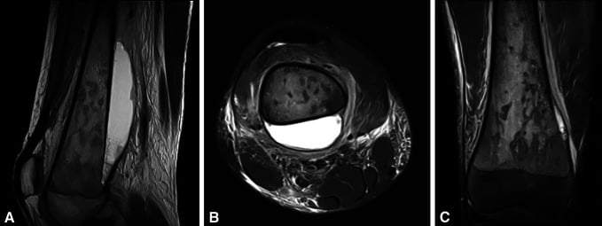
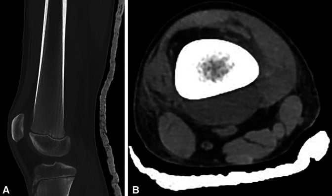
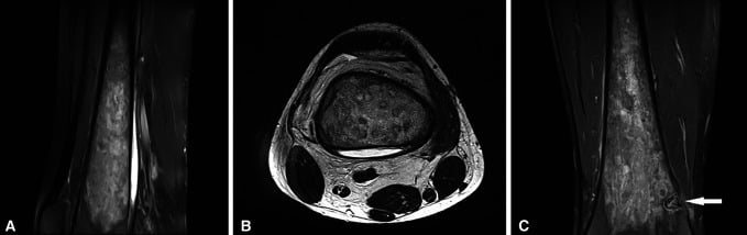
LEAD GNOME
Hook:The established radiological mnemonic for the Erlenmeyer flask differential.
The Differential
- Typical age / setting
- Infantile form presents in the first year; benign autosomal dominant form in adolescence or adulthood
- Discriminating feature
- Diffusely DENSE bone with bone-in-bone vertebrae, sandwich (rugger-jersey) spine, and marrow failure with anaemia, hepatosplenomegaly and cranial nerve compression
- What confirms it
- Radiographic density plus FBC; CLCN7/TCIRG1 genetics; bone marrow transplant is curative in the infantile form
- Typical age / setting
- Ashkenazi Jewish predominance; presents in childhood to young adult life
- Discriminating feature
- OSTEOPENIC flask deformity with avascular necrosis of the femoral head, bone infarcts and painless massive splenomegaly with normal neurology
- What confirms it
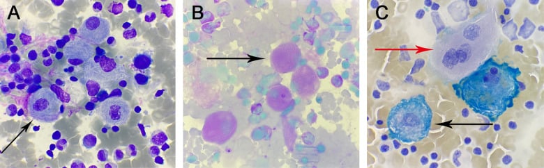
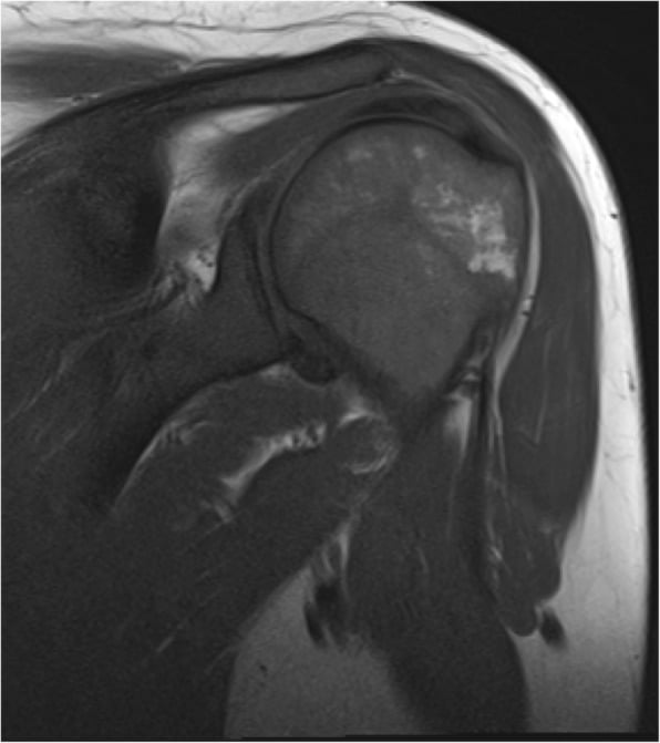
- Leucocyte beta-glucocerebrosidase assay (low) and GBA1 mutation; MRI shows marrow infiltration replacing fatty marrow
- Typical age / setting
- Any childhood age, unwell child
- Discriminating feature
- Metaphyseal LUCENT bands, permeative destruction and periosteal reaction accompany the under-modelling — the bone looks aggressive, not merely wide
- What confirms it
- FBC with blast cells, blood film and bone marrow aspirate
- Typical age / setting
- Mediterranean, Middle Eastern, South Asian; transfusion-dependent from infancy
- Discriminating feature
- Hair-on-end skull, obliterated maxillary sinuses, coarse widened trabeculae from marrow hyperplasia, and osteopenia
- What confirms it
- Haemoglobin electrophoresis / HPLC; transfusion history
- Typical age / setting
- African, Afro-Caribbean, Middle Eastern; from early childhood
- Discriminating feature
- H-shaped (Lincoln-log) vertebrae, bone infarcts, dactylitis in infancy, and avascular necrosis of the humeral and femoral heads
- What confirms it
- Sickle solubility test and haemoglobin electrophoresis
- Typical age / setting
- Recognised in childhood; often incidental in a well child
- Discriminating feature
- Extreme metaphyseal flaring with a genuinely NORMAL skull and normal bone density; genu valgum and limited elbow extension are the clinical clues
- What confirms it
- Clinical and radiographic diagnosis; SFRP4 mutation — no laboratory abnormality
- Typical age / setting
- Presents in infancy or early childhood with facial change
- Discriminating feature
- Same metaphyseal flaring PLUS a thick sclerotic skull base and paranasal bossing causing cranial nerve palsy, nasal obstruction and hypertelorism
- What confirms it
- Skull radiograph or CT showing craniofacial hyperostosis; ANKH mutation
- Typical age / setting
- Infancy to childhood; often neuronopathic
- Discriminating feature
- Flask deformity with generalised osteopenia PLUS a cherry-red macular spot and neurodegeneration — the neurology separates it from Gaucher
- What confirms it
- Sphingomyelinase assay; foam cells on marrow aspirate; SMPD1 / NPC1 genetics
- Typical age / setting
- Childhood, autosomal dominant family history
- Discriminating feature
- The metaphysis is broad because sessile and pedunculated osteochondromas arise from it — cortex and medulla flow continuously into the lesion
- What confirms it
- Radiographs of multiple sites; EXT1/EXT2 mutation. Watch for malignant transformation to chondrosarcoma in adults
- Typical age / setting
- Toddlers with pica; occupational exposure in adults
- Discriminating feature
- DENSE transverse metaphyseal bands wider than the normal zone of provisional calcification, at multiple metaphyses, with band width tracking the exposure
- What confirms it
- Serum lead level; abdominal radiograph may show radiodense paint flakes
- Typical age / setting
- Short stature, presents in childhood
- Discriminating feature
- Dense bone with acro-osteolysis of the terminal phalanges, obtuse mandibular angle and persistent open fontanelles — dense but SHORT, unlike osteopetrosis
- What confirms it
- Cathepsin K (CTSK) mutation; characteristic hand and skull radiographs
- Typical age / setting
- Childhood to young adult
- Discriminating feature
- Ground-glass matrix with endosteal scalloping, shepherd's crook proximal femur; typically ASYMMETRICAL
- What confirms it
- Ground glass on radiograph or CT; café-au-lait macules with jagged coast-of-Maine borders; GNAS mutation
- Typical age / setting
- Childhood, unilateral predominance
- Discriminating feature
- Columnar lucent chondroid streaks extending from the physis into the metaphysis with rings-and-arcs calcification; limb-length discrepancy
- What confirms it
- Radiographic appearance; MRI for extent; monitor for chondrosarcoma
- Typical age / setting
- Childhood; male predominance (X-linked)
- Discriminating feature
- Metaphyseal flaring with a prominent supraorbital ridge, absent frontal sinuses, deafness and progressive joint contractures
- What confirms it
- Skull radiograph plus FLNA mutation analysis
- Typical age / setting
- Toddler; nutritional or renal
- Discriminating feature
- Metaphyseal cupping and fraying with a dense reappearing zone of provisional calcification — the deformity is post-inflammatory, not a primary modelling failure
- What confirms it
- Serum calcium, phosphate, alkaline phosphatase, 25-OH vitamin D and PTH
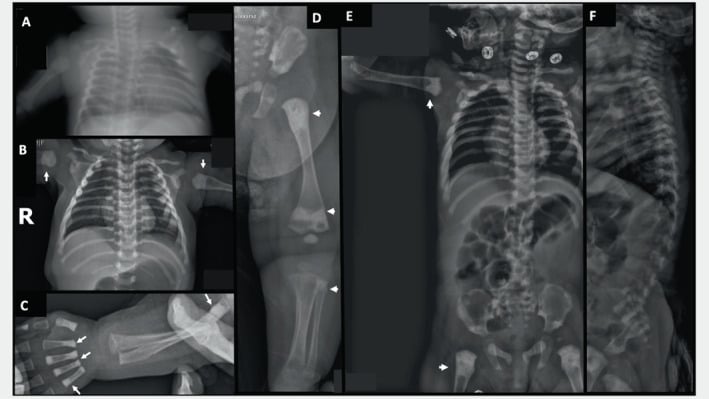
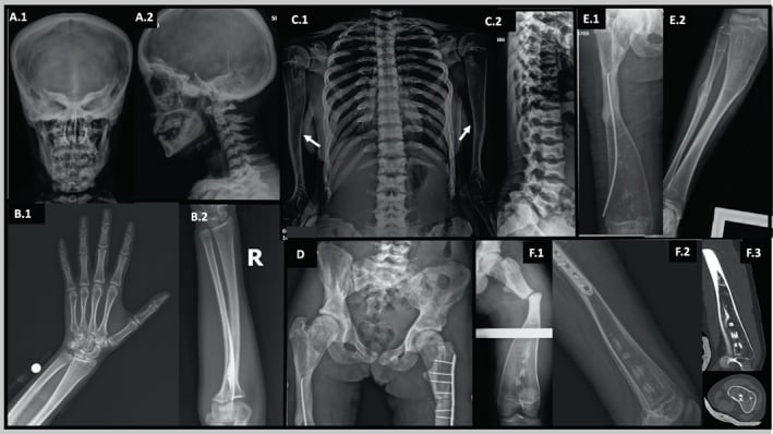
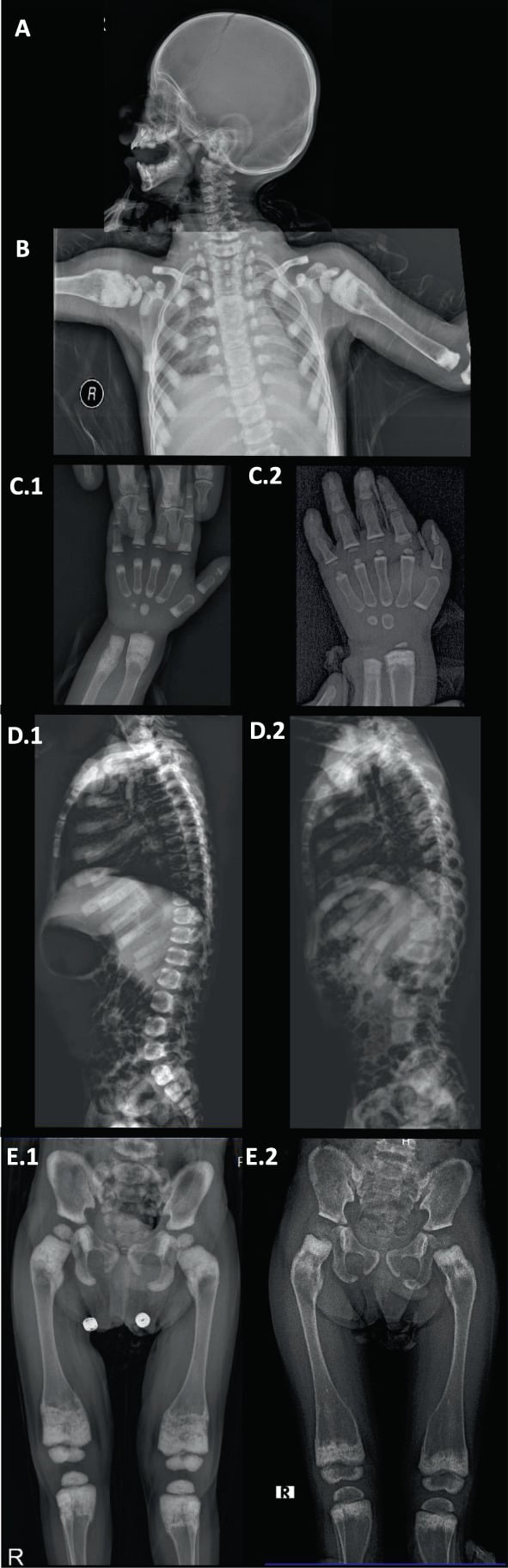
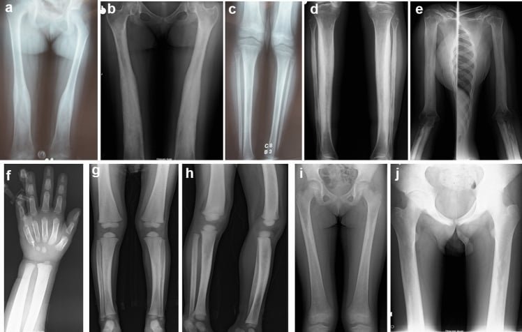
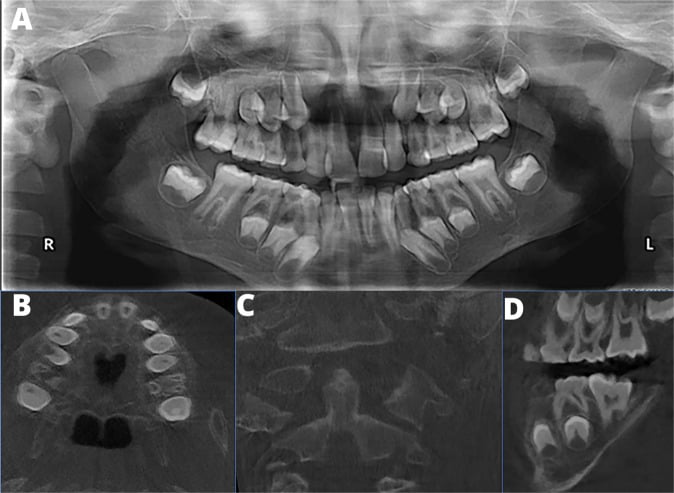
Narrowing It Down
- 11. Is the sign real, and is it bilateral?
Confirm intact smooth cortices, an abrupt metaphyseal-to-diaphyseal transition, and a second involved site. Bilateral symmetrical involvement means a systemic or genetic cause and effectively excludes fibrous dysplasia, Ollier disease and post-infective deformity. A unilateral flask-shaped metaphysis should send you looking for a local lesion.
Bilateral and symmetrical means systemic; unilateral means look for a local lesion
- 22. What is the bone density?
This single question halves the list. DENSE: osteopetrosis, pyknodysostosis, craniometaphyseal dysplasia, lead poisoning (banding). OSTEOPENIC: Gaucher, Niemann-Pick, thalassaemia, sickle cell, leukaemic infiltration. NORMAL: Pyle metaphyseal dysplasia and diaphyseal aclasis.
Faden's three groups map onto this: abnormal-dense trabeculae, marrow-expansion, or normal trabeculae
- 33. Look at the skull and spine on the same day
Hair-on-end diploic widening with obliterated maxillary sinuses equals a chronic haemoglobinopathy. Bone-in-bone vertebrae and a sandwich spine equal osteopetrosis. H-shaped vertebrae equal sickle cell. Craniofacial hyperostosis with hypertelorism equals craniometaphyseal dysplasia. A conspicuously normal skull with massive metaphyseal flaring equals Pyle disease.
The skull and spine usually name the disease when the femur only suggests it
- 44. Look at the femoral head and for infarcts
Avascular necrosis or serpiginous medullary infarcts alongside osteopenia point strongly to Gaucher or sickle cell disease. In Gaucher the spleen is huge and the child is neurologically normal; in sickle cell there is a crisis history and the spleen is small and autoinfarcted.
Osteonecrosis plus an unmodelled femur in a young adult means assay for Gaucher BEFORE any arthroplasty
- 55. Take the systemic history
Hepatosplenomegaly and easy bruising suggest Gaucher or marrow infiltration. Neurodegeneration with a cherry-red spot suggests Niemann-Pick. Deafness, nasal obstruction or facial palsy suggests craniometaphyseal dysplasia or osteopetrosis. Pica in a toddler on a housing estate with old paint suggests lead. Transfusion dependence suggests thalassaemia.
The history distinguishes causes the radiograph cannot
- 66. Check the blood
FBC and film for anaemia, cytopenias and blast cells; haemoglobin electrophoresis for the haemoglobinopathies; serum lead; calcium, phosphate and alkaline phosphatase for rickets; and — if the picture is osteopenic flask plus splenomegaly plus avascular necrosis — leucocyte beta-glucocerebrosidase for Gaucher.
One assay settles the commonest orthopaedically relevant cause
- 77. Decide whether the deformity itself needs treatment
In most cases the flask deformity is a marker, not a problem. Treat the disease: enzyme replacement or substrate reduction for Gaucher, transfusion and chelation for thalassaemia, haematopoietic stem cell transplant for infantile malignant osteopetrosis, chelation for lead. Orthopaedic input is for avascular necrosis, pathological fracture, deformity correction and cranial nerve decompression.
Treat the disease, not the flask - but let the flask change how you plan the operation
MCQ Practice Points
Q: Which single radiographic parameter most efficiently divides the Erlenmeyer flask differential? A: Bone density. Sclerotic points to osteopetrosis, pyknodysostosis and craniometaphyseal dysplasia; osteopenic points to Gaucher, Niemann-Pick, thalassaemia and sickle cell disease; normal points to Pyle metaphyseal dysplasia and diaphyseal aclasis.
Q: How is Gaucher disease confirmed? A: Low leucocyte beta-glucocerebrosidase activity, supported by GBA1 mutation analysis. Bone marrow biopsy showing Gaucher cells is neither necessary nor desirable, and biopsy of bone risks fracture and infection.
Q: What distinguishes Pyle metaphyseal dysplasia from craniometaphyseal dysplasia? A: The skull. Pyle disease has marked metaphyseal flaring with an essentially normal skull and no cranial nerve involvement; craniometaphyseal dysplasia has skull-base and facial hyperostosis causing hypertelorism, nasal obstruction, deafness and facial palsy.
Q: A toddler has dense transverse metaphyseal bands and mild metaphyseal widening but otherwise normal bone density. What is the diagnosis and the test? A: Lead poisoning. The bands are wider and denser than the normal zone of provisional calcification and appear at multiple metaphyses. Confirm with a serum lead level; an abdominal radiograph may show ingested radiodense material.
Q: Which secondary radiographic sign most reliably indicates a chronic haemoglobinopathy as the cause? A: Hair-on-end diploic widening of the skull with obliteration of the maxillary sinuses, reflecting marrow hyperplasia. H-shaped vertebrae favour sickle cell disease specifically.
Q: When operating on a patient with osteopetrosis, what are the key technical precautions? A: Use sharp new drill bits at low speed with copious irrigation to avoid thermal necrosis and drill breakage, anticipate difficult reaming and implant failure, plan for prolonged operative time, and be alert to associated anaemia and the risk of osteomyelitis, particularly of the mandible.
Exam Viva Scenarios
Practise clinical reasoning and management decisions out loud
“You are shown a frontal radiograph of the knees of a 19-year-old man with bilateral distal femoral metaphyseal widening. A second film of the pelvis shows subchondral sclerosis and flattening of the right femoral head. He has a large spleen and normal neurology.”
“You are shown the radiograph of a 9-month-old with diffusely dense bones, widened distal femoral metaphyses and dense transverse bands. The child has anaemia, hepatosplenomegaly and a facial nerve palsy.”
“You are shown the knee radiograph of an 8-year-old imaged after a minor fall. There is striking bilateral distal femoral metaphyseal flaring, normal bone density and no fracture. She is otherwise well with mild genu valgum.”
The sign
- Failure of metaphyseal tubulation: wide metaphysis, narrow diaphysis, abrupt transition
- Best seen at the distal femur; also proximal tibia, proximal humerus, anterior rib ends
- Cortex smooth and intact — a modelling defect, never a destructive lesion
- Bilateral and symmetrical in systemic causes
The words for the viva
- 'Symmetrical loss of metaphyseal tubulation of the distal femora with an abrupt transition to a narrow diaphysis — an Erlenmeyer flask deformity.'
- 'I would assess bone density, look at the skull and spine, and examine the femoral heads.'
Density splits the list
- Dense: osteopetrosis, pyknodysostosis, craniometaphyseal dysplasia, lead banding
- Osteopenic: Gaucher, Niemann-Pick, thalassaemia, sickle cell, leukaemic infiltration
- Normal: Pyle metaphyseal dysplasia, diaphyseal aclasis
One-line discriminators
- Gaucher — osteopenia + AVN + huge spleen + normal neurology
- Niemann-Pick — same plus neurodegeneration and cherry-red spot
- Thalassaemia — hair-on-end skull, obliterated maxillary sinuses, transfusion history
- Sickle cell — H-shaped vertebrae, infarcts, dactylitis
- Osteopetrosis — bone-in-bone vertebrae, sandwich spine, cytopenias, cranial nerve palsy
- Pyknodysostosis — dense but short, acro-osteolysis, obtuse mandibular angle
- Pyle — massive flaring, normal skull, well child
- Craniometaphyseal dysplasia — flaring plus craniofacial hyperostosis
- Lead — dense metaphyseal bands, toddler with pica
- Diaphyseal aclasis — osteochondromas with continuous cortex and medulla
- Fibrous dysplasia — ground glass, asymmetrical, shepherd's crook
- Ollier — columnar chondroid lucencies, unilateral, limb-length discrepancy
Mnemonic
- LEAD GNOME: Lead, Enchondromatosis, Anaemias, Diaphyseal aclasis, Gaucher, Niemann-Pick, Osteopetrosis, Metaphyseal dysplasia, Extra (fibrous dysplasia, rickets)
Investigation
- Skeletal survey for the secondary signs; FBC and film for marrow disease
- MRI for marrow replacement, avascular necrosis and infarcts
- Targeted assay: beta-glucocerebrosidase, sphingomyelinase, haemoglobin electrophoresis, serum lead
- CT skull base if cranial nerve signs
- No biopsy of an under-modelled metaphysis; no further imaging in a stable, diagnosed patient
Orthopaedic consequences
- Gaucher — AVN, bone crisis, pathological fracture, bleeding risk at surgery
- Osteopetrosis — brittle transverse fractures, coxa vara, mandibular osteomyelitis, hard drilling
- Thalassaemia and sickle cell — AVN, osteopenic fracture, growth disturbance
- Diaphyseal aclasis — deformity, impingement, malignant transformation in adults
- Pyle — genu valgum, occasionally requiring guided growth
Evidence Base
The Erlenmeyer Flask Bone Deformity in the Skeletal Dysplasias
- Defines the sign precisely: lack of modelling of the DI-METAPHYSIS with abnormal cortical thinning and loss of the concave di-metaphyseal curve.
- Literature review plus a cohort of 12 disorders, interrogating the International Skeletal Dysplasia Registry radiographic database from 1988 to 2007; 20 distinct disorders were associated with the deformity.
- EFD-T (TYPICAL) - absent di-metaphyseal modelling with RELATIVELY NORMAL trabecular bone: frontometaphyseal dysplasia, craniometaphyseal dysplasia, craniodiaphyseal dysplasia, Engelmann diaphyseal dysplasia, Pyle metaphyseal dysplasia, Melnick-Needles osteodysplasty and otopalatodigital syndrome type I.
- EFD-A (ATYPICAL) - absent modelling with ABNORMAL trabecular bone: dysosteosclerosis and osteopetrosis.
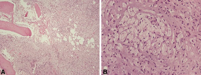
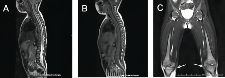
- EFD-ME (MARROW EXPANSION) - marrow hyperplasia or infiltration drives the abnormal modelling, for example Gaucher disease.
- Radiographic review found the deformity was NOT always a consistent finding, and varied in both appearance and location within the skeleton.
Gaucher Disease - Outcome Following Total Hip Replacement and the Effect of Enzyme Replacement Therapy
- 12 total hip replacements in 9 patients with Gaucher disease, mean age 39 years at surgery.
- Four hips (33.3 per cent) required revision for aseptic loosening at a mean of 11.5 years - better survival than the historical literature, which reported high rates of EARLY loosening.
- FIVE OF THE 12 REPLACEMENTS WERE PERFORMED WHEN THE DIAGNOSIS OF GAUCHER DISEASE WAS UNKNOWN, AND ALL FIVE HAD EARLY COMPLICATIONS.
- Enzyme replacement therapy at the time of surgery did not significantly change survival, but that group had fewer peri-operative complications and a trend to better outcome scores.
- Every patient not on enzyme replacement at surgery was started on it later, at a mean of 8.6 years afterwards.
Management of Proximal Femoral Shaft Fractures in Osteopetrosis - a Case Series Using Internal Fixation
- Nine osteopetrotic femoral fractures in three patients, managed and followed long-term.
- States the mechanical problem exactly: osteopetrotic bone CAN be penetrated with a drill bit, but the flutes fill with bone immediately.
- That renders the drill ineffective and the frictional heat generated can break the drill.
- Fractures occur from relatively low-energy mechanisms in characteristically brittle 'marble bone'.
- The difficulties encountered prompted the authors to develop a technique for intramedullary fixation in these femora.