The Nerve You Cut Without Knowing
- It is the terminal SENSORY branch of the radial nerve, separating at the radiocapitellar joint - it supplies no muscle.
- It runs deep to brachioradialis for most of the forearm, then pierces the deep fascia between the tendons of brachioradialis and extensor carpi radialis longus a mean 7.8 cm proximal to the radial styloid.
- By the level of the radial styloid it has divided into a mean of 5 branches lying in subcutaneous fat directly over the first dorsal compartment.
- Wartenberg syndrome is compression at the fascial exit, where the two tendons scissor across the nerve in forearm pronation.
- It is the commonest nerve injured in wrist and thumb base surgery, and the resulting neuroma is more disabling than the numbness.
- “A positive Finkelstein test does NOT distinguish Wartenberg from de Quervain - both stretch the same territory.
- “The distinguishing feature is a Tinel sign 4 to 8 cm proximal to the radial styloid plus altered sensation.
- “Snuffbox wire placement injured a branch of the nerve in 22 per cent of cadaveric specimens - open, do not percutaneous.
- “The nerve travels with the cephalic vein - if you see the vein in the field, the nerve is there too.
Wartenberg mistaken for de Quervain.
- Both hurt on the radial side of the wrist.
- Both are positive on Finkelstein testing.
- Releasing the first dorsal compartment in a patient with Wartenberg syndrome does not help, and places the already irritated nerve directly in the wound.
- Look for a Tinel sign well proximal to the styloid and for altered sensation - de Quervain has neither.
Blind wires and stab incisions.
- Snuffbox K-wire placement injured a branch of the nerve in 22 per cent of cadaveric specimens, and the cephalic vein in 33 per cent.
- External fixator pins placed through stab incisions injure the nerve in the same way.
- Open, spread bluntly to bone, use a protective sleeve. Every time.
- Where the Nerve Lies
- Divides from the radial nerve, between brachialis and brachioradialis
- Relation to Radial Artery
- Not yet related
- What Threatens It
- Radial head fracture, radial tunnel surgery
- Where the Nerve Lies
- Deep surface of brachioradialis
- Relation to Radial Artery
- Lateral to the artery
- What Threatens It
- Anterior (Henry) approach to the radius
- Where the Nerve Lies
- Deep to brachioradialis, running alongside the artery
- Relation to Radial Artery
- Directly lateral, closely applied
- What Threatens It
- Volar plating, radial forearm flap harvest
- Where the Nerve Lies
- Pierces the deep fascia between the BR and ECRL tendons
- Relation to Radial Artery
- Diverges from the artery
- What Threatens It
- Wartenberg compression, external fixator pins
- Where the Nerve Lies
- Subcutaneous, in fat, over the first dorsal compartment
- Relation to Radial Artery
- Superficial to the artery in the snuffbox floor
- What Threatens It
- Compartment release, snuffbox wires, thumb base surgery
BR over ECRLWhere the Nerve Leaves the Fascia
Hook:Two tendons, one nerve between them, and pronation closes the blades.
RADIAL, ULNAR, LATERALThree Nerves on the Dorsum of the Hand
Hook:The fingertips on the back of the hand are MEDIAN, not radial - a classic examination trap.
TINEL DECIDESWartenberg Versus de Quervain
Hook:Both are tender on the radial side. Only one has a Tinel sign and numbness.
Overview
The superficial branch of the radial nerve is the purely sensory terminal branch of the radial nerve. It carries fibres predominantly from C6 and C7, and it supplies no muscle at all. Everything examinable about it flows from where it lies: for most of the forearm it is protected under a muscle belly, and then, in the distal third, it emerges into subcutaneous fat and spreads over exactly the structures that hand surgeons cut.
It separates from the radial nerve at the level of the radiocapitellar joint, in the interval between brachialis and brachioradialis, at the same moment the posterior interosseous nerve is given off. From there the two nerves take entirely different lives: the deep branch dives into the supinator and becomes pure motor, and the superficial branch runs distally on the deep surface of brachioradialis, hugging the radial side of the forearm.
Patients tolerate an anaesthetic patch on the dorsoradial hand remarkably well. The territory is not a working sensory surface, and the lateral antebrachial cutaneous nerve overlaps a good deal of it.
What patients do not tolerate is a painful neuroma. A divided or tethered superficial radial nerve sits in subcutaneous fat directly over the radial styloid and the first dorsal compartment, where every watch strap, sleeve and handshake loads it. This is why the operative principle for this nerve is prevention, and why an accidentally divided stump should never simply be left - it should be repaired, or resected and transposed into muscle away from a pressure point.
The corollary for consent is that discussion of first dorsal compartment release, snuffbox wiring or thumb base surgery must name painful neuroma, not just numbness.
The nerve is also useful to know as an investigative and reconstructive resource: superficial radial sensory nerve conduction is one of the most sensitive early studies in vasculitic neuropathy, the nerve is a standard biopsy site when a sensory nerve biopsy is required, and it serves as a small-calibre donor graft when a sural nerve is unavailable.
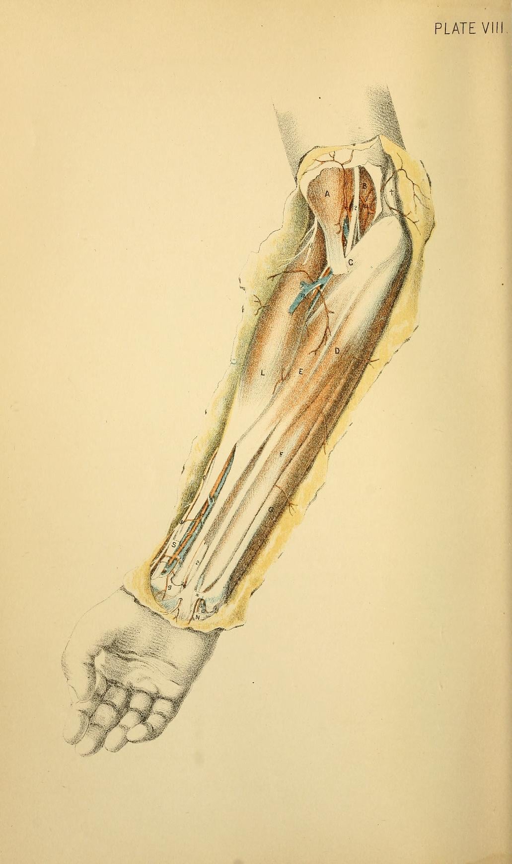
Course, Supply and Relations
Origin
- Terminal sensory division of the radial nerve, arising at the level of the radiocapitellar joint, in the interval between brachialis and brachioradialis.
- Root value predominantly C6 and C7.
- Its sibling, the deep branch, becomes the posterior interosseous nerve; the two separate here and never meet again.
Forearm course
- Runs distally beneath the brachioradialis muscle belly, on the supinator, pronator teres and flexor digitorum superficialis in sequence.
- In the proximal third it lies lateral to the radial artery; in the middle third the two run closely applied, with the nerve lateral to the artery. This is the relationship divided in a radial forearm flap harvest and encountered in a volar Henry approach.
- In the distal third the nerve diverges laterally from the artery, which continues towards the wrist volarly, while the nerve turns dorsally.
Fascial exit - the critical point
- The nerve pierces the deep fascia by passing between the tendons of brachioradialis and extensor carpi radialis longus.
- Leroux and colleagues found this pattern in 9 of 10 cadaveric specimens, at a mean 7.8 cm proximal to the radial styloid.
- This is the site of Wartenberg compression and the reason a Tinel sign here is diagnostic.
Terminal divisions
- The nerve divides into a dorsolateral (radial) branch to the thumb and a dorsomedial branch to the index, middle and radial ring digits, though the branching pattern is genuinely variable.
- In the same cadaveric series, the first division occurred a mean 4.5 cm proximal to the radial styloid and the second a mean 3.0 cm proximal.
- At the level of the radial styloid there were a mean of 5.1 branches crossing the field.
- Practically, this means there is no safe zone in the distal 5 cm - the nerve is not one cord to be avoided, it is a fan of branches to be found.
Relations at the wrist
- It crosses superficial to the tendons of the first dorsal compartment (abductor pollicis longus and extensor pollicis brevis) and then extensor pollicis longus.
- Its branches run with and around the cephalic vein and its tributaries. A vein encountered in a dorsoradial wrist incision is a reliable warning that a nerve branch is within millimetres.
- In the anatomical snuffbox the radial artery lies deep, on the floor of the snuffbox against the scaphoid and trapezium; the nerve branches lie superficial, in the roof.
The parent trunk is developed in radial nerve anatomy and its motor twin in posterior interosseous nerve anatomy. The pathologies it is confused with are covered in de Quervain tenosynovitis and distal radius fractures.
Surface Anatomy and Examination
Making the nerve visible before you cut
This is a skill worth practising on yourself and on every patient before a radial-sided wrist incision.
- Position: forearm fully pronated, wrist in ulnar deviation, thumb flexed and adducted across the palm.
- Look and palpate over the radial border of the distal forearm, 4 to 8 cm proximal to the styloid. In thin patients the branches stand out as fine subcutaneous cords rolling under the fingertip.
- Confirm with a Tinel sign: tap along the line of the nerve. A reproducible electric paraesthesia radiating into the thumb and index maps the branch precisely.
- Mark the branches on the skin before scrubbing and plan the incision to cross them at right angles under direct vision rather than parallel to them.
- In an obese or oedematous wrist the branches will not be visible. In that case assume they are everywhere and dissect accordingly.
Named tests around the radial wrist
- How to Perform
- Percuss 4-8 cm proximal to the radial styloid between the BR and ECRL tendons
- Positive Finding
- Electric paraesthesia radiating into the thumb and index
- What It Means / False Positives
- Most specific sign of Wartenberg syndrome; absent in de Quervain disease
- How to Perform
- Hold the forearm in maximal pronation for 30 to 60 seconds
- Positive Finding
- Reproduction of the dorsoradial pain and paraesthesia
- What It Means / False Positives
- Closes the tendon scissor; a normal wrist tolerates this comfortably
- How to Perform
- Thumb in palm, fist closed, wrist ulnar deviated
- Positive Finding
- Sharp radial wrist pain
- What It Means / False Positives
- POSITIVE IN BOTH de Quervain and Wartenberg - never use it alone to decide
- How to Perform
- Press directly over the first dorsal compartment at the tip of the styloid
- Positive Finding
- Localised tenderness, sometimes with crepitus or a thickened sheath
- What It Means / False Positives
- Points to de Quervain disease rather than the nerve
- How to Perform
- Light touch and two-point discrimination, compared side to side
- Positive Finding
- Reduced or altered sensation
- What It Means / False Positives
- Present in Wartenberg; entirely normal in de Quervain
- How to Perform
- Small volume around the nerve at the fascial exit, well proximal to the compartment
- Positive Finding
- Abolition of the pain and transient numbness
- What It Means / False Positives
- Confirms the nerve as the pain generator; a large volume floods the compartment and confuses the picture
- How to Perform
- Scratch over the site, then test resisted shoulder external rotation
- Positive Finding
- Transient loss of resistance
- What It Means / False Positives
- Adjunctive only; operator dependent and not diagnostic in isolation
- Superficial radial sensory nerve action potentials are recorded over the dorsum of the hand with stimulation in the distal forearm. Reduced amplitude or absent response with a normal median and ulnar study supports a focal lesion.
- Always compare side to side, since absolute values vary widely with age, temperature and limb size. A cold hand alone can abolish the response.
- In Wartenberg syndrome the study may be entirely normal, and a normal study does not exclude the diagnosis.
- The superficial radial sensory response is one of the earliest studies to become abnormal in vasculitic neuropathy and is a standard site for sensory nerve biopsy when tissue diagnosis is required - a reminder that not every abnormal result here is mechanical.
Complications
- Mechanism
- Blade or blind scissors through subcutaneous fat
- How to Avoid It
- Cut skin only, spread longitudinally, protect each branch
- What to Do
- Repair immediately under magnification
- Mechanism
- Percutaneous wire through the roof of the snuffbox - 22 per cent in cadaveric study
- How to Avoid It
- Open mini-incision and blunt spread to bone, protective sleeve
- What to Do
- Remove and reposition; explore if a Tinel sign persists
- Mechanism
- Stab incision and freehand drilling on the radial border of the radius
- How to Avoid It
- Formal open incision, dorsoradial pin placement between ECRL and ECRB
- What to Do
- Reposition the pin; most traction lesions recover
- Mechanism
- Divided stump left in subcutaneous fat over a bony prominence
- How to Avoid It
- Repair, or resect and bury in muscle proximally
- What to Do
- Desensitise, block to confirm, then excise and transpose
- Mechanism
- Self-retaining retractor in subcutaneous fat
- How to Avoid It
- Use loops and skin hooks, release retraction periodically
- What to Do
- Usually recovers in weeks - reassure and desensitise
- Mechanism
- Incomplete fascial release, or the wrong diagnosis
- How to Avoid It
- Confirm with a Tinel sign and a diagnostic block first
- What to Do
- Re-examine for de Quervain disease and for a proximal cause
- Mechanism
- Any injury to this nerve in a susceptible patient
- How to Avoid It
- Prompt mobilisation, effective analgesia, early recognition
- What to Do
- Treat the regional pain syndrome before any further surgery
- Mechanism
- Prominent radial column wire or screw head
- How to Avoid It
- Bury or trim prominent metalwork on the radial column
- What to Do
- Remove the implant and neurolyse
Consequences of permanent loss
- Sensory loss alone is well tolerated. The dorsoradial hand is not a working sensory surface, and lateral antebrachial cutaneous overlap limits the anaesthetic patch.
- Functionally there is no motor loss and no need for tendon transfer.
- The disability, when it occurs, is pain. A neuroma over the radial styloid prevents watch wearing, tool use and handshakes, and can be far more disabling than a comparable motor deficit elsewhere in the hand.
- Counsel accordingly: consent for radial wrist surgery should name painful neuroma and altered sensation explicitly.
Clinical Relevance
Cheiralgia paraesthetica
Compression of the superficial branch of the radial nerve at the point where it emerges through the deep fascia between the brachioradialis and extensor carpi radialis longus tendons.
Presentation
- Pain and paraesthesia over the dorsoradial wrist, thumb and first web space.
- Worse with repetitive pronation and supination, with wrist flexion and ulnar deviation, and at night.
- Often a clear history of external compression: a tight watch strap, a wrist band, handcuffs, a cast, or a new work tool.
- No weakness - if there is weakness, reconsider the diagnosis.
Examination
- Tinel sign 4 to 8 cm proximal to the radial styloid over the fascial exit - the most useful single sign.
- Altered sensation in the dorsoradial territory.
- Positive hyperpronation test.
- Positive Finkelstein manoeuvre - present but non-discriminating.
- Tenderness over the nerve rather than over the compartment.
Management
- Non-operative first, and it usually works. Remove the offending compression, splint the wrist in slight extension with the forearm out of pronation, use a short course of anti-inflammatory medication, and modify activity. In one series of 22 patients with Wartenberg neuritis, 19 responded to conservative treatment.
- Corticosteroid injection around the nerve at the fascial exit can be both diagnostic and therapeutic - inject small and inject accurately.
- Surgical decompression for refractory cases: a longitudinal incision over the fascial exit, release of the fascia between the two tendons over several centimetres, and neurolysis. Do not skeletonise the nerve; leave a bed of soft tissue under it.
- Warn the patient that operating on an already irritable subcutaneous sensory nerve can produce a scar-related neuroma, which is worse than the original complaint.
Surgical Relevance
From cadaveric measurement, referenced to the radial styloid:
- 7.8 cm proximal - mean point where the nerve pierces the deep fascia between the brachioradialis and extensor carpi radialis longus tendons.
- 4.5 cm proximal - mean point of the first division.
- 3.0 cm proximal - mean point of the second division.
- At the styloid - a mean of 5.1 branches crossing the operative field.
Read those four numbers in order and the conclusion is unavoidable: the further distal you operate, the more branches there are and the smaller each one is. Proximal to 8 cm you are looking for one nerve under fascia. Distal to 3 cm you are looking for five nerves in fat.
Universal principles for any dorsoradial incision
- Mark the branches before you scrub, using pronation, ulnar deviation and thumb adduction to make them visible, and a Tinel sign to map them.
- Cut skin only. The dermis is the last thing you divide with a blade in this region.
- Spread longitudinally with fine scissors through the subcutaneous fat, parallel to the expected course of the branches.
- Treat every vein as a warning. The cephalic vein and its tributaries travel with the nerve.
- Hold branches with moist loops or a skin hook, never with a self-retaining retractor placed in fat.
- Cross the branches at right angles, with the branch mobilised and visible, rather than dissecting along them.
- Release the tourniquet and inspect before closure. An intact-looking branch under tension from a stitch will still form a neuroma.
- Document the nerve findings in the operation note - both for the patient and for whoever sees them next.
Anaesthetic technique as a protective measure
- A wrist block or regional block placed proximally avoids the local infiltration of subcutaneous fat around the branches, which distorts the tissue planes exactly where you need to see them.
- If local infiltration is used, place it intradermally along the incision line only, not into the subcutaneous plane.
The differential for a patient with continuing pain after a first dorsal compartment release is short and specific: a missed separate extensor pollicis brevis subcompartment, a superficial radial nerve neuroma or tethering, volar subluxation of the released tendons, and an unrecognised Wartenberg syndrome that was there before the operation. Examine for a Tinel sign, block the nerve diagnostically, and re-image the compartment before assuming the release was inadequate.
Guidelines, Registries & Global Practice
Anatomical variation across populations
- The fascial exit between the brachioradialis and extensor carpi radialis longus tendons is the dominant pattern, found in 9 of 10 specimens in the referenced cadaveric series, but the remainder emerge through brachioradialis itself or more dorsally. A surgeon who cannot find the nerve in the expected interval should not assume it is absent.
- The branching pattern distal to the exit is genuinely variable in number, level and calibre. The mean of five branches at the styloid should be read as a warning rather than as a number to count to.
- Communicating branches between the superficial radial nerve and the lateral antebrachial cutaneous nerve are frequently described, and they blur the sensory boundary. This is one reason a divided nerve can leave a smaller anaesthetic patch than the textbook territory predicts.
- Anomalous muscle slips, particularly accessory abductor pollicis longus tendons, cross the nerve at the wrist and are encountered during first compartment release.
Differences in described technique
- Emphasis
- Formal open incision and soft tissue protection sleeves for all external fixator and percutaneous pin placement
- Practical Point
- Never drill freehand through skin on the radial side of the forearm
- Emphasis
- Identify and protect every branch during first dorsal compartment release; seek a separate EPB subcompartment
- Practical Point
- A separate subcompartment was present in 55 per cent of one operated cohort
- Emphasis
- Non-operative management of Wartenberg syndrome first, with documented sensory examination before any radial wrist operation
- Practical Point
- Documentation before operation is as important as the technique during it
- Emphasis
- Prevention over treatment for cutaneous neuroma; transposition into muscle rather than excision alone
- Practical Point
- Targeted muscle reinnervation and regenerative peripheral nerve interfaces where available
- Emphasis
- Open or mini-open snuffbox start point rather than blind percutaneous wiring
- Practical Point
- Cadaveric branch injury rate of 22 per cent with percutaneous wires
Registry, service and resource considerations
- There is no registry that captures iatrogenic cutaneous nerve injury. Its true incidence after common hand operations is therefore unknown, and published rates come from small single-centre series where the complication was actively sought. The honest position is that the rate is higher than the literature suggests.
- Medicolegal weight is disproportionate to the anatomy. A painful neuroma over the radial styloid after an elective operation for a benign condition is one of the more common sources of complaint in elective hand surgery in every health system. The defence is a documented pre-operative sensory examination and an operation note that records how the branches were identified and protected.
- High-resource settings increasingly use high-resolution ultrasound to map the branches pre-operatively and to guide diagnostic blocks, and have access to targeted muscle reinnervation and regenerative peripheral nerve interface techniques for established neuromas.
- Limited-resource settings lose nothing important here. The critical steps - marking the nerve on the skin, cutting skin only, spreading longitudinally, and protecting branches with loops - require no equipment. Diagnostic blocks require only local anaesthetic.
- Occupational and cultural context shapes presentation: tight wrist ornaments, watch straps and bangles worn habitually on the dominant wrist are a recognised cause of this compression, and simply asking the patient to change the wrist or loosen the strap is often curative.
MCQ Practice Points
Q: Where does the superficial branch of the radial nerve arise? A: At the radiocapitellar joint, in the interval between brachialis and brachioradialis, at the same point the deep branch is given off. Root value predominantly C6 and C7.
Q: Between which two tendons does the nerve pierce the deep fascia, and how far above the styloid? A: Between brachioradialis and extensor carpi radialis longus, a mean 7.8 cm proximal to the radial styloid.
Q: How many branches of the nerve cross the level of the radial styloid? A: A mean of 5.1 in cadaveric measurement - which is why there is no safe zone over the first dorsal compartment.
Q: Which muscles does the superficial radial nerve supply? A: None. It is purely sensory. Any weakness places the lesion proximal to the radial nerve division.
Q: Which nerve supplies the dorsum of the index fingertip? A: The median nerve, through the dorsal branches of the proper digital nerves. The superficial radial nerve reaches only to the level of the proximal interphalangeal joints.
Q: What is the mechanism of compression in Wartenberg syndrome? A: Scissoring of the extensor carpi radialis longus tendon beneath the brachioradialis tendon in forearm pronation, trapping the nerve at its fascial exit.
Q: Does a positive Finkelstein test distinguish de Quervain disease from Wartenberg syndrome? A: No. Both are positive. The discriminators are a proximal Tinel sign and altered sensation, both present only in Wartenberg syndrome.
Q: What proportion of cadaveric specimens sustained a branch injury from Kirschner wires placed in the anatomical snuffbox? A: 22 per cent, with the cephalic vein injured in 33 per cent. The recommendation is an open rather than percutaneous approach.
Q: Why is dorsoulnar hand sensation preserved in compression within Guyon canal? A: The dorsal cutaneous branch of the ulnar nerve leaves the ulnar nerve 5 to 8 cm proximal to the pisiform, above the canal. Its sparing localises the lesion distally - the same logic applied to the superficial radial nerve localises radial lesions.
Q: Why does simple excision of a superficial radial nerve neuroma fail? A: A new neuroma forms in the same subcutaneous plane over a bony prominence. The proximal stump must be transposed into muscle well away from any pressure point.
Exam Viva Scenarios
Practise clinical reasoning and management decisions out loud
“A 34-year-old cyclist has three months of pain over the radial side of the right wrist, with tingling into the thumb. She wears a fitness tracker tightly on that wrist. Finkelstein testing is positive. She has been referred for a first dorsal compartment release. What do you do?”
“Four months after an uncomplicated first dorsal compartment release performed elsewhere, a 46-year-old woman has burning pain over the radial wrist that stops her wearing a watch, and altered sensation over the dorsum of her thumb. Her original pain has gone. How do you assess and manage her?”
“A 28-year-old man had a bridging external fixator applied for a comminuted distal radius fracture. On day two he reports burning numbness over the dorsum of the thumb and index. The fixator is well positioned radiographically. What has happened, and what will you do?”
Anatomy High Yield
- Roots: C6, C7
- Origin: radiocapitellar joint, from the radial nerve
- Course: deep to brachioradialis, lateral to the radial artery
- Exit: between BR and ECRL tendons, 7.8 cm above the styloid
- Supplies: NO muscle - purely sensory
Key Numbers
- 7.8 cm: mean fascial exit above the radial styloid
- 4.5 cm: mean first division above the styloid
- 3.0 cm: mean second division above the styloid
- 5.1: mean branches crossing the styloid level
- 22 per cent: branch injury from snuffbox wires (cadaveric)
- 55 per cent: separate EPB subcompartment in one operated series
Wartenberg Syndrome
- Scissoring of ECRL under BR in pronation
- Tinel 4-8 cm proximal to the styloid
- Altered sensation in the first dorsal web
- Positive hyperpronation test
- Finkelstein positive in this AND de Quervain
- Most settle non-operatively
Iatrogenic Risk Points
- First dorsal compartment release
- Snuffbox and radial styloid wires
- Distal radius external fixator pins
- Thumb base and trapeziectomy incisions
- Radial forearm flap harvest
- Prominent radial column metalwork
Sensory Boundaries
- Dorsoradial hand to the PIP joints only
- Distal to the PIP joints: MEDIAN dorsal digital branches
- Dorsoulnar hand: dorsal cutaneous branch of ulnar
- Overlap with lateral antebrachial cutaneous nerve
- Autonomous zone: dorsal first web space
Evidence Base
Anatomy of the Superficial Radial Nerve and the Danger of Snuffbox Wires
- FORTY Kirschner wires were placed in the anatomical snuffbox of TEN cadaveric forearms - testing a safe zone previously described by Steinberg - then assessed radiologically and by dissection
- The nerve emerged between the brachioradialis and extensor carpi radialis longus tendons in 9 of 10 specimens, a mean 7.8 cm proximal to the radial styloid; first and second divisions occurred a mean 4.5 cm and 3.0 cm proximal to the styloid, with a mean of 5.1 branches at styloid level
- A branch of the nerve was injured in 22 per cent of specimens and the cephalic vein in 33 per cent; the radial artery was never transfixed but a wire lay within 1 mm in 22 per cent
Neuritis and Neuroma of the Sensory Branches of the Radial Nerve
- Series of 44 patients with sensory disturbance in the radial nerve territory, comprising 22 with Wartenberg neuritis, 18 with pure painful neuroma and 9 with combined pathology
- Wartenberg neuritis responded favourably to conservative treatment in 19 of 22 patients
- Of 18 pure painful neuromas, 11 achieved excellent results by Herndon criteria using various techniques; the authors highlight the medicolegal problem when Wartenberg neuritis is not recognised before release of the first extensor compartment
Surgical Release of the First Extensor Compartment - Findings and Outcomes
- 33 patients (3 men and 30 women) operated 2003-2009 for refractory de Quervain disease under LOCAL anaesthesia through a 2 cm transverse incision, with the retinaculum over extensor pollicis brevis incised longitudinally; all had a positive Finkelstein test and localised first-compartment tenderness preoperatively
- A separate extensor pollicis brevis subcompartment was present in 18 of 33 patients (55 per cent), and intracompartmental ganglia in 8
- DASH improved from 53.2 to 3.45 and the visual analogue score from 7.42 to 1.33 (both p less than 0.05), all but one patient had a negative Finkelstein test at final follow-up, and there were no injuries to the sensory branch of the radial nerve and no tendon subluxation
Radial Sensory Neuropathy in the Context of Posterior Interosseous Compression
- 110 patients operated 1992-1997 for posterior interosseous nerve compression, diagnosis made on clinical examination alone; 69 of 103 radial tunnel operations were reviewed at a mean 41 months, with a mean DASH of 32
- Only 60 per cent achieved an excellent or good result and recompression by scarring occurred in 17 per cent after direct decompression
- 52 per cent of patients also had lateral epicondylitis, leading the authors to argue that radial tunnel syndrome is a specific FORM of tennis elbow, linked anatomically by the inseparable origin of the radial wrist extensors and supinator
- The practical consequence they drew is a change of operation: they ABANDONED direct decompression because of the 17 per cent recompression rate and now use Wilhelm denervation, which decompresses the nerve indirectly by releasing the superficial origin of supinator and relaxing the arcade of Frohse
Wartenberg Superficial Radial Neuropathy - Surgical Decompression
- Case report of surgical decompression of the superficial branch of the radial nerve for Wartenberg superficial radial neuropathy
- Documents the entrapment at the point where the nerve emerges from the deep fascia in the distal forearm
- Symptomatic improvement followed decompression after failure of non-operative measures
The Anatomical Relationship of the Superficial Radial Nerve and the Lateral Antebrachial Cutaneous Nerve: A Possible Factor in Persistent Neuropathic Pain
- 20 arms dissected and mapped with Computer Assisted Surgical Anatomy Mapping, with the distance between the two nerves measured at 5 mm increments along their whole course
- IN 95 PER CENT OF ARMS THE SUPERFICIAL RADIAL AND LATERAL ANTEBRACHIAL CUTANEOUS NERVES ACTUALLY INTERSECTED - they do not merely lie near one another
- They intersected on average 2.25 TIMES per arm
- The authors' mechanism: when one nerve is transected, nerve growth factor released along the whole distal segment binds TrkA on the ADJACENT uninjured nerve, driving sprouting and morphological change in fibres that were never cut
- This is offered as an anatomical explanation for why superficial radial nerve injury so often produces persistent neuropathic pain that is hard to localise and hard to treat
The Cutaneous Innervation of the Dorsal Hand: Detailed Anatomy with Clinical Implications
- 30 cadaver forearms dissected to map the lateral antebrachial cutaneous nerve, the superficial branch of the radial nerve and the dorsal branch of the ulnar nerve together
- Innervation of the dorsal hand VARIES, but recognisable patterns exist - the authors derived two classification systems from them
- The territory is shared EVENLY between the superficial radial and dorsal ulnar nerves, and DUAL INNERVATION BETWEEN THE TWO IS FREQUENT
- The lateral antebrachial cutaneous nerve is a COMMON contributor to innervation of the THUMB - a territory usually attributed wholly to the superficial radial nerve
- The stated purpose was to define an area of dorsal wrist free of major nerve branches for safe incision placement
Anatomical Relationships and Branching Patterns of the Dorsal Cutaneous Branch of the Ulnar Nerve
- 28 unmatched fresh-frozen cadavers dissected from the origin of the dorsal cutaneous branch of the ulnar nerve to the metacarpophalangeal joints
- THE NERVE IS NOT ONE STRUCTURE: on average 2 branches were present at the distal ulnar articular surface (range 1 to 4) and 2.2 branches 2 cm distal to it
- At least one longitudinal branch crossed DORSAL TO THE EXTENSOR CARPI ULNARIS TENDON in 23 of 28 specimens (82 per cent)
- In 27 of 28 (96 PER CENT) all longitudinal branches lay between the dorsal-volar midpoint of the subcutaneous border of the ulna and the pisotriquetral joint - a definable at-risk corridor in the pronated wrist
- A transverse branch to the distal radioulnar joint was present in 20 of 28 (71 per cent). The authors conclude that identifying and protecting a SINGLE branch is unlikely to make dissection safe, and that the 6U, 6R and ulnar midcarpal arthroscopy portals put these branches at risk