Familial avascular necrosis of the phalangeal epiphyses | Typically affects adolescents | Presents with painless or mildly painful PIP joint swelling
- Classified as an idiopathic avascular necrosis (osteochondrosis) of the phalangeal epiphyses, most frequently at the proximal interphalangeal (PIP) joints of the hands. The label is inherited from the radiographic appearance, not a demonstrated mechanism.
- Typically presents in adolescence with broad, swollen digits that are often painless or only mildly painful, with range of motion preserved initially.
- Radiographs show sclerosis, flattening and fragmentation of the affected phalangeal epiphyses, which later fuse prematurely, leading to brachydactyly.
- Often familial with an autosomal dominant pattern, though sporadic cases occur. No gene variant has been shown to cause it; one family referred as Thiemann disease carried a TRPV4 variant already known to cause FDAB.
- Management is primarily conservative: observation, activity modification and NSAIDs for pain. Surgery is rarely indicated unless severe deformity or osteoarthritis develops later in life.
- “Unlike inflammatory arthritis, Thiemann disease often presents with painless or mildly painful joint swelling without systemic inflammatory markers.
- “The hallmark radiological finding is the 'fragmented epiphysis' of the phalanx.
- “Premature physeal closure is a common sequela, resulting in shortened digits (brachydactyly).
- “Always differentiate from juvenile idiopathic arthritis to avoid unnecessary immunosuppressive therapy.
Overview
Thiemann disease is a rare osteochondrosis of the phalangeal epiphyses, conventionally described as avascular necrosis. It most commonly affects the proximal interphalangeal (PIP) joints of the hands, though the distal interphalangeal (DIP) joints and the toes can also be involved.
Who gets it. It typically presents in adolescence, between 11 and 19 years of age, with broad, swollen digits. Unlike many other arthropathies, the swelling is often painless or accompanied by only mild discomfort, and range of motion is initially preserved.
Inheritance. The condition can occur sporadically, but it frequently shows a strong familial tendency, with an autosomal dominant pattern of inheritance and high penetrance.
Why it matters. The skill being examined is distinguishing this benign, self-limiting osteochondrosis from more destructive inflammatory conditions such as juvenile idiopathic arthritis (JIA).
Where Thiemann Sits Among the Osteochondroses
The osteochondroses are a heterogeneous group of disorders of the growing skeleton caused by disturbed endochondral ossification, many of them representing avascular necrosis of an ossification centre. They are conventionally divided by the site involved:
- Articular (epiphyseal), involving the joint surface or epiphysis: Legg-Calvé-Perthes (femoral head), Freiberg (2nd metatarsal head), Kienböck (lunate), Köhler (tarsal navicular), Panner (capitellum) and Thiemann (phalangeal epiphyses)
- Non-articular (apophyseal), at a traction apophysis or tendon insertion: Osgood-Schlatter (tibial tubercle), Sever (calcaneal apophysis), Sinding-Larsen-Johansson (inferior pole of patella)
- Physeal, at the growth plate: Scheuermann (vertebral) and Blount (proximal tibial physis)
Thiemann is an articular (epiphyseal) "crushing" osteochondrosis of the phalangeal epiphyses. That is why it behaves like the rest of the articular group: the weakened epiphysis flattens and fragments, and the physis may close prematurely, leaving a short digit.
Genetics: TRPV4 & the Thiemann-FDAB Spectrum
What was found, in one family. A father and son referred with a diagnosis of Thiemann disease were found to carry a heterozygous missense variant, TRPV4 c.809G>T (Damseh 2019). TRPV4 (Transient Receptor Potential Vanilloid 4) is a calcium-permeable cation channel, and that identical variant had already been reported to cause familial digital arthropathy-brachydactyly (FDAB).
What it does not establish. As the same authors state, no gene variant has ever been shown to be causative for Thiemann disease. The finding is equally consistent with those two patients having had FDAB misdiagnosed as Thiemann.
One spectrum or two. The authors hypothesise that the two entities may be the same, and they are explicit about why it is unproven: despite many attempts, they could not obtain DNA from a single one of the other reported Thiemann cases. Their own comparison also cuts both ways, because of 20 individuals with Thiemann disease only 5 had features that also fitted FDAB, so three-quarters did not. Roughly 33 cases of Thiemann disease have ever been described, which is the real reason this cannot be settled.
How they differ. This part is solid and examinable.
- Thiemann disease initially affects the PIP joints, primarily the bases of the middle phalanges
- FDAB affects the distal joints first, with the heads of the middle phalanges as the primary site, and has more extensive joint involvement and more pronounced brachydactyly
- Radial deviation has been described only in FDAB
Counselling. TRPV4-related digital arthropathy is inherited in an autosomal dominant pattern, so where a variant is found, testing guides reproductive counselling. The authors' point is that genetic testing of an affected individual is what would make such counselling accurate, because a clinical label of "Thiemann disease" currently carries no confirmed genotype. Until then, counsel on the phenotype in front of you: the arthropathy is non-inflammatory and the digital changes are largely self-limiting.
A pleiotropic gene. Other TRPV4 variants cause skeletal dysplasias and neuropathies, but the digital-arthropathy phenotype is the relevant one here.
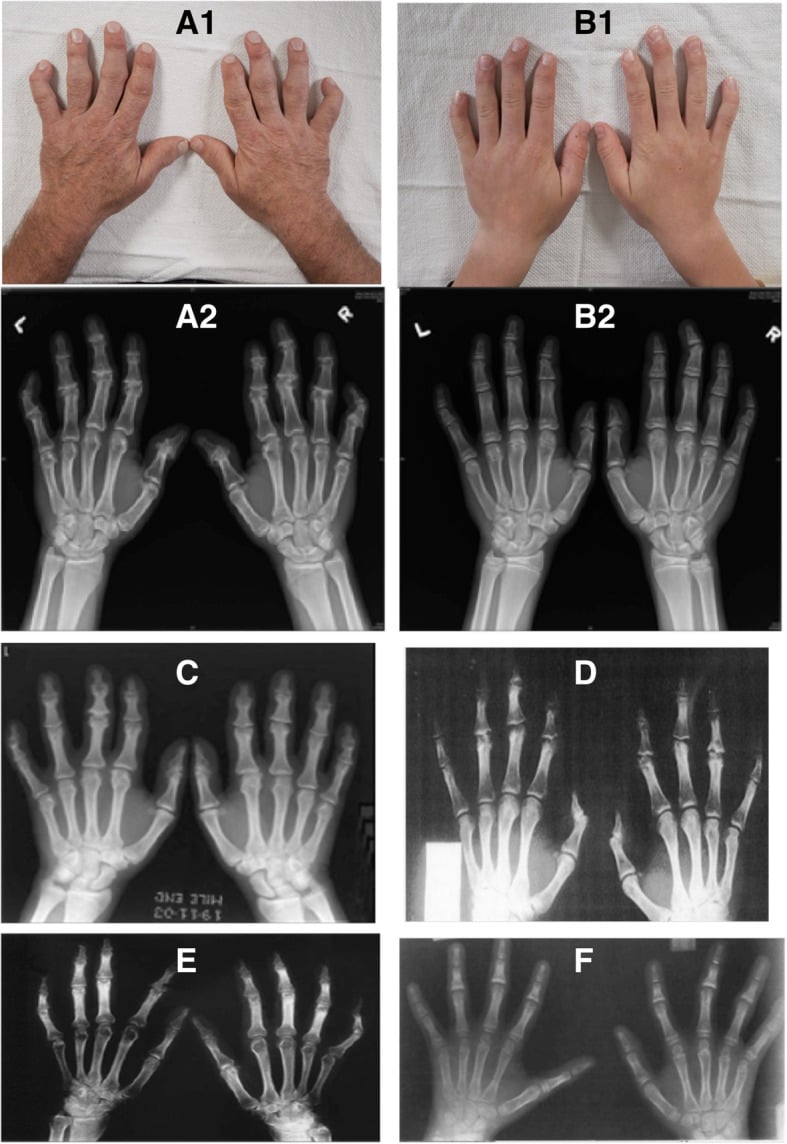
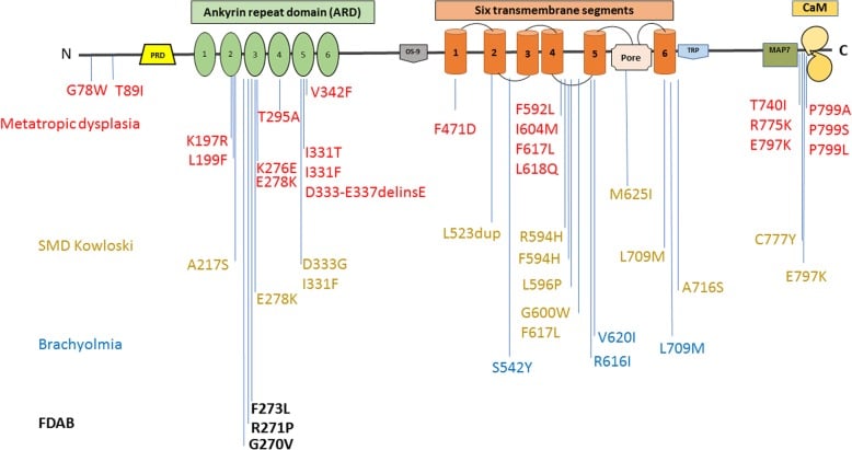
One family, no DNA from any other reported case, and no gene ever proven. The honest answer is that the two may be one spectrum and that this is an unverified hypothesis; then give the discriminator, PIP joints and middle-phalanx bases for Thiemann against DIP joints and middle-phalanx heads for FDAB.
Pathophysiology
What the label means. Thiemann disease is classified as idiopathic avascular necrosis (osteonecrosis) of the secondary ossification centres of the phalanges. Read that as a traditional descriptor inherited from its radiographic appearance, not a demonstrated mechanism. No perfusion study, biopsy series or histological description has established ischaemia in this condition; it was grouped with the osteochondroses because the film looks like the others.
The genetic finding points the other way. The only one is a TRPV4 variant, which would make this a chondrocyte channelopathy and a genetic dysplasia rather than an infarct.
The conventional account. The cascade below is inference from the radiographic sequence. It is worth knowing because it is the account an examiner will expect; state the uncertainty alongside it and you are ahead of the textbook.
Vascular insufficiency. During the adolescent growth spurt, the expanding epiphyses have high metabolic demands. A mismatch between blood supply and demand, possibly exacerbated by microtrauma or genetic predisposition, leads to ischaemia and necrosis of the epiphyseal bone.
Repair and collapse. The necrotic bone is resorbed and replaced by new bone (creeping substitution). During this vulnerable phase, normal mechanical stresses across the joint can cause the weakened epiphysis to flatten, fragment and deform.
The physis. The adjacent growth plate is often damaged by the ischaemic process. The result is premature physeal closure, which arrests longitudinal growth of the affected phalanx and produces brachydactyly.
The joint. The articular cartilage is initially spared, but the altered shape of the subchondral bone eventually makes the joint incongruous, which predisposes it to secondary osteoarthritis.
Classification
There is no universally adopted staging system for Thiemann disease alone. Its progression follows the typical radiological stages of the osteochondroses, much as in Legg-Calvé-Perthes disease.
- Radiographic findings
- Normal, or a slight increase in radiodensity (sclerosis) of the epiphysis
- Clinical correlation
- Mild swelling, often painless
- Radiographic findings
- Epiphysis fragmented, flattened and irregular; clefts may be visible
- Clinical correlation
- Peak of swelling and potential discomfort
- Radiographic findings
- New bone forms and coalesces the fragments; the epiphysis remains deformed (flattened, broad)
- Clinical correlation
- Symptoms resolve; swelling persists because of bony enlargement
- Radiographic findings
- Premature physeal closure, short phalanx (brachydactyly), broadened joint surface, early osteoarthritic changes (osteophytes, joint-space narrowing)
- Clinical correlation
- Short digits; risk of joint stiffness and arthritis in adulthood
Clinical Presentation
The diagnosis relies heavily on recognising the classic phenotype.
The adolescent. A healthy adolescent notices gradual swelling of the PIP joints. The swelling is remarkably painless or causes only a mild ache after exertion, and there are no systemic symptoms: no fever, rash or malaise.
Signs. The PIP joints look broadened and knobbly, often on both hands though not necessarily symmetrically. The overlying skin is normal, without erythema or warmth. Range of motion is often fully preserved early on, though mild restriction may develop as the bony deformity progresses.
The adult who had it. Adults who had the disease in adolescence present with enlarged joints, the symptoms of secondary osteoarthritis (pain, stiffness, loss of motion) and shortened digits, brachydactyly being the classic late finding.
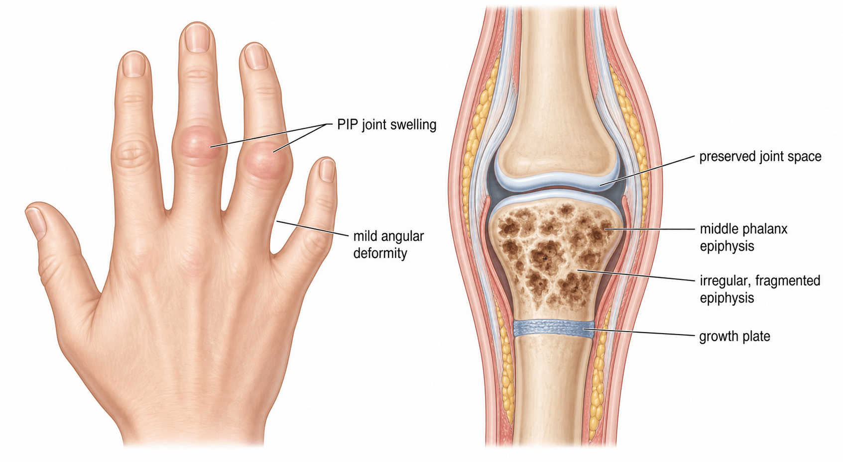
Differential diagnosis. Do not jump straight to JIA without considering the osteochondroses. The alternatives for swollen digits in an adolescent:
- Juvenile idiopathic arthritis: an inflammatory arthritis with painful, warm, swollen joints and systemic signs
- Infection: septic arthritis or osteomyelitis, acute and hot, with systemic signs
- Frostbite: a history of cold exposure, with cold injury damaging the epiphyses
- Trauma: direct injury or repetitive stress to the epiphyses
Investigations
Diagnosis is primarily clinical and radiological.
Radiographs. Plain films of the hands or feet are the gold standard. They move through the stages above, from sclerosis and flattening of the epiphyses to fragmentation, and finally to premature fusion of the growth plate with broad, flattened articular surfaces and shortened phalanges. The joint space is typically preserved while the epiphysis fragments, which differentiates it from inflammatory arthritis.
Blood tests. These are typically completely normal: ESR, CRP, rheumatoid factor and anti-CCP antibodies are negative. This is crucial for excluding juvenile idiopathic arthritis.
MRI. Rarely required. It would show bone marrow oedema and the typical changes of avascular necrosis (loss of normal marrow signal on T1-weighted images) early in the disease, before plain-film changes are obvious.
Management
Management is overwhelmingly conservative. The goal is to manage symptoms while the epiphysis revascularises and heals, on the conventional account, and to avoid unnecessary intervention. Before settling on that plan, rule out inflammatory or infectious causes.
Non-operative care. Reassurance and education are the cornerstone: explain to the patient and family the self-limiting nature of the acute phase, but warn about the potential for short digits and early arthritis. Do not prescribe aggressive systemic treatment. The rest is symptomatic:
- Activity modification, avoiding heavy lifting, contact sports and activities that place excessive axial load on the fingers during the active fragmentation phase
- Simple analgesics or NSAIDs for pain during flares
- Temporary splinting for symptomatic relief if pain is significant, though prolonged immobilisation should be avoided to prevent stiffness
Surgery. Surgery is rarely indicated during the acute adolescent phase, and do not plan early surgical intervention unless significant deformity dictates it. It is generally reserved for severe late sequelae in adults:
- Corrective osteotomy for severe angular deformities that impair function
- Arthrodesis of the PIP joint, which may be required for end-stage, painful secondary osteoarthritis
- Arthroplasty (small joint replacement), an option in low-demand adults with severe osteoarthritis, though less common for the PIP joints
Complications
The complications follow from the mechanical damage to the growing bone and the joint surface.
Brachydactyly. Premature physeal closure leaves noticeably shortened fingers or toes. This is mostly a cosmetic issue but can occasionally affect grip mechanics.
Secondary osteoarthritis. The deformed, flattened articular surface makes the joint incongruous, and it wears early. This is the most significant long-term clinical problem, causing pain and stiffness in early to mid-adulthood.
Deformity and stiffness. The joints remain broad and knobbly, and may develop flexion contractures or lose terminal flexion.
Guidelines, Registries and Global Practice
- Diagnostic Approach: Global practice emphasises the clinical differentiation of Thiemann disease from Juvenile Idiopathic Arthritis (JIA). The presence of painless epiphyseal fragmentation with negative inflammatory markers is universally accepted as diagnostic, avoiding unnecessary rheumatological workups.
- Genetic Counseling: In cases with a strong family history, genetic counseling may be offered to discuss the autosomal dominant inheritance pattern and the potential link to broader skeletal dysplasias (such as familial digital arthropathy-brachydactyly).
- Surgical Indications: There are no formal guidelines advocating for early surgical intervention in Thiemann disease. International consensus supports conservative management during adolescence.
- Nomenclature: While eponyms are becoming less favored in some medical communities, "Thiemann disease" remains the standard term used in orthopaedic literature and examinations worldwide to describe this specific epiphyseal avascular necrosis.
Related pages: Juvenile Idiopathic Arthritis is the misdiagnosis this condition exists to be distinguished from, and the distinction is worth money to the patient - a child labelled JIA may be started on immunosuppression for a non-inflammatory dysplasia; Psoriatic Arthritis of the Hand is the other differential that attacks interphalangeal joints and, unlike Thiemann, erodes; Interphalangeal Arthritis for the adult endpoint these fingers reach, and for the degenerative pattern that a burnt-out Thiemann hand is mistaken for decades later; Kohler Disease, Freiberg Disease, Panner Disease, Kienbock's Disease, Legg-Calve-Perthes Disease, Sever Disease, Osgood-Schlatter Disease, Scheuermann Kyphosis and Blount Disease are the rest of the osteochondrosis family this page positions Thiemann within - and the useful exam point is that the label "osteochondrosis" groups conditions by radiographic appearance and age, not by a shared mechanism, so several of them have turned out to be traction, dysplastic or genetic rather than ischaemic; Skeletal Dysplasias for the TRPV4 pleiotropy that puts digital arthropathy in the same gene as the metatropic dysplasia spectrum and some hereditary neuropathies; and Congenital Hand Overview for the wider differential of a short, deformed digit in a child.
Exam Viva Scenarios
Practise clinical reasoning and management decisions out loud
“A 14-year-old boy presents with a 6-month history of swelling in the PIP joints of his middle and ring fingers bilaterally. He denies any significant pain, morning stiffness, or systemic symptoms. Blood tests (ESR, CRP, RF) are normal. Radiographs show fragmentation of the PIP epiphyses. What is the diagnosis and how do you manage it?”
“The examiner shows you a radiograph of a 15-year-old's hand with flattening and sclerosis of the PIP epiphyses. They ask you to outline your differential diagnosis for avascular necrosis in the hand and wrist, and how you differentiate them clinically.”
“A 45-year-old man presents with chronic pain and stiffness in his PIP joints. Examination shows broad, shortened fingers (brachydactyly). He mentions his fingers have looked 'knobbly' since he was a teenager. Radiographs show severe PIP joint osteoarthritis with flattened articular surfaces. What is the likely underlying etiology and what are your management options?”
Core Concepts
- Avascular necrosis (osteochondrosis) of phalangeal epiphyses
- Peak incidence in adolescence (11-19 years)
- Familial cases show autosomal dominant inheritance
- Most commonly affects PIP joints of the hands
Clinical Presentation
- Broad, swollen, 'knobbly' PIP joints
- Painless or only mildly aching
- No systemic symptoms; normal inflammatory blood markers
- Late presentation: short digits (brachydactyly) and early OA
Radiographic Findings
- Early: Epiphyseal sclerosis and flattening
- Active: Epiphyseal fragmentation
- Late: Premature physeal closure, broad joint surface, OA changes
- Joint space is initially preserved
Management Strategy
- Conservative: Reassurance, activity modification, NSAIDs
- Self-limiting acute phase
- Avoid aggressive interventions
- Surgery only for end-stage secondary osteoarthritis in adults
Evidence: Key Papers in Thiemann Disease
Because Thiemann disease is rare, the literature consists primarily of case series and case reports. These papers highlight the genetic links and the diagnostic challenges.
Thiemann disease and familial digital arthropathy - brachydactyly: two sides of the same coin?
- A father and son REFERRED WITH A DIAGNOSIS of Thiemann disease were found to carry TRPV4 c.809G>T - a variant already reported to cause FDAB
- The authors HYPOTHESISE that Thiemann disease and FDAB may be one entity; no gene variant has ever been shown to cause Thiemann disease
- On their review of 20 Thiemann cases, only 5 had features that also fitted FDAB - so three-quarters did not
- Discriminators: Thiemann affects PIP joints and middle-phalangeal BASES first; FDAB affects distal joints and middle-phalangeal HEADS, and radial deviation is described only in FDAB
- Roughly 33 cases of Thiemann disease have been reported in total
Thiemann's disease: a very rare bone disorder.
- Case report detailing the presentation of a patient with Thiemann disease.
- Emphasised the diagnostic challenge, as patients often present to rheumatology clinics with suspected inflammatory arthritis.
- Confirmed the classic radiographic findings of epiphyseal fragmentation and subsequent early fusion without erosions typical of inflammatory disease.
Thiemann disease.
- Reported on the clinical and radiographic progression of Thiemann disease.
- Reiterated the hallmark features of painless or mildly painful swelling in the PIP joints during adolescence.
- Documented the typical conservative management strategy and the natural history leading to brachydactyly.