Evaluation and Management of Unhealed Tibial Fractures | Exchange Nailing | Bone Grafting | Ilizarov
- Definition: Fracture that has not healed by 9 months, or shows no progression for 3 months.
- Workup: Verify INFECTION status (ESR, CRP, aspiration) and METABOLIC status (Vit D, Ca, PTH).
- Hypertrophic: Needs STABILITY (Exchange nailing usually successful).
- Atrophic: Needs BIOLOGY (Bone graft, RIA) and STABILITY.
- Infected: Needs DEBRIDEMENT first (Masquelet technique or Bone Transport).
- “The 'Diamond Concept' covers: Mechanical Stability, Osteogenesis, Osteoconduction, Osteoinduction.
- “Exchange nailing allows reaming (autograft) and larger nail (stability).
- “Fibulectomy may be needed to allow compression of the tibial nonunion site.
Overview
Definition. Two definitions are in use. The FDA definition is a fracture that has not healed at 9 months; the clinical definition is a fracture showing no radiographic progression over 3 consecutive months.
Why the tibia. The tibial shaft is the most common location for long-bone nonunion. The reasons are its poor blood supply, with the anteromedial surface subcutaneous, and high-energy trauma.
The Diamond Concept. Giannoudis's framework considers the biological and mechanical environment together. Its four pillars:
- Osteogenesis - cells (osteoblasts), supplied by bone marrow aspirate or graft
- Osteoconduction - a scaffold (matrix), such as allograft or ceramic
- Osteoinduction - signals, the growth factors such as BMPs
- Mechanical stability - the strain environment
Biology also needs vascularity, which depends on a healthy soft-tissue envelope.
Anatomy and Pathophysiology
The blood supply. The tibia is subcutaneous anteromedially, which leaves its blood supply vulnerable. The nutrient artery, from the posterior tibial artery, enters through the posterior cortex in the proximal third and provides 30-40% of the supply; it is often disrupted in the initial fracture. The periosteal supply provides 60-70% and depends on the surrounding soft-tissue envelope: the anterior tibial artery and muscles laterally, the posterior tibial artery and muscles posteriorly. Once the nutrient artery is disrupted, it is the sole supply.
The watershed. The distal third has the poorest blood supply and limited collateral circulation, and the highest nonunion rate.
Strain. Micro-motion promotes callus, too much motion prevents it, and absolute rigid stability gives primary healing without callus. The thresholds:
- Strain under 2% - primary healing under absolute stability, no callus
- Strain 2-10% - secondary healing, micro-motion and callus formation
- Strain over 10% - nonunion, the motion is excessive and prevents healing
Hypertrophic nonunion. The biology is good, with abundant callus, but the stability is poor. The body is trying to heal, and excess motion, strain over 10%, lets shear forces prevent the callus bridging.
Atrophic nonunion. A biological failure: poor vascularity and dead bone, no callus and sclerotic ends, with no biological drive. Stability is variable.
Infected nonunion. Biofilm prevents biological healing, so the biology is poor; stability is variable.
Classification Systems
Weber and Cech classify by the vascularity of the bone ends, originally on a strontium scan and now on radiographic appearance:
- Hypertrophic - high vascularity, the "elephant foot"; the problem is instability
- Atrophic - low vascularity, the "pencil point"; the problem is biology
- Oligotrophic - intermediate, the "horse hoof"
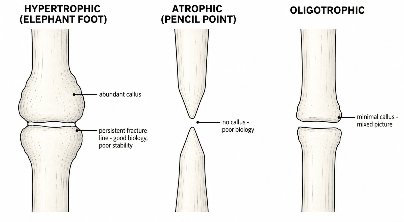
Paley classifies by bone loss:
- Type A - bone loss less than 1cm
- Type B - bone loss greater than 1cm
- B1 - with bony contact (shortening)
- B2 - without bony contact (a defect)
Clinical Assessment
Pain. Pain on weight bearing is the hallmark of nonunion: persistent pain despite adequate time for healing. Pain at rest suggests infection.
Constitutional symptoms. Fevers, chills and night sweats point to infection; weight loss to infection or malignancy. Ask about previous drainage from the wound.
Risk factors. Smoking is the most important modifiable risk factor, with slower union and more secondary surgery. Then the rest of the history:
- Diabetes - poor healing and infection risk
- NSAIDs and steroids - both inhibit bone healing
- Previous open fracture - infection risk
- Metabolic disease - vitamin D deficiency, hyperparathyroidism and other thyroid or parathyroid disorders
NICE MDNonunion Risk Factors
Hook:NICE MDs prevent nonunions.
Look. The skin records the history: flaps and scars from previous surgery or an open fracture, swelling from infection or chronic inflammation, erythema, and deformity from malalignment or shortening. A sinus tract is pathognomonic of infection.
Feel. Palpate for tenderness at the fracture site, and for warmth, which suggests infection. Motion at the fracture site indicates gross instability; crepitus indicates motion at the nonunion.
Neurovascular. Record the posterior tibial and dorsalis pedis pulses, sensation in the saphenous, sural and superficial peroneal territories, and ankle dorsiflexion and plantarflexion.
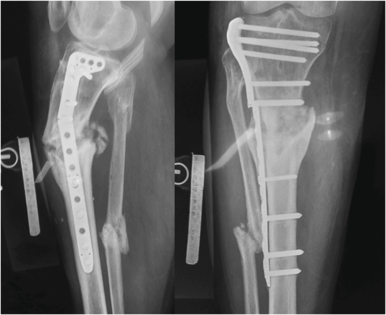
Investigations
Any nonunion is infected until proven otherwise, even when the skin is normal.
Bloods. Two screens:
- Infection - full blood count, ESR and CRP; if they are raised, aspiration or biopsy
- Metabolic - calcium, phosphate, ALP, vitamin D (25-OH), PTH, TSH, HbA1c and albumin
Radiographs. AP, lateral and oblique views show the callus, any implant failure and the alignment.
CT is the gold standard for bony bridging, which metal often obscures on plain films. Look for less than 50% bridging on the cuts.
Nuclear medicine. A bone scan has high sensitivity and low specificity. A tagged white cell scan (Indium/Sulfur colloid) helps to separate infection from remodelling.
Scoring Union Objectively: RUST and mRUST
How RUST is scored. RUST turns a subjective "united or not united" read into a reproducible number. Score four cortices on orthogonal radiographs, two on the AP and two on the lateral:
- 1 - fracture line visible, no callus
- 2 - callus present, fracture line still visible
- 3 - bridging callus, fracture line no longer visible
The sum runs from 4 (no healing at any cortex) to 12 (complete union at all four). mRUST scores each cortex out of 4, adding a top tier for a bridged cortex that has remodelled with no visible line. Its range of 4-16 discriminates more finely, particularly around metaphyseal or plated fractures where callus is subtle.
Why it matters in nonunion. RUST is not only a fresh-fracture tool. In operatively treated tibial nonunions a low preoperative RUST predicts failure of nonunion surgery, and both pre- and postoperative RUST correlate with time to union. Scoring the cortices at presentation therefore risk-stratifies the nonunion itself and calibrates how aggressive the reconstruction must be: biology plus stability, not stability alone.
The threshold, and its provenance. A preoperative RUST below 7 has been reported to flag a high risk of persistent nonunion (Christiano/Egol). The cut-off was derived from seven failures in 68 patients, in the same cohort it was tested on, and was never externally validated, so treat it as a prompt to look harder rather than as a decision threshold.
Serial scoring. A score that plateaus and fails to climb over 3 months supports the nonunion diagnosis and the case for intervention. CT remains the arbiter when metal obscures cortices or the plain-film score is borderline.
RUST Predicts Success of Tibial Nonunion Surgery
- RUST applies to the nonunion itself, not only to fresh fractures
- A preoperative RUST below 7 flags a high risk of persistent nonunion
- Serial RUST helps time and judge the success of nonunion surgery
Differential Diagnosis
The clinical picture of persistent pain and failure to heal overlaps with several entities. The single most important fork is infected versus aseptic, because it changes the whole reconstructive plan.
- Distinguishing Features
- Mechanical pain on loading, normal inflammatory markers, no sinus
- Key Discriminator
- Normal ESR/CRP, negative cultures
- Distinguishing Features
- Rest/night pain, sinus, warmth, raised ESR/CRP
- Key Discriminator
- Positive deep cultures / sinus tract
- Distinguishing Features
- Healing progressing but slow; callus advancing on serial films
- Key Discriminator
- Progression over 3 months (vs none in nonunion)
- Distinguishing Features
- Deformity/limb-length issue, pain may be adjacent joint
- Key Discriminator
- Bridging callus present on CT
- Distinguishing Features
- Implant breakage or backout, mechanical pain
- Key Discriminator
- Radiographic implant failure
- Distinguishing Features
- No clear preceding adequate fracture healing window; lytic lesion
- Key Discriminator
- MRI/biopsy if neoplasm suspected
Management Algorithm
The pathway. Work up and optimise first; the type of nonunion then decides the operation:
- Classify and work up - Weber and Cech type; rule out infection (ESR, CRP, aspiration); metabolic status (vitamin D, calcium, PTH); CT for bony bridging
- Optimise - smoking cessation is mandatory for elective nonunion surgery, because nicotine is a potent vasoconstrictor and inhibitor of healing, and smokers have slower union and more secondary surgery (Adams 2001). Supplement vitamin D if deficient, control diabetes and stop NSAIDs if possible
- Treat the type - stability for the hypertrophic nonunion, biology and stability for the atrophic one, infection first for the infected one
- Callus
- Abundant (Elephant Foot)
- Vascularity
- Excellent
- Treatment Principles
- Stability (Stiffen fixation)
- Callus
- None (Pencil Tip)
- Vascularity
- Poor
- Treatment Principles
- Biology (Graft) + Stability
- Callus
- Variable
- Vascularity
- Poor (Biofilm)
- Treatment Principles
- Debridement first to Reconstruction
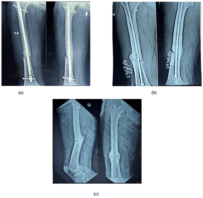
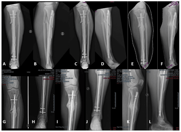
Hypertrophic: add stability. The biology is already there, so the treatment improves stability rather than adding bone graft. Exchange nailing is the gold standard: remove the old nail, ream the canal, which deposits internal graft, and insert a larger-diameter, stiffer nail, considering a fibulectomy if the fibula is holding the fracture apart. Compression plating across the nonunion is the alternative if nailing is not suitable. If the tibia is already nailed and the gap is less than 5mm, dynamise: remove the locking screws to allow compression, and monitor with serial radiographs.
Atrophic: biology and stability. Resect the dead bone ends back to bleeding bone (the "paprika sign"), removing the fibrous tissue to leave a healthy bone bed. Then graft: iliac crest autograft is the gold standard, with the RIA (Reamer Irrigator Aspirator) for large volumes, and graft provides osteogenic cells, osteoinductive factors and an osteoconductive scaffold. Finally fix it, with compression plating or nailing, which must provide a stable environment. Atrophic nonunions need both: the graft for biology and the fixation for stability.
Infected: infection first. Eradicate the infection, with debridement and antibiotics, before reconstructing; infected nonunions need staged reconstruction:
- Stage 1 - hardware removal, radical debridement of all infected bone, an antibiotic spacer (Masquelet) or beads, cultures including sonication of the hardware, and an external fixator or plate for stability
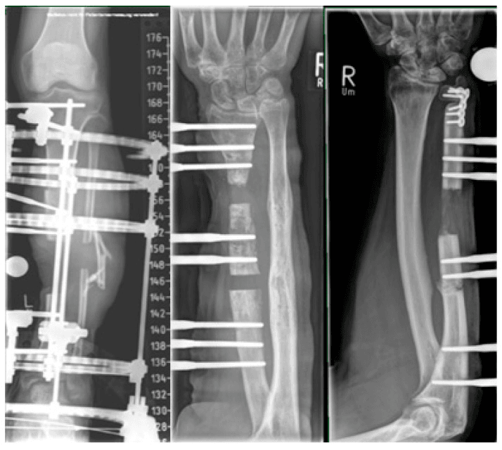
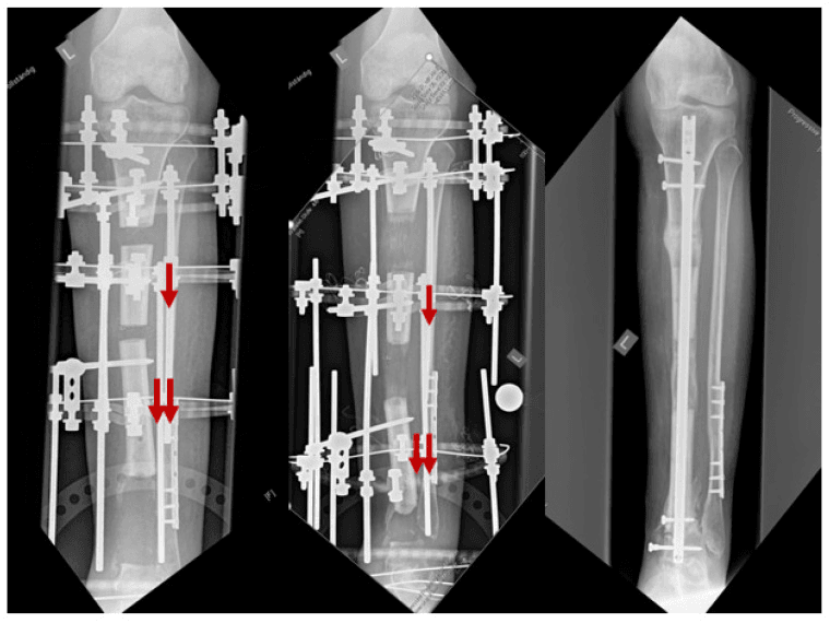
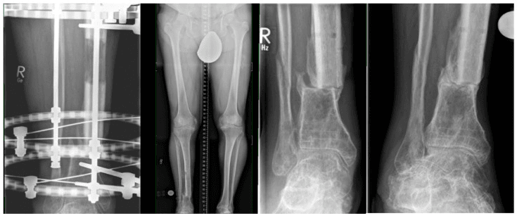
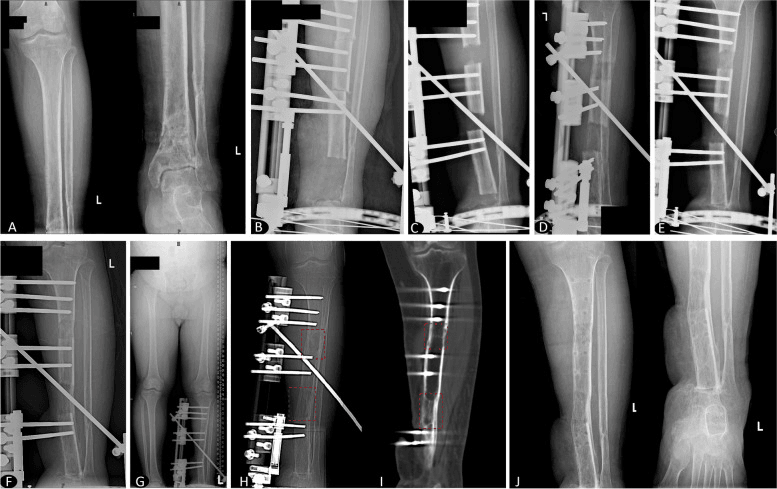
- Stage 2, 6-8 weeks later - bone grafting if a membrane has formed (the Masquelet technique), or bone transport (Ilizarov) if the defect is large, over 5cm
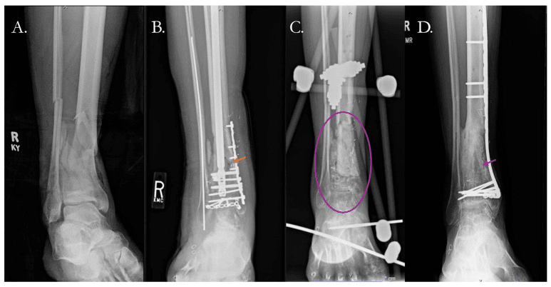
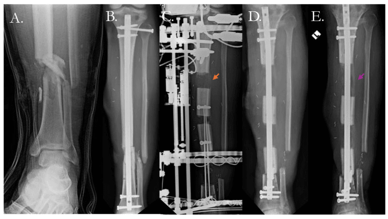
Surgical Technique
Indications.
- Hypertrophic nonunion (good biology, poor stability)
- Aseptic nonunion
- Previous intramedullary nailing
Technique.
- Removal - extract the old nail and send the tip for sonication and culture to rule out infection
- Reaming - ream the canal aggressively, often 1-2mm larger than previously; the reamings act as autograft
- Insertion - insert a nail 1-2mm larger than the largest reamer
- Locking - static locking, or dynamic if axial stability is assured
- Fibulectomy - consider it if the fibula is holding the fracture apart
Check the distal pulses after reaming: there is a risk of vascular injury.
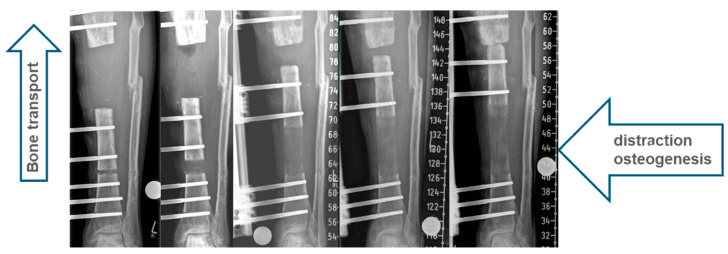
Fibular Osteotomy: Releasing the 'Hold-Open' Strut
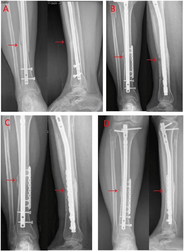
The mechanical problem. In the leg the tibia and fibula share length. A fibula that has healed, or was never broken, behaves as a rigid lateral strut. As the tibia tries to compress across its nonunion, under weight bearing, dynamisation or a compression device, the fibula "holds it open", distracting the tibial bone ends and defeating the axial compression and callus-generating micromotion the treatment relies on. A persistent tibial gap beside a united fibula on the radiographs is the classic clue.
When to release it.
- A hypertrophic (or oligotrophic) tibial nonunion with an intact or healed fibula where compression is the goal
- Before dynamising a nailed tibia: removing the locking screws will not compress a fracture the fibula is strutting
- When planning acute shortening of a segmental defect, where the fibula would otherwise resist limb-length adjustment
Technique. Perform a segmental fibular osteotomy or ostectomy through a small direct lateral incision, removing a short segment, about 1cm, enough that the fibula cannot simply re-strut by bridging. Site it offset from the tibial nonunion, so the two do not consolidate into a single mass and the exposure is safe.
Pitfalls. Stay away from the common peroneal nerve at the fibular neck proximally. Preserve the distal syndesmosis and lateral malleolus: a resection too distal destabilises the ankle mortise.
An adjunct, not a stand-alone. Fibular release enables the mechanical solution, whether compression plating, nailing, dynamisation or frame compression. It does not substitute for biology in an atrophic nonunion, and it is unnecessary when the fibula is itself un-united or already absent.
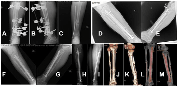
Complications
- Incidence
- 10-20%
- Risk Factors
- Previous open fracture, infected nonunion, biofilm
- Prevention/Management
- Thorough debridement, antibiotics, staged approach; if it recurs, radical debridement, hardware removal, antibiotics and reconstruction
- Incidence
- 5-10%
- Risk Factors
- Through graft site, screw holes; premature weight bearing
- Prevention/Management
- Protected weight bearing, gradual progression; revision fixation if needed
- Incidence
- 10-20%
- Risk Factors
- Iliac crest harvest (cluneal nerve injury), RIA (femur fracture)
- Prevention/Management
- Careful technique, consider RIA for large volumes; pain management, nerve blocks if needed
- Incidence
- Less than 5%
- Risk Factors
- Bleeding into tight compartments after surgery
- Prevention/Management
- Monitor, careful haemostasis; four-compartment fasciotomy if needed
- Incidence
- 10-15%
- Risk Factors
- Smoking, infection, poor biology
- Prevention/Management
- Address all Diamond Concept components
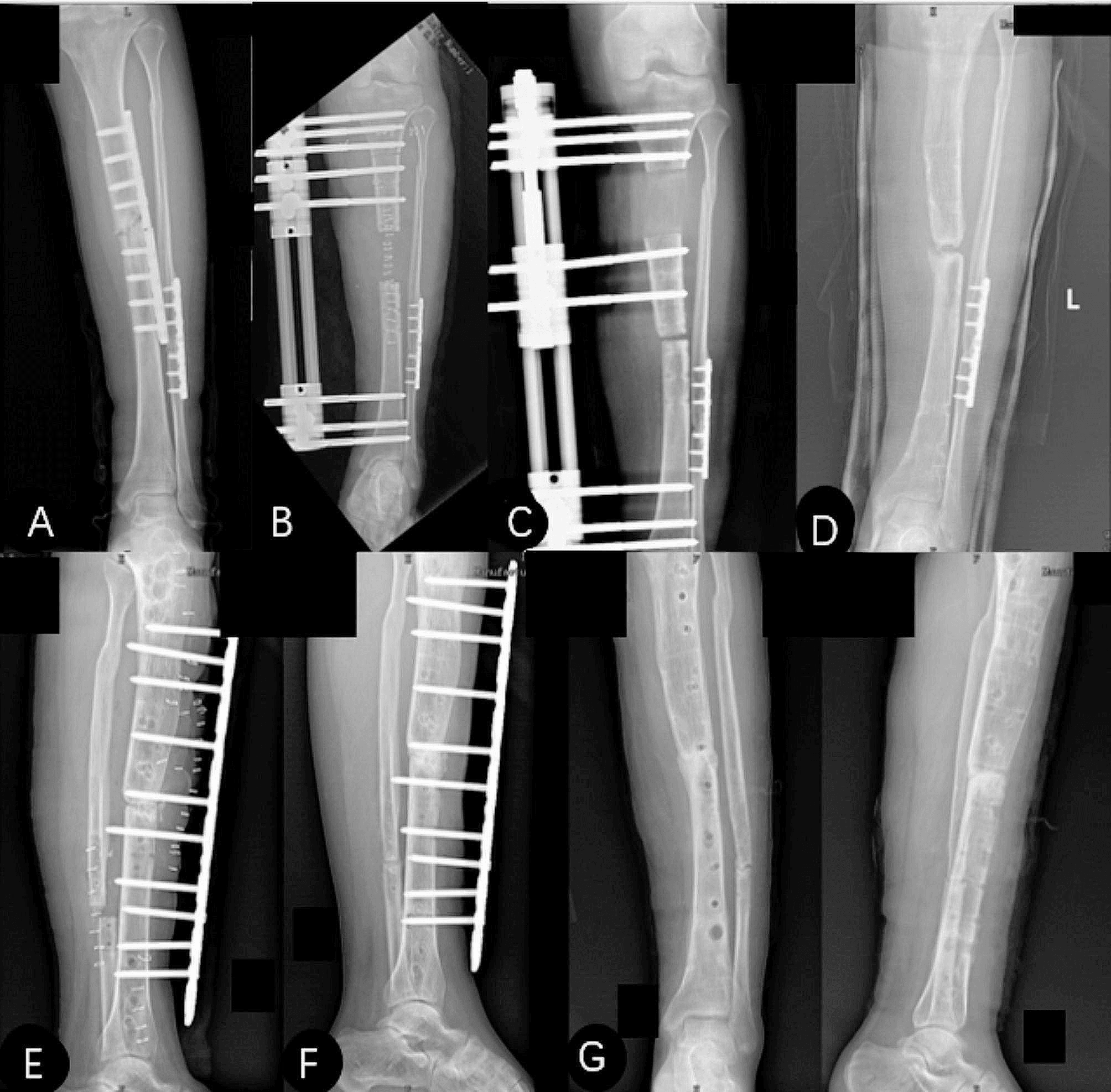
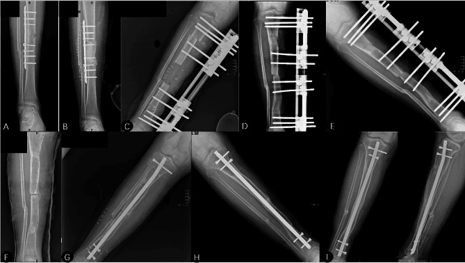
Postoperative Care
Immediately. No immobilisation: mobilise early, with immediate knee and ankle range of motion. Weight bearing starts partial, how much depending on the stability of the fixation, and physiotherapy covers ambulation training and strengthening.
Rehabilitation protocol.
- Weeks 0-2 - partial weight bearing (20-50% body weight), knee and ankle range of motion, quadriceps and calf strengthening, ice and elevation
- Weeks 2-6 - progressive weight bearing (50-75% body weight), continued range of motion and strengthening, balance and proprioception
- Weeks 6-12 - progress to full weight bearing, full range of motion, progressive activity
- Weeks 12+ - full weight bearing; return to activity when union is confirmed, still monitored with serial radiographs
Slow healing. If healing is slow at 3-4 months, dynamise: remove the static locking screws, proximal or distal, to allow localised compression at the nonunion, and follow it with serial radiographs.
LIPUS. Regulatory approval of low-intensity pulsed ultrasound covers established nonunion and fresh fracture, depending on jurisdiction. TRUST is the trial everyone cites, and it does not answer the question this page asks. It randomised 501 patients with acute tibial fractures after nailing and found no effect on radiographic healing or function, but it explicitly excluded fractures with less than 25% cortical contact or a gap over 1cm: exactly the mechanical situations that become a nonunion.
What that leaves. TRUST argues firmly against routine LIPUS after acute nailing. It provides no evidence for or against LIPUS in an established nonunion, where no adequately powered trial exists. It is not a substitute for correcting mechanics or biology, and should never delay a definitive operation.
Outcomes and Prognosis
- Success rate
- Greater than 90% for hypertrophic nonunions
- Time to union after revision
- 6-9 months
- Complications
- 10-15% (infection recurrence, re-fracture)
- Success rate
- 85-90%
- Time to union after revision
- 6-9 months
- Complications
- 10-15% (infection, hardware failure)
- Success rate
- Greater than 90% for infected nonunions, including those with large defects
- Time to union after revision
- 9-12 months (longer for large defects)
- Complications
- 20-30% (pin site infection, joint stiffness)
Union by type. The hypertrophic figure is the exchange-nailing row above. Atrophic nonunions unite in 85-90% with bone graft and fixation, and infected nonunions in 80-90% with staged reconstruction.
Function. Return to activity takes 6-12 months after surgery, and 80-85% return to their pre-injury level; the nonunion type, the treatment method, smoking status and compliance with rehabilitation all bear on it. On functional testing, strength reaches 90% or more of the other side, range of motion is full if there are no complications, and there is no pain or instability.
What predicts the result. Outcome is better with a hypertrophic nonunion, no infection, smoking cessation, good bone quality and completed rehabilitation. Failure is more likely with:
- Continued smoking - prolongs union and increases the need for secondary surgery
- Infection - a higher failure rate
- Poor bone quality - osteoporosis, metabolic disease
- An atrophic nonunion, and large defects greater than 5cm
Guidelines, Registries & Global Practice
Global Epidemiology
- Tibial shaft fracture is the most common long-bone fracture; nonunion complicates roughly 5-15% of shaft fractures and is higher (up to ~30-50%) after high-energy open (especially Gustilo IIIB/IIIC) injuries.
- The distal third is the highest-risk segment due to a precarious blood supply and thin soft-tissue envelope.
- Modifiable drivers (smoking, diabetes, malnutrition, NSAIDs) and injury factors (open wound, segmental loss, infection) dominate risk across all populations.
Guideline & Registry Landscape (Side by Side)
- Region
- USA
- Key Position on Nonunion
- Optimise mechanics + biology; reserve BMP for selected high-risk cases; routine adjuncts (LIPUS) not supported
- Region
- UK
- Key Position on Nonunion
- Early ortho-plastic combined care, definitive soft-tissue cover within 72h to reduce deep infection and nonunion
- Region
- Global
- Key Position on Nonunion
- Diamond Concept framework; absolute vs relative stability matched to nonunion type and strain
- Region
- Europe
- Key Position on Nonunion
- Non-union work-up must exclude low-grade infection; staged reconstruction for infected/segmental defects
- Registry evidence: trauma and limb-reconstruction registries (e.g. UK BOA open-fracture audits, national fracture databases) consistently link delayed soft-tissue cover, higher Gustilo grade and smoking to nonunion and deep infection.
High- vs Limited-Resource Practice Variation
- Ortho-plastic teams, free-flap cover, RIA grafting
- CT for union assessment, advanced imaging for infection
- Access to BMP, induced-membrane and circular-frame reconstruction
- Reliance on iliac-crest autograft and standard internal fixation
- Greater role for low-cost external fixation and bone transport
- Infection and delayed presentation more common; staged debridement central
Tibial nonunion is a common viva topic. Know that hypertrophic = needs stability (exchange nailing, high union rates), atrophic = needs biology + stability (debridement, bone graft + stable fixation), always rule out infection (ESR, CRP, aspiration/biopsy), smoking cessation is mandatory (slower union and more secondary surgery in smokers - Adams 2001), and the Diamond Concept (cells, scaffold, factors, stability). Be prepared to discuss exchange nailing technique and fibulectomy indications.
Related pages: Tibial Shaft Fractures is where the nonunion is made or avoided — reaming, gap, cortical contact and the soft-tissue envelope at the index operation predict most of what happens here; Nonunion Management carries the general long-bone principles and the atrophic/oligotrophic/hypertrophic reasoning applied across segments; Malunion and Delayed Union and Tibial Malunion for the adjacent failure of healing — and note the two coexist often enough that a correction osteotomy and a nonunion takedown are frequently the same operation; Bone Healing for the biology the diamond concept is a framework over, and Growth Factors in Bone Healing for the osteoinductive pillar including the BMP evidence in full; Masquelet Induced Membrane Technique is the dedicated page for the two-stage reconstruction summarised above, including the membrane maturation interval this page's original-description card cannot supply; Bone Transport Techniques, Distraction Osteogenesis and Ilizarov External Fixation for the segmental-defect pathway and the external fixation index the Tetsworth comparison turns on; Osteomyelitis Pathophysiology is the page to read before calling any nonunion aseptic, because occult infection is the commonest reason a technically sound revision fails; Open Fracture Management for the Gustilo grading that predicts whether exchange nailing will hold; Electrical Stimulation for Bone Healing is the other physical adjunct with the same evidential problem as LIPUS; and Bioabsorbable Materials for the scaffold pillar.
Controversies and Areas of Uncertainty
Both reliably heal aseptic, stable nonunions. Exchange nailing re-reams (autograft + larger nail) but exposes to canal contamination spread; augmentation plating retains the nail and controls rotation with lower morbidity. No definitive RCT favours one.
For infected segmental defects, acute shortening/lengthening had fewer complications than transport in comparative data (Tetsworth 2017), but transport avoids acute soft-tissue and neurovascular compromise. Choice remains case-specific.
rhBMP-2 reduces secondary interventions in open fractures (BESTT), but high cost, off-label nonunion use and heterotopic ossification temper routine use. BMAC and RIA graft are popular yet less rigorously proven.
The TRUST RCT found LIPUS did not accelerate union or improve function after tibial nailing, undermining routine use despite earlier enthusiasm and regulatory clearance.
The FDA 9-month definition is arbitrary; many advocate intervening earlier when no progression is seen for 3 months rather than waiting for an absolute time threshold.
Up to 20% of presumed aseptic nonunions yield positive cultures. The threshold for tissue sampling and the value of routine implant sonication are debated.
MCQ Practice Points
Q: What is the primary cause of a hypertrophic nonunion (Elephant Foot)? A: Inadequate Stability - The biology is sufficient (callus formation), but motion prevents bridging. Treatment is exchange nailing with larger diameter nail (greater than 90% success).
Q: What is the primary cause of an atrophic nonunion (Pencil Point)? A: Poor Biology - No callus formation, sclerotic bone ends, indicating biological failure. Treatment requires bone grafting (biology) + stable fixation (stability). Success rate 85-90%.
Q: What is the primary biological function of the induced membrane in the Masquelet technique? A: Secretes growth factors (VEGF, BMP-2) and provides vascularity - The membrane prevents soft tissue interposition and vascularizes the graft. Wait 6-8 weeks for membrane formation before Stage 2.
Q: When performing exchange nailing for a tibial nonunion, by how much should you over-ream? A: 1.5mm to 2mm larger than the existing canal/nail - This ensures fresh bleeding bone surfaces (autograft from reamings) and allows insertion of larger diameter nail for increased stability.
Q: When is a fibulectomy indicated in tibial nonunion surgery? A: When the fibula has healed and is strutting the tibia - The intact fibula prevents compression at the fracture site ('hold-open' effect). Excision of a small section allows compression and dynamization.
Q: What are the four pillars of the Diamond Concept for fracture healing? A: Osteogenic cells, Osteoconductive scaffolds, Growth factors, Mechanical environment - Systematic framework for analyzing why a nonunion occurred and how to treat it. Address all four pillars for success.
Exam Viva Scenarios
Practise clinical reasoning and management decisions out loud
“A 35-year-old smoker presents with a distal third tibial fracture treated with an IM nail 6 months ago. He has pain with weight bearing. X-rays show a transverse fracture line with sclerosis and no callus. He continues to smoke.”
“A 40-year-old man presents 8 months after tibial shaft fracture treated with IM nail. He has persistent pain with weight bearing. X-rays show abundant callus formation ('Elephant Foot') but the nail is broken at the nonunion site. The fracture line is still visible.”
“A 52-year-old man had a Gustilo IIIB open distal tibial fracture nailed 9 months ago. He now has a discharging sinus, pain at rest, and X-rays showing a lucent fracture line with a sequestrum. CRP is elevated. There is an estimated 4cm of devitalised bone.”
Classification
- Hypertrophic = Lack of stability
- Atrophic = Lack of biology
- Infected = Biofilm presence
- Oligotrophic = Mixed picture
Workup
- ESR / CRP (Infection screen)
- Vitamin D / Calcium / PTH (Metabolic)
- CT Scan (Assess bony bridging)
- WBC Scan (If infection ambiguous)
Treatment Choice
- Hypertrophic to Exchange Nail
- Atrophic to Bone Graft + Plate/Nail
- Infected to Debridement + Abx + Staged reconstruction
- Defect to Bone Transport or Masquelet
Key Concepts
- Dynamization (remove locking screws)
- Fibulectomy (remove 'hold-open' strut)
- Reamer Irrigator Aspirator (RIA) harvest
- Diamond Concept (Cells, Scaffold, Factors, Stability)
Evidence Base
Exchange Nailing for Aseptic Tibial Nonunion
- Reliable union in closed and lower-grade open aseptic nonunions
- Fails in type IIIb fractures with significant bone loss
- Wound infection is the commonest complication
Augmentation Plating Over a Retained Nail
- Plate added over a retained nail controls rotational instability nails resist poorly
- Tibial union 84.6% (11/13); femoral union 100% (25/25)
- Avoids the morbidity of nail removal and re-reaming
The Diamond Concept Applied to Long-Bone Nonunion
- Four pillars: cells, scaffold, growth factors, mechanical stability
- 98.4% union when all deficient pillars are addressed
- Provides a systematic diagnostic and treatment framework
Masquelet Induced-Membrane Technique
- Two-stage technique for critical-sized diaphyseal defects (4-25cm)
- Induced membrane is vascular and prevents graft resorption
- Preserve the membrane at stage 2 - it is the active biological element
Bone Transport vs Acute Shortening for Infected Defects
- Both techniques reliably achieve union and limb salvage
- Acute shortening/lengthening had fewer complications than transport
- Plan for docking-site grafting in most cases
rhBMP-2 in Open Tibial Fractures (BESTT Trial)
- rhBMP-2 1.50 mg/mL reduced secondary interventions by 44%
- Faster fracture and wound healing, fewer hardware failures
- Evidence base for osteoinductive augmentation in high-risk open fractures
LIPUS Does Not Accelerate Tibial Healing (TRUST Trial)
- No effect on radiographic union or functional recovery
- No reduction in nonunion rate vs sham
- Challenges routine LIPUS use after tibial nailing
Smoking and Open Tibial Fracture Healing
- Smoking prolongs union and increases need for secondary surgery
- Higher rates of flap failure, delayed union and nonunion
- Cessation advised to minimise complications