Age, site, zone of transition, matrix, periosteal reaction — in that order, every time
- Describe in a fixed order: patient age, which bone, which part of the bone (epiphysis/metaphysis/diaphysis), transverse position (central/eccentric/cortical/juxtacortical), size, zone of transition, margin (Lodwick), matrix, cortical integrity, periosteal reaction, soft tissue mass.
- Age is the most powerful single discriminator in bone tumour work: under 5 favours metastatic neuroblastoma, Langerhans cell histiocytosis and leukaemia; 5-25 favours osteosarcoma and Ewing; over 40 favours metastasis, myeloma and chondrosarcoma.
- Zone of transition trumps everything else: a narrow zone with sclerotic rim is Lodwick IA and almost always benign; a permeative or moth-eaten pattern (Lodwick III) is aggressive.
- Periosteal reaction type reflects speed, not histology — Codman triangle, sunburst and onion-skin all say 'fast', not 'osteosarcoma'.
- A soft tissue mass with the lesion is the strongest single sign of a malignant or infective process; benign lesions expand bone but rarely produce extraosseous mass.
- Never biopsy an aggressive bone lesion locally — refer to the sarcoma unit; a badly placed tract converts a limb-salvage case into an amputation.
- “Say the words: 'This is a well-defined lytic lesion with a narrow zone of transition and a sclerotic margin, no matrix mineralisation, intact cortex and no periosteal reaction — features of a non-aggressive lesion.'
- “Chondroid matrix = rings-and-arcs / popcorn. Osteoid matrix = cloud-like, fluffy, amorphous. Fibrous matrix = ground glass.
- “Epiphyseal lytic lesion in a skeletally mature patient = giant cell tumour; in a child = chondroblastoma; with pain and a nidus = osteoid osteoma variant. Do not forget infection.
- “Multiple lytic lesions over 40: metastasis, myeloma, or brown tumours of hyperparathyroidism — check calcium and serum electrophoresis before biopsy.
- “Cortical destruction can be absent in myeloma on radiographs — the skeletal survey is dead; whole-body MRI or low-dose CT is the modern standard.
Codman triangle and sunburst indicate rate of growth. Osteomyelitis, Ewing sarcoma, osteosarcoma and even eosinophilic granuloma in a child can all produce an aggressive-looking periosteum. Say "aggressive periosteal reaction" and then use age, site and matrix to narrow it.
A solitary lytic lesion changes to a metastatic or polyostotic differential the moment a second is found. Always ask for the whole bone and consider a skeletal screen. Fibrous dysplasia, enchondromatosis, LCH and metastases are all frequently multiple.
Cortical desmos (avulsive cortical irregularity of the distal medial femur), fibrous cortical defect, bone island and healing fracture callus can look alarming. Their discriminators are classic site and lack of progression — biopsy is harmful and unnecessary.
Haematoma, oedema and tract seeding from biopsy corrupt MRI staging. Sequence is always radiograph, then MRI of the whole involved bone, then staging CT chest, then biopsy performed by or in discussion with the treating sarcoma surgeon.
Recognising the Pattern
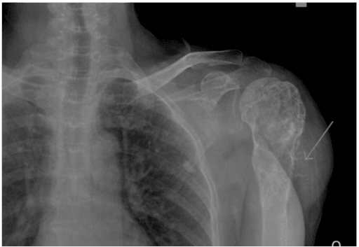
What the pattern is. A focal abnormality of bone density — lytic, sclerotic or mixed — on a plain radiograph. The task in the viva is not to name it in the first sentence but to build a description that constrains the differential to two or three possibilities, and then to state whether the lesion is aggressive.
Confirming the finding is genuine. Before describing, exclude the mimics of a lesion:
- Overlying soft tissue or bowel gas projecting as a lucency — reproduce it on the orthogonal view. A true lesion is present on both views.
- Normal anatomical lucencies — the pseudocyst of the humeral head, the femoral neck Ward's triangle, the greater trochanteric trabecular void, the calcaneal neutral triangle. All are trabecular, not destructive, and have no cortical involvement.
- Nutrient foramen and vascular channels — thin, oblique, cortically based lines with sclerotic walls.
- Healing fracture and stress fracture — a linear sclerotic band with solid periosteal reaction, often at a classic site (proximal femur, tibial diaphysis, second metatarsal). A stress fracture with florid periosteum is the classic false-positive for osteosarcoma.
- Osteomyelitis and Brodie abscess — the great impersonator of both benign and malignant lesions; a penumbra sign (T1 hyperintense rim on MRI) supports abscess.
- Bone infarct — serpiginous sclerotic border enclosing normal-density marrow, distinguishing it from a chondroid lesion which contains rings-and-arcs internally.
The exact words to use out loud. Recite in this fixed order:
- "This is a radiograph of the [bone], [AP/lateral], in a patient stated to be [age]."
- "There is a [lytic / sclerotic / mixed] lesion measuring approximately [x] cm."
- "It lies in the [epiphysis / metaphysis / diaphysis], [centrally / eccentrically / cortically / juxtacortically / surface]."
- "The zone of transition is [narrow / wide]; the margin is [sclerotic / well-defined non-sclerotic / moth-eaten / permeative] — Lodwick grade [IA / IB / IC / II / III]."
- "There is [no matrix / chondroid rings-and-arcs / cloud-like osteoid / ground-glass fibrous] mineralisation."
- "The cortex is [intact / thinned and expanded / breached]."
- "There is [no / solid / lamellated / spiculated / Codman triangle] periosteal reaction."
- "There is [no] associated soft tissue mass."
- "Overall these features are [non-aggressive / indeterminate / aggressive]. My differential is..."
Lodwick grading, verbatim. IA geographic with sclerotic rim; IB geographic well-defined without sclerosis; IC geographic with ill-defined margin; II moth-eaten; III permeative. IA and IB are essentially benign; IC onwards is indeterminate to aggressive.
What the grade is actually worth — and where it stops helping. Lodwick's grades express rate of growth, not histology, and he proposed them to decide which lesions need a biopsy and which can safely be followed. Caracciolo and colleagues applied a three-grade modification to 183 lesions and correlated it with the final pathology, which puts numbers on that judgement:
- Lodwick equivalent
- IA + IB
- Result
- 76 of 81 (94%) benign
- Lodwick equivalent
- IC
- Result
- 29 of 54 benign, 28 of 54 malignant - a coin flip
- Lodwick equivalent
- changing margination, moth-eaten/permeative, radiographically occult
- Result
- 39 of 48 (81%) malignant
So the system is powerful at the two ends and decides nothing in the middle. That is the honest thing to say in a viva: an ill-defined geographic margin is not a "probably benign" finding to be watched, it is a genuinely indeterminate one, and it is the grade at which the description stops answering the question and the next investigation has to.
Next Investigation
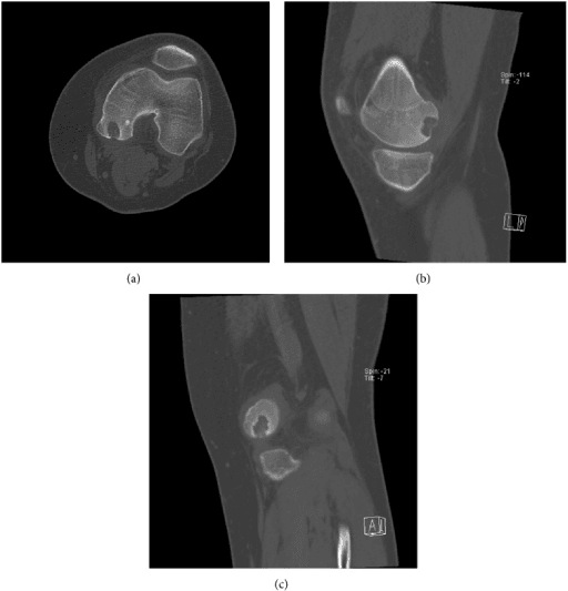
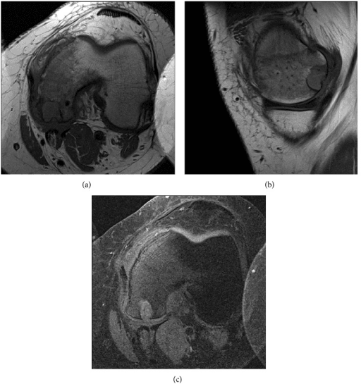
FEGNOMASHIC
Hook:The established Helms mnemonic for lytic bone lesions. Use it as a safety net at the END of your differential, not as the differential itself - the examiner wants age, site and zone of transition applied first, then this list used to check nothing has been omitted.
The Differential
- Typical age / setting
- 10-20 years; second peak over 60 with Paget or prior radiation
- Discriminating feature
- Metaphyseal, around the knee, cloud-like osteoid matrix WITHIN the soft tissue mass plus Codman triangle or sunburst
- What confirms it
- MRI whole bone, staging CT chest, sarcoma-unit core biopsy; raised ALP
- Typical age / setting
- 5-25 years; may present with fever, raised inflammatory markers
- Discriminating feature
- Diaphyseal, permeative destruction with a soft tissue mass disproportionately large for the bone change; onion-skin periosteum
- What confirms it
- MRI showing large extraosseous mass; biopsy with EWSR1 rearrangement (t(11;22))
- Typical age / setting
- Over 40; pelvis, proximal femur, proximal humerus, scapula
- Discriminating feature
- Chondroid rings-and-arcs matrix WITH deep endosteal scalloping greater than two-thirds of cortical thickness, size greater than 5 cm and pain at rest
- What confirms it
- MRI for scalloping depth and soft tissue mass; biopsy — but grade is often underestimated on core
- Typical age / setting
- Over 40; breast, lung, prostate, renal, thyroid primaries
- Discriminating feature
- Multiple lesions, axial and proximal-limb distribution, rarely distal to elbow or knee; renal and thyroid deposits are expansile and blow-out lytic
- What confirms it
- CT chest/abdomen/pelvis, bone scan or whole-body MRI, myeloma screen; biopsy the most accessible lesion
- Typical age / setting
- Over 50
- Discriminating feature
- Endosteal scalloping with punched-out lucencies and NO reactive sclerosis; often bone scan negative — the classic photopenic mismatch
- What confirms it
- Serum and urine electrophoresis, free light chains, whole-body low-dose CT or MRI, marrow aspirate
- Typical age / setting
- Any age; children metaphyseal, adults diabetic foot or spine
- Discriminating feature
- Aggressive appearance but with a serpiginous tract, sequestrum or involucrum; systemic features and raised CRP
- What confirms it
- MRI with contrast (penumbra sign, rim enhancement), aspiration and culture before antibiotics
- Typical age / setting
- Over 40; femur, pelvis, spine
- Discriminating feature
- Extensive permeative marrow infiltration with a soft tissue mass but strikingly preserved cortical outline on radiograph
- What confirms it
- MRI showing marrow replacement out of keeping with plain film; core biopsy and immunohistochemistry
- Typical age / setting
- 20-45, skeletally mature
- Discriminating feature
- Eccentric, subarticular, extends to within 1 cm of the articular surface; lytic with NO matrix and NO sclerotic rim
- What confirms it
- Radiograph is often sufficient; MRI for soft tissue extension, biopsy shows H3F3A mutation
- Typical age / setting
- 5-15 years; proximal humerus and proximal femur
- Discriminating feature
- Central, metaphyseal, mildly expansile, cortex thinned but not breached; fallen-fragment sign after fracture
- What confirms it
- Radiograph diagnostic; no further imaging needed if classic
- Typical age / setting
- Under 20; metaphysis of long bones, posterior elements of spine
- Discriminating feature
- Markedly expansile 'blown-out' lytic lesion with fine septations; fluid-fluid levels on MRI
- What confirms it
- MRI fluid-fluid levels; exclude secondary ABC within GCT or telangiectatic osteosarcoma
- Typical age / setting
- 5-20 years; distal femur, proximal tibia
- Discriminating feature
- Eccentric, CORTICALLY based, lobulated with a thick sclerotic scalloped margin; asymptomatic incidental
- What confirms it
- Radiograph alone — a 'don't touch' lesion; sclerosis and involution with age
- Typical age / setting
- 20-50; hands and feet most commonly
- Discriminating feature
- Central chondroid matrix with punctate/ring calcification, endosteal scalloping less than two-thirds of cortex, painless
- What confirms it
- Radiograph; MRI if pain, size greater than 5 cm or deep scalloping to exclude chondrosarcoma
- Typical age / setting
- 10-25; femoral neck, tibial diaphysis, spinal posterior elements
- Discriminating feature
- Lucent nidus less than 1.5 cm with dense surrounding reactive sclerosis; night pain relieved by NSAIDs
- What confirms it
- Thin-slice CT to demonstrate the nidus — CT is superior to MRI here
- Typical age / setting
- Any age, often 10-30; femoral neck, ribs, craniofacial
- Discriminating feature
- Ground-glass matrix, long lesion in a long bone, no periosteal reaction; shepherd's crook deformity
- What confirms it
- Radiograph typically diagnostic; GNAS mutation if biopsied
- Typical age / setting
- Under 15
- Discriminating feature
- Skull with bevelled edge / hole-within-a-hole; vertebra plana; can look permeative with aggressive periosteum yet heals spontaneously
- What confirms it
- Skeletal survey, biopsy CD1a/langerin positive
- Typical age / setting
- Adults with renal failure or parathyroid adenoma
- Discriminating feature
- Lytic expansile lesions PLUS subperiosteal resorption of radial aspect of middle phalanges and distal clavicle erosion
- What confirms it
- Serum calcium, phosphate, PTH — biochemistry, not biopsy
Narrowing It Down
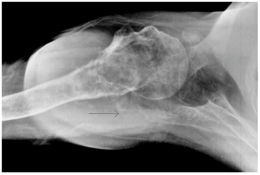
- 1Step 1 — How old is the patient?
Under 5: metastatic neuroblastoma, leukaemia, LCH, osteomyelitis. 5-25: osteosarcoma, Ewing, simple cyst, ABC, NOF, chondroblastoma, osteoid osteoma. 25-40: giant cell tumour, enchondroma, fibrous dysplasia. Over 40: metastasis, myeloma, chondrosarcoma, lymphoma. Age alone removes roughly half the list and should be the first sentence of your differential.
Age alone removes roughly half the list - open with it
- 2Step 2 — Is it solitary or are there multiple lesions?
Multiple over 40 means metastasis, myeloma or brown tumours — and the next step becomes biochemistry and staging CT, not biopsy. Multiple in a child means LCH, leukaemia, neuroblastoma metastases or osteomyelitis. Multiple with a growth-plate distribution suggests enchondromatosis or hereditary multiple exostoses.
Multiple changes the pathway from biopsy to biochemistry and staging
- 3Step 3 — Where in the bone, longitudinally and transversely?
Epiphysis in a mature skeleton: giant cell tumour, then clear cell chondrosarcoma and infection. Epiphysis in a child: chondroblastoma. Metaphysis: the vast majority — osteosarcoma, simple cyst, ABC, NOF. Diaphysis: Ewing, lymphoma, LCH, osteoid osteoma, adamantinoma (anterior tibial cortex). Transversely, eccentric favours NOF, GCT and ABC; central favours simple cyst, enchondroma and fibrous dysplasia; surface lesions are osteochondroma and parosteal osteosarcoma.
Longitudinal site names the shortlist; transverse position ranks it
- 4Step 4 — What is the zone of transition and margin?
Narrow zone with sclerotic rim (Lodwick IA) = a slow, benign process — the lesion has been there long enough for bone to wall it off. Ill-defined, moth-eaten or permeative (Lodwick IC-III) = fast growth: sarcoma, infection, lymphoma, myeloma, metastasis. This single feature is the strongest predictor of aggressiveness and should be stated explicitly.
Grade I is 94% benign, grade III 81% malignant, grade II a coin flip
- 5Step 5 — Is there matrix, and what type?
Rings-and-arcs or popcorn calcification = chondroid: enchondroma, chondroblastoma, chondrosarcoma. Fluffy, cloud-like amorphous density = osteoid: osteosarcoma, osteoblastoma, bone island. Ground glass = fibrous dysplasia. No matrix at all keeps giant cell tumour, simple cyst, ABC, myeloma and metastasis in play.
Matrix names the tissue of origin; absent matrix excludes nothing
- 6Step 6 — What is the cortex and periosteum doing, and is there a soft tissue mass?
Intact expanded cortex with solid periosteal reaction = slow. Cortical breach, spiculated or interrupted periosteum (Codman triangle, sunburst, onion-skin) plus an extraosseous soft tissue mass = aggressive. A soft tissue mass is the discriminator that should trigger urgent sarcoma referral regardless of the rest of the description.
A soft-tissue mass triggers sarcoma referral whatever else you found
- 7Step 7 — What are the systemic features and bloods?
Fever with raised CRP and WCC pushes towards osteomyelitis or Ewing. Weight loss, smoking history or a known primary pushes towards metastasis. Anaemia, renal impairment and raised ESR push towards myeloma. Raised calcium with subperiosteal resorption means hyperparathyroidism — and the lesion is a brown tumour requiring no biopsy at all.
A brown tumour needs a corrected calcium, not a biopsy
MCQ Practice Points
Q: Which single radiographic feature best predicts the aggressiveness of a bone lesion?
A: The zone of transition. A narrow zone with a sclerotic rim indicates the host bone has had time to wall the lesion off — a slow, usually benign process. A wide, imperceptible zone with moth-eaten or permeative destruction indicates rapid growth, and is seen in sarcoma, lymphoma, myeloma, metastasis and infection.
Q: A 12-year-old has a diaphyseal permeative femoral lesion with onion-skin periosteal reaction, a large soft tissue mass, fever and a raised CRP. What are the two diagnoses and how are they separated?
A: Ewing sarcoma and osteomyelitis. Both cause fever and raised inflammatory markers. Ewing typically produces a soft tissue mass disproportionately large relative to the bone destruction, whereas osteomyelitis produces a sequestrum, involucrum and cloaca with rim-enhancing collections on contrast MRI. Both require tissue — aspiration for culture and core biopsy through a planned tract.
Q: What features distinguish an enchondroma from a low-grade chondrosarcoma in a long bone?
A: Chondrosarcoma is favoured by pain at rest not attributable to another cause, size greater than 5 cm, endosteal scalloping deeper than two-thirds of cortical thickness, cortical destruction or periosteal reaction, an axial or proximal-limb location such as pelvis or proximal femur, and interval growth. Enchondromas of the hands and feet may look active without being malignant.
Q: Which lesions are safe to diagnose on plain radiograph alone and require no biopsy?
A: The "don't touch" lesions: non-ossifying fibroma and fibrous cortical defect, bone island (enostosis), cortical desmoid of the distal medial femoral metaphysis, myositis ossificans in its mature phase, avulsion injuries at the ischium or lesser trochanter, and a classic simple bone cyst of the proximal humerus in a child. Biopsy of these causes harm without gain.
Q: Why must the biopsy of a suspected primary bone sarcoma be performed at the treating centre?
A: The tract is contaminated with tumour and must be excised en bloc with the specimen. A tract placed transversely, through a second compartment, or through a neurovascular plane cannot be excised without sacrificing the limb or the reconstruction. Poorly planned biopsy is documented to alter treatment and worsen outcome, which is why staging MRI and CT precede tissue sampling.
Exam Viva Scenarios
Practise clinical reasoning and management decisions out loud
“You are shown this radiograph of the knee in a 14-year-old boy with 3 months of increasing pain, now waking him at night. There is a mixed lytic and sclerotic lesion of the distal femoral metaphysis with cortical breach and spiculated periosteal reaction.”
“You are shown this radiograph of the knee in a 28-year-old woman with 6 weeks of pain and a sensation of giving way. There is a lucent lesion in the proximal tibia reaching the subchondral bone.”
“You are shown this pelvic radiograph in a 68-year-old man with back pain and fatigue. There are several small, well-defined lucencies in the ilium and proximal femora, and the vertebral bodies appear osteopenic.”
The Description Sequence
- Age and bone
- Lytic / sclerotic / mixed, and size
- Longitudinal site: epiphysis, metaphysis, diaphysis
- Transverse site: central, eccentric, cortical, juxtacortical, surface
- Zone of transition and margin (Lodwick IA to III)
- Matrix: chondroid, osteoid, fibrous, none
- Cortex, periosteal reaction, soft tissue mass
- Conclude: non-aggressive, indeterminate or aggressive
Age Buckets
- Under 5: neuroblastoma metastases, leukaemia, LCH, infection
- 5-25: osteosarcoma, Ewing, simple cyst, ABC, NOF, chondroblastoma, osteoid osteoma
- 25-40: giant cell tumour, enchondroma, fibrous dysplasia
- Over 40: metastasis, myeloma, chondrosarcoma, lymphoma
Site Shortcuts
- Epiphysis mature = giant cell tumour; immature = chondroblastoma
- Diaphysis = Ewing, lymphoma, LCH, adamantinoma (anterior tibia)
- Posterior spinal elements = osteoblastoma, ABC, osteoid osteoma
- Vertebra plana in a child = LCH
- Bevelled-edge skull lucency = LCH
Matrix Language
- Rings and arcs / popcorn = chondroid
- Cloud-like, fluffy, amorphous = osteoid
- Ground glass = fibrous dysplasia
- Serpiginous sclerotic rim = bone infarct, not a tumour
Red Flags Demanding Sarcoma Referral
- Wide zone of transition or permeative destruction
- Cortical breach with interrupted periosteal reaction
- Any associated soft tissue mass
- Night pain, rest pain, or a lesion increasing in size
- Any lesion you cannot confidently name as a 'don't touch' lesion
Investigation Order
- Two-view radiograph of the whole bone plus old films
- Bloods including calcium, PTH, ALP, electrophoresis and light chains
- MRI whole involved bone
- CT chest for staging; CT for matrix or nidus detail
- Biopsy last, by or with the sarcoma unit
Evidence Base
Determining Growth Rates of Focal Lesions of Bone from Radiographs
- Sets out the five grades in the presence of bone destruction, with the rules for establishing each and the radiologic signs needed to apply them.
- States the purpose plainly: rate of growth divides focal bone lesions into two largely mutually exclusive classes.
- The stated clinical aim is triage - 'not all focal lesions require biopsy, and grading is especially helpful in deciding which should be biopsied and which may be safely followed'.
A Modified Lodwick-Madewell Grading System for the Evaluation of Lytic Bone Lesions
- 183 bone lesions selected to span a broad range of benign and malignant tumours, graded on radiographs by consensus and correlated with the final pathological diagnosis.
- Collapses Lodwick into three grades and adds two Madewell patterns: grade I = Lodwick IA and IB; grade II = Lodwick IC; grade III = changing margination, moth-eaten and permeative patterns, and radiographically occult lesions.
- Grade I: 76 of 81 (94 per cent) were BENIGN.
- Grade III: 39 of 48 (81 per cent) were MALIGNANT.
- Grade II: essentially even - 29 of 54 benign and 28 of 54 malignant. The middle grade does not discriminate.
Bone Tumors and Tumorlike Conditions - Analysis with Conventional Radiography
- Sets out the organised search pattern this page is built on: location, margins and zone of transition, periosteal reaction, mineralisation, size and number of lesions, and the presence of a soft-tissue component.
- Names patient age as an important clinical factor because lesions have predilections for specific age groups.
- The organising claim is that the analysis must be done in a fixed order rather than by pattern recognition.
Radiography in the Initial Diagnosis of Primary Bone Tumors
- Argues the position this page depends on: the radiograph remains the single most useful investigation in the initial diagnosis of a primary bone tumour, and cross-sectional imaging supplements rather than replaces it.
- From the same group whose patterns (changing margination, radiographically occult) Caracciolo later folded into the modified grading system.
Classifications for Radiographic Evaluation of Radiolucent Bone Lesions Have Poor Inter- and Intra-observer Agreement
- 48 case sets of radiolucent lesions, each with two orthogonal views, graded TWICE at least two weeks apart by 20 observers, using the original Lodwick, modified Lodwick and Enneking systems
- Inter-observer reliability was POOR for all three: agreement 39 per cent for Lodwick (kappa 0.23), 39 per cent for modified Lodwick (kappa 0.25), 53 per cent for Enneking (kappa 0.28)
- Intra-observer reproducibility was no better than moderate (kappa 0.42 to 0.45), with individual self-agreement between 39 and 78 per cent
- TRAINING LEVEL HAD NO EFFECT on reproducibility
- Benign versus malignant - the classification that actually matters - was correct in only 73.3 per cent of cases using Lodwick, 59.0 per cent with modified Lodwick and 62 per cent with Enneking
- Reproducibility was best at the EXTREMES, for the highest and lowest grades; it was worst in the middle, which is where the decision is hard