A lucent lesion crossed by internal ridges or septa producing a soap-bubble or trabeculated appearance
- Most 'loculations' are not true septa — they are ridges on the endosteal surface of an expanded cortex. Say this out loud.
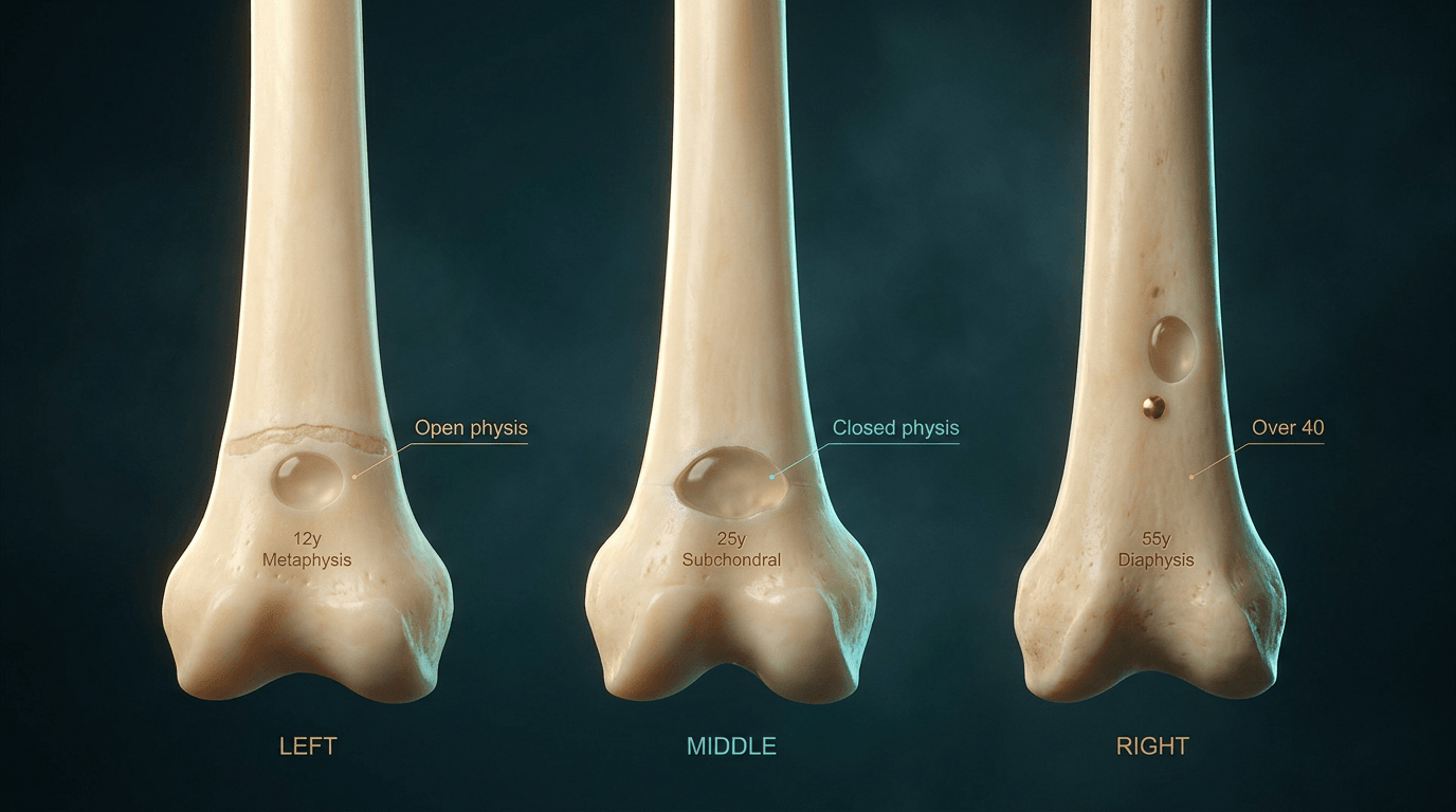
- Age plus zone of the bone (epiphysis / metaphysis / diaphysis) narrows the list to two or three before you mention any lesion by name.
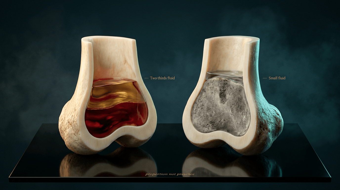
- Fluid-fluid levels are NOT specific for aneurysmal bone cyst — telangiectatic osteosarcoma, secondary ABC in GCT, chondroblastoma and a fractured simple bone cyst all show them. What discriminates is the PROPORTION: at two thirds or more of the lesion, 89 per cent are benign; a SMALL fluid component in a mostly solid lesion is the malignant pattern, most often conventional osteosarcoma.
- A secondary ABC sits on a parent lesion in a third of cases: always hunt for GCT, chondroblastoma, fibrous dysplasia or osteoblastoma underneath.
- Giant cell tumour requires a closed physis and a subarticular epiphysial epicentre reaching within 1 cm of the subchondral plate.
- In an adult over 40 with a bubbly lucent lesion, brown tumour and metastasis (renal, thyroid) must be actively excluded before benign labels are applied.
- “Non-ossifying fibroma is eccentric, cortically based, has a sclerotic scalloped rim and needs no follow-up if typical and asymptomatic.
- “Chondromyxoid fibroma: eccentric metaphysial lesion of the proximal tibia in the second and third decades, sharply sclerotic margin, no calcified matrix on radiograph despite being cartilage.
- “Fibrous dysplasia is ground-glass, not truly bubbly; the pseudoloculation comes from endosteal scalloping without periosteal reaction.
- “Multiple bubbly lesions in the hands with subperiosteal resorption equals hyperparathyroidism until the calcium is back.
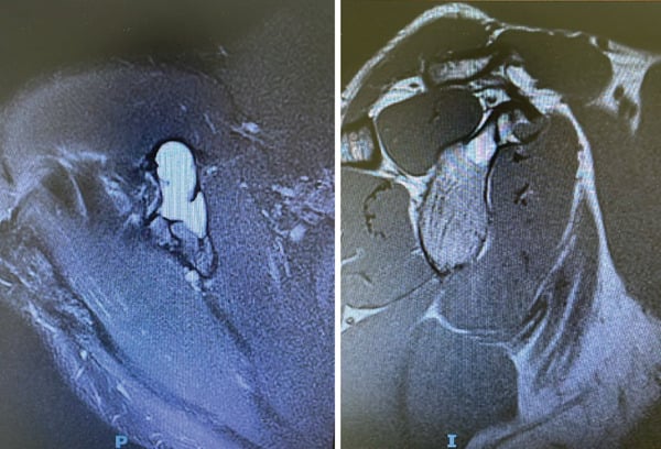
Fluid-fluid levels reflect blood of differing age in cystic spaces, not a diagnosis. Telangiectatic osteosarcoma shows identical levels. The discriminator is thick, nodular, enhancing solid tissue around the septa and cortical destruction with a wide zone of transition.
A secondary aneurysmal bone cyst is a reactive phenomenon. If the surrounding bone contains chondroid calcification, ground-glass matrix or an epiphysial subarticular component, the ABC is the passenger, not the driver.
Giant cell tumour in an open physis is very rare; chondroblastoma occupies the epiphysis before fusion. Reversing these two in a viva is the classic fail.
Brown tumours of hyperparathyroidism and lytic renal or thyroid metastasis both produce expansile trabeculated lucencies. Check serum calcium, PTH and consider whole-body imaging before biopsy in the over-40s.
Recognising the Pattern
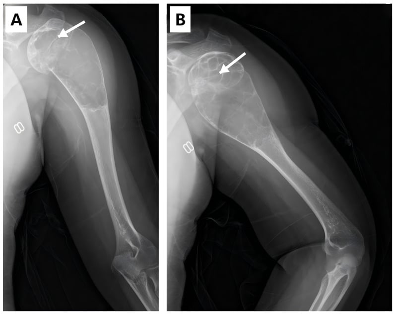
Definition. A multiloculated or "soap-bubble" lesion is a lucent, expansile bone lesion whose interior is crossed by linear densities dividing it into compartments. Radiographically it looks septated; pathologically the majority of these lines are not free-standing septa but ridges of residual endosteal cortex on an expanded, remodelled shell. A minority — aneurysmal bone cyst above all — do contain genuine fibro-osseous septa between blood-filled cavities.
Confirming the pattern is genuine.
- Obtain two orthogonal views. Apparent loculation on one projection alone is usually overlapping cortical ridge.
- Ask whether the "septa" arise from the cortex and taper inwards (endosteal ridging) or cross the lesion completely (true septation).
- Confirm expansion: the outer cortical margin bulges beyond the normal contour, with a thin but continuous shell. Loss of that shell moves you towards aggression.
- Assess the zone of transition. A narrow transition with a sclerotic rim supports a slow-growing benign process even when floridly bubbly.
The words to use out loud. "This is a frontal radiograph of the skeletally mature right knee. There is a well-defined, eccentric, expansile lucent lesion centred on the subarticular proximal tibial epiphysis, extending to within a few millimetres of the subchondral plate. It has a narrow zone of transition, a non-sclerotic margin, and internal ridging giving a multiloculated soap-bubble appearance. The cortex is thinned and expanded but appears intact, and there is no periosteal reaction, no matrix mineralisation and no soft-tissue mass. My leading diagnosis is a giant cell tumour."
Mimics — the false positives.
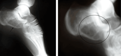
- Overlapping trabeculae in normal cancellous bone, especially the calcaneal neutral triangle and the greater trochanter, mimicking a bubbly lucency.
- Intraosseous ganglion or subchondral cyst near a joint with an internal septum.
- Healing fracture or post-traumatic cortical remodelling producing a pseudo-septated lucency.
- Fibrous dysplasia in which ground-glass matrix over a thinned endosteally scalloped cortex is misread as loculation.
- Chronic osteomyelitis with a Brodie abscess and a serpiginous channel simulating septa.
- Poor radiographic technique — an underpenetrated or obliqued film exaggerates internal lines.
Next Investigation
The Differential
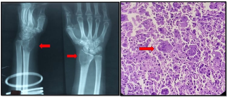
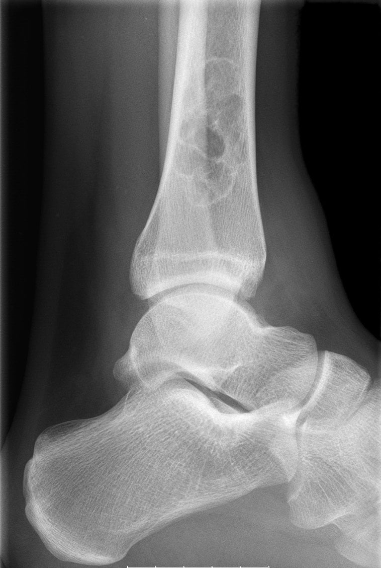
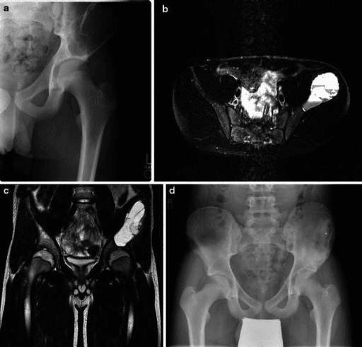
- Typical age / setting
- 10-25 years, distal femur/proximal tibia metaphysis
- Discriminating feature
- Fluid-fluid levels PLUS thick nodular enhancing solid tissue, cortical destruction and wide transition zone; often a soft-tissue mass
- What confirms it
- MRI showing solid nodules; raised ALP; biopsy with malignant osteoid at septal margins
- Typical age / setting
- Over 40, proximal femur, humerus, pelvis, spine
- Discriminating feature
- Expansile blow-out lucency with destroyed rather than thinned cortex; often multiple; pulsatile if renal
- What confirms it
- CT chest/abdomen/pelvis, thyroid ultrasound, biopsy of an accessible lesion
- Typical age / setting
- Adults; dialysis or parathyroid adenoma
- Discriminating feature
- Multiple bubbly lucencies with subperiosteal resorption of radial phalangeal margins, distal clavicle erosion, salt-and-pepper skull
- What confirms it
- Serum calcium, phosphate, PTH; sestamibi scan — no biopsy needed if biochemistry diagnostic
- Typical age / setting
- Over 50, pelvis, spine, proximal long bones
- Discriminating feature
- 'Mini brain' trabeculated vertebral lesion; endosteal scalloping without sclerosis and no reactive rim
- What confirms it
- Serum and urine electrophoresis, free light chains, skeletal survey or whole-body MRI, biopsy
- Typical age / setting
- Under 20, eccentric metaphysis of long bone or posterior spinal elements
- Discriminating feature
- Rapidly expansile, ballooned with a thin eggshell shell; fluid-fluid levels with NO solid enhancing nodule; septal and rim enhancement only
- What confirms it
- MRI fluid-fluid levels plus curettage histology; USP6 rearrangement in primary ABC
- Typical age / setting
- 20-45, physis CLOSED, subarticular epiphysis of distal femur, proximal tibia, distal radius
- Discriminating feature
- Epicentre in the epiphysis abutting subchondral bone; narrow transition but NON-sclerotic margin; low signal on T2 from haemosiderin
- What confirms it
- MRI plus biopsy; H3F3A G34W mutation; check chest CT for benign pulmonary implants
- Typical age / setting
- 5-20 years, distal femoral or proximal tibial metaphysis
- Discriminating feature
- Eccentric, cortex-based, elongated along the shaft with a scalloped SCLEROTIC rim; migrates away from physis with growth
- What confirms it
- Radiograph alone is diagnostic when typical — no biopsy, no follow-up if asymptomatic
- Typical age / setting
- Children to young adults; femoral neck, ribs, craniofacial, tibia
- Discriminating feature
- Ground-glass matrix, long lesion in a long bone, endosteal scalloping, shepherd's crook deformity; no periosteal reaction
- What confirms it
- Radiograph often sufficient; intense uptake on bone scan; GNAS mutation if biopsied
- Typical age / setting
- 5-15, proximal humerus and proximal femur
- Discriminating feature
- Central, symmetric, does NOT expand beyond the physeal width; fallen-fragment sign after fracture
- What confirms it
- Radiograph with fallen fragment; MRI shows uniform fluid unless fractured
- Typical age / setting
- 10-30, proximal tibial metaphysis, eccentric
- Discriminating feature
- Sharply lobulated with a THICK sclerotic scalloped margin; despite cartilage origin, calcification rarely visible on radiograph
- What confirms it
- MRI very high T2 signal lobules; biopsy showing lobular chondromyxoid tissue
- Typical age / setting
- Any age; hands and feet, proximal humerus, femur
- Discriminating feature
- Rings-and-arcs or punctate chondroid calcification within the lucency; endosteal scalloping less than two-thirds of cortical thickness
- What confirms it
- Radiograph diagnostic in typical hand lesions; MRI/CT if pain or deep scalloping suggests chondrosarcoma
- Typical age / setting
- 10-20, physis OPEN, epiphysis or apophysis of femur, humerus, tibia
- Discriminating feature
- Epiphysial lucency with fine punctate calcification, thin sclerotic rim and marked surrounding marrow oedema on MRI
- What confirms it
- MRI oedema pattern; biopsy showing chondroid matrix with chicken-wire calcification
- Typical age / setting
- 10-30, posterior spinal elements, sacrum, long bones
- Discriminating feature
- Greater than 2 cm expansile lucency with mineralised matrix and painful scoliosis; may harbour secondary ABC
- What confirms it
- CT for nidus size and matrix; biopsy
- Typical age / setting
- Second and third decades; mandible, metaphysis of long bones, pelvis
- Discriminating feature
- Trabeculated expansile lucency with prominent coarse pseudoseptation and no matrix; low T2 signal fibrous tissue
- What confirms it
- MRI low signal on all sequences; biopsy — locally aggressive, wide excision
- Typical age / setting
- Osteofibrous dysplasia under 10; adamantinoma 20-40; ANTERIOR TIBIAL DIAPHYSIS
- Discriminating feature
- Multiple lucencies in a bubbly soap-bubble line along the anterior tibial cortex with anterior bowing
- What confirms it
- MRI and biopsy — adamantinoma requires wide resection, osteofibrous dysplasia observation
Narrowing It Down
- 1Step 1 — How old is the patient and is the physis open?
Establish age and physeal status from the radiograph before looking at the lesion itself.
Under 20 with an open physis: simple bone cyst, ABC, non-ossifying fibroma, chondroblastoma (epiphysis), osteofibrous dysplasia. Aged 20-45 with a closed physis: giant cell tumour tops the list for subarticular lesions. Over 40: metastasis, myeloma and brown tumour must be excluded before any benign label. This single question removes half the differential.
- 2Step 2 — Solitary or multiple?
Image the whole bone, then screen the rest of the skeleton before committing to a single-lesion diagnosis.
Multiple bubbly lucencies point away from a primary bone tumour: metastasis, myeloma, brown tumours of hyperparathyroidism, polyostotic fibrous dysplasia, multiple enchondromatosis (Ollier, Maffucci), Langerhans cell histiocytosis. Order a skeletal survey or whole-body MRI before biopsy.
- 3Step 3 — Where in the bone, longitudinally and axially?
Locate the epicentre in two planes: epiphysis, metaphysis or diaphysis, and central, eccentric, cortical or juxtacortical.
Epiphysis subarticular with closed physis equals GCT; epiphysis with open physis equals chondroblastoma. Metaphysis eccentric equals NOF, ABC or chondromyxoid fibroma; metaphysis central equals simple bone cyst. Diaphysis anterior tibial cortex equals osteofibrous dysplasia or adamantinoma. Posterior spinal elements equal ABC or osteoblastoma.
- 4Step 4 — Is there matrix, and what kind?
Look inside the lucency on a bright light or CT before deciding it is empty.
Rings-and-arcs or punctate calcification equals chondroid: enchondroma, chondroblastoma, chondrosarcoma. Ground-glass haze equals fibrous dysplasia. Cloud-like or ill-defined density equals osteoid - think osteoblastoma or osteosarcoma. Absence of matrix keeps ABC, GCT, NOF and desmoplastic fibroma alive.
- 5Step 5 — How aggressive is the margin?
Grade the zone of transition, then look SEPARATELY for cortical breach, periosteal reaction and soft-tissue mass - any one of those overrides a reassuring margin.
Sclerotic scalloped rim with narrow transition equals slow benign growth (NOF, chondromyxoid fibroma, simple bone cyst). Narrow transition WITHOUT sclerosis is characteristic of GCT. Wide transition, cortical breach, periosteal reaction or a soft-tissue mass demands staging and referral before biopsy - telangiectatic osteosarcoma, metastasis, infection.
- 6Step 6 — What PROPORTION of the lesion is fluid-fluid, and is there solid tissue?
Get the MRI, and estimate how much of the lesion the fluid-fluid levels occupy - not merely whether they are present.
The extent is inversely related to malignancy (O'Donnell 2004): where two thirds or more of the lesion is fluid-fluid change, 89 per cent are benign, and where the whole lesion is, all 11 in that series were benign. The dangerous pattern is a SMALL fluid-fluid component in a largely solid lesion - two thirds of the malignancies fell in that band, and half of those were CONVENTIONAL osteosarcoma. Thin rim and septal enhancement only supports ABC; thick nodular enhancing solid tissue mandates staging and sarcoma referral. Look also for a parent lesion generating a secondary ABC.
- 7Step 7 — Are there systemic or biochemical features?
Send calcium, phosphate, PTH, ALP, full blood count and serum electrophoresis in any adult BEFORE biopsy.
Renal failure, dialysis or hypercalcaemia points to brown tumour - and if the biochemistry is diagnostic, no biopsy is needed. Anaemia, renal impairment and raised ESR point to myeloma. Fever, raised CRP and a sequestrum point to subacute osteomyelitis.
MCQ Practice Points
Q: Which single radiographic feature most reliably separates giant cell tumour from chondroblastoma?
A: Physeal status. Chondroblastoma arises in the epiphysis or apophysis of a skeletally immature patient with an open physis, showing fine punctate chondroid calcification and florid surrounding marrow oedema; giant cell tumour occurs after physeal closure, extends to the subchondral plate and shows no matrix. Where the physis is closing and the sign fails, the molecular answer is decisive: chondroblastoma carries H3F3B K36M (73 of 77 cases) and giant cell tumour H3F3A G34W (49 of 53), and the two are mutually exclusive.
Q: A fluid-fluid level on MRI is diagnostic of aneurysmal bone cyst — true or false, and what is the real discriminator?
A: False — and the discriminator is HOW MUCH of the lesion is fluid, not whether fluid is present. Fluid-fluid levels occur in telangiectatic osteosarcoma, secondary ABC within a giant cell tumour, chondroblastoma, fibrous dysplasia and a fractured simple bone cyst; in 738 consecutive bone lesions they appeared in 11.2 per cent. O'Donnell showed the extent is inversely related to malignancy: at two thirds or more fluid-fluid change, 89 per cent were benign, and where the entire lesion was fluid-fluid all 11 were benign. The malignant pattern is a small fluid component in a largely solid lesion — and the commonest tumour in that band was conventional intramedullary osteosarcoma, not the telangiectatic variant. Thick nodular enhancing solid tissue is the corroborating sign, not the primary one.
Q: Which bubbly lesion characteristically involves the anterior cortex of the tibial diaphysis with anterior bowing?
A: Osteofibrous dysplasia in the first decade, and adamantinoma in the third and fourth. They are radiographically similar, and the distinction is the whole point: adamantinoma is a low-grade malignancy requiring wide resection, whereas osteofibrous dysplasia is observed to skeletal maturity.
Q: Which lucent metaphysial lesion requires no further imaging and no follow-up?
A: A typical asymptomatic non-ossifying fibroma — eccentric, cortically based, elongated along the shaft with a scalloped sclerotic rim in a child or adolescent. It resolves with skeletal maturity; intervention is considered only for very large lesions with fracture risk. Counsel on the real timescale: in Yanagawa's series of spontaneously regressing lesions, resolution took a mean of about 25 months and up to 79 — a lesion unchanged at the one-year review has not failed to behave, it has not finished.
Q: In an adult over 40 with a solitary expansile bubbly lesion of the proximal femur, what comes before biopsy?
A: Bloods and staging. Serum calcium, phosphate, PTH, ALP, full blood count and protein electrophoresis; plus CT chest, abdomen and pelvis to identify an occult renal or thyroid primary. Two of the three diagnoses you are excluding do not need a biopsy at all — a brown tumour is diagnosed biochemically, and myeloma on electrophoresis and marrow. Only then is biopsy performed, targeted and planned along an excisable track.
Exam Viva Scenarios
Practise clinical reasoning and management decisions out loud
“You are shown this radiograph of the knee of a 28-year-old woman with three months of medial knee pain. There is an eccentric expansile lucent lesion of the proximal tibia extending to the subchondral plate, with internal ridging and a non-sclerotic margin.”
“You are shown this radiograph of a 14-year-old boy with six weeks of increasing thigh pain. There is a markedly expansile lucent lesion of the distal femoral metaphysis with a thin shell and internal septation. MRI shows multiple fluid-fluid levels.”
“You are shown hand radiographs of a 52-year-old man on haemodialysis with several expansile trabeculated lucencies in the phalanges and a lytic lesion in the pelvis.”
Age brackets
- Under 20, open physis: simple bone cyst, ABC, NOF, chondroblastoma, osteofibrous dysplasia
- 20-45, closed physis: giant cell tumour, chondromyxoid fibroma, adamantinoma
- Over 40: metastasis, myeloma, brown tumour — exclude before benign labels
Site within the bone
- Subarticular epiphysis, closed physis: giant cell tumour
- Epiphysis, open physis: chondroblastoma
- Eccentric metaphysis: NOF, ABC, chondromyxoid fibroma
- Central metaphysis: simple bone cyst
- Anterior tibial cortex: osteofibrous dysplasia / adamantinoma
- Posterior spinal elements: ABC, osteoblastoma
Matrix clues
- Rings and arcs or punctate: chondroid — enchondroma, chondroblastoma
- Ground glass: fibrous dysplasia
- Cloud-like osteoid: osteoblastoma, osteosarcoma
- No matrix: ABC, GCT, NOF, desmoplastic fibroma
Red flags for malignancy
- Wide zone of transition and cortical destruction
- Nodular solid enhancement within a cystic lesion
- Aggressive periosteal reaction or soft-tissue mass
- Night pain, weight loss, raised ALP or LDH
Investigation order
- Two orthogonal radiographs — may be sufficient alone
- Contrast-enhanced MRI for fluid levels, solid tissue and extent
- CT for matrix, cortex and sequestrum
- Biochemistry in all adults before biopsy
- Staging CT chest, then planned image-guided biopsy at the tumour unit
Evidence Base
The Prevalence and Diagnostic Significance of Fluid-Fluid Levels in Focal Lesions of Bone
- 738 consecutive patients with a focal bone lesion imaged by MRI at a supraregional bone tumour service; fluid-fluid levels were present in 83 (11.2 per cent)
- THE DISCRIMINATOR IS THE PROPORTION OF THE LESION OCCUPIED BY FLUID, NOT THE PRESENCE OF FLUID
- Where fluid-fluid levels occupied LESS THAN A THIRD of the lesion, 22 of the 32 malignant tumours (68.8 per cent) fell in that band - and HALF of that group were CONVENTIONAL intramedullary osteosarcomas (16), not telangiectatic
- Where fluid-fluid change occupied more than two thirds but not all of the lesion, 13 of 16 (81 per cent) were benign
- Where the ENTIRE lesion showed fluid-fluid change, histology was benign in 11 of 11 (100 per cent)
- The authors' conclusion: the extent of fluid-fluid change is INVERSELY related to malignancy; at two thirds or more, 89 per cent of diagnoses are benign
Five-Year Evolution of a Telangiectatic Osteosarcoma Initially Managed as an Aneurysmal Bone Cyst
- A 20-year-old man with a femoral neck lesion diagnosed and curetted as an aneurysmal bone cyst
- First local recurrence at 4 years 8 months - again called recurrent ABC, again curetted and grafted
- Second recurrence 8 months later with a destroyed lesser trochanter and a large medial soft-tissue mass; only then diagnosed as high-grade telangiectatic osteosarcoma, requiring proximal femoral resection and replacement
- ON RETROSPECTIVE REVIEW the original histology resembled ABC apart from 'a few bizarre cells without mitosis' in the cyst wall - recoverable in principle, easily missed in practice