The Only Two-Joint Head of the Quadriceps
- Two heads: the direct (straight) head from the anterior inferior iliac spine and the reflected head from a groove above the superior acetabular rim.
- It is the ONLY head of the quadriceps that crosses the hip — therefore the only biarticular quadriceps head, and the only one that can produce a two-joint contracture.
- Innervated by the femoral nerve (posterior division), root values L2, L3, L4; the motor branch enters the proximal third of the muscle on its deep surface.
- Blood supply from the descending branch of the lateral circumflex femoral artery — the same pedicle exploited in anterolateral thigh and quadriceps-based flaps.
- Adolescent apophyseal avulsion of the AIIS is a rectus femoris direct-head injury; skeletally mature athletes instead tear the intramuscular central tendon.
- “Ely test: prone passive knee flexion causing ipsilateral pelvic rise or hip flexion is a positive test for rectus femoris spasticity or contracture — the hip cannot be blinded to a two-joint muscle.
- “Reflected-head tears look confusing on MRI because the tendon is intra-capsular in position and the oedema is deep, adjacent to the hip capsule, not in the muscle belly.
- “AIIS morphology (Hetsroni classification) tracks with hip motion IN PATIENTS WHO ALREADY HAVE IMPINGEMENT - mean flexion 120, 107 and 93 degrees and internal rotation 21, 11 and 8 degrees for Types I, II and III. Both of Hetsroni's cohorts were symptomatic, so there is no asymptomatic comparator and the study cannot show that a Type III spine CAUSES impingement; the authors' own wording is that it supports considering decompression.
- “In stiff-knee gait, rectus femoris transfer works only if the hamstrings are competent and the knee has passive flexion — it converts a knee extensor into a flexion-phase-friendly structure but does NOT lengthen the vasti.
Overview
The rectus femoris is the most anterior and most superficial of the four quadriceps heads. It is fusiform, bipennate, and unique among the extensor group because it originates from the pelvis rather than the femur. That single fact drives almost every clinically relevant behaviour of the muscle: it flexes the hip as well as extending the knee, it becomes taut when the hip is extended and the knee flexed simultaneously, and it is the only quadriceps head that can be injured by an avulsion of a pelvic apophysis.
Surgically, the rectus femoris is the roof over the anterior hip. Both the Smith-Petersen and the direct anterior (Hueter) approaches pass alongside it; the reflected head is the last soft-tissue veil over the anterosuperior capsule, and the rectus origin is the structure a surgeon takes down (or works around) to reach the acetabular rim.
A muscle crossing two joints can never be at optimal length for both. This is the concept examiners will push on.
- Length-tension coupling: the rectus femoris shortens when the hip flexes and when the knee extends. In swing phase the hip flexes (rectus shortens proximally) while the knee must flex (rectus lengthens distally). If the muscle is spastic or contracted, the distal demand cannot be met and the knee fails to flex — stiff-knee gait.
- Active insufficiency: the rectus femoris is a weak knee extensor when the hip is already flexed (it is short and cannot generate force) and a weak hip flexor when the knee is extended. Test the quadriceps at 90 degrees of hip flexion and you are largely testing the vasti.
- Passive insufficiency and injury: simultaneous hip extension plus knee flexion — the football kicking wind-up, the sprinting late-swing — puts the muscle at maximum length while it contracts eccentrically. This is the mechanism of both the adolescent AIIS avulsion and the adult central-tendon strain.
- The clinical corollary: any test, contracture, transfer, or lengthening involving the rectus femoris MUST control the position of the hip. A test performed with the hip flexed will falsely reassure.
DIRECT / REFLECTEDRectus Femoris — Two Heads
Hook:Direct is superficial and avulses; Reflected is deep and confuses the MRI.
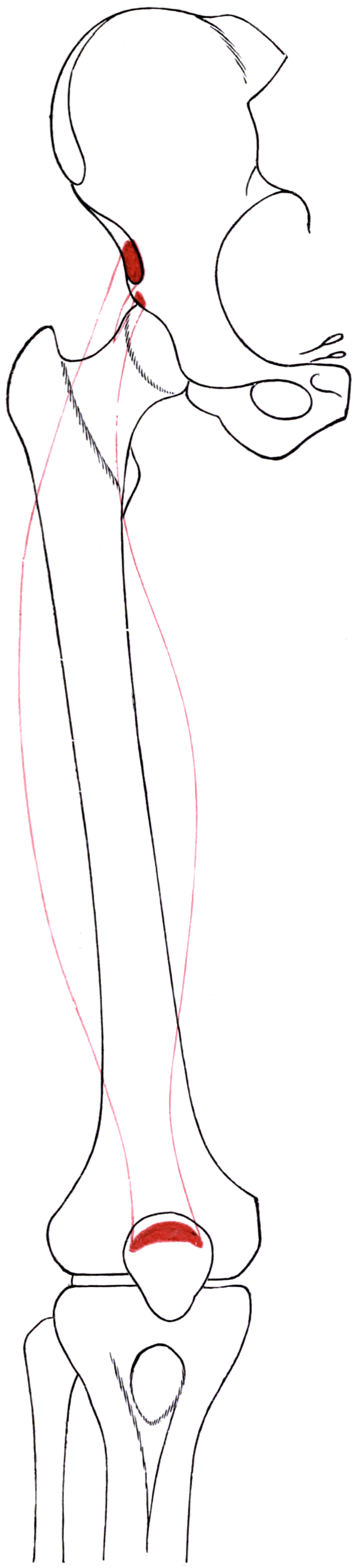
Attachments, Innervation and Relations
Origin — Two Distinct Heads
1. Direct (straight) head
- From the anterior inferior iliac spine (AIIS).
- Short, thick, and anterosuperficial; its fibres run almost vertically down the front of the muscle.
- This is the head that fails through the apophysis in the skeletally immature athlete.
2. Reflected (indirect) head
- From a shallow groove immediately above the superior (posterosuperior) acetabular rim, blending with the hip joint capsule and the iliofemoral ligament.
- Runs posterolateral and deep, then turns forward (hence reflected) to join the direct head at an acute angle roughly 2 cm distal to the AIIS.
- Its tendon continues distally within the muscle belly as the central (intramuscular) tendon — an aponeurotic septum that penetrates deep into the muscle, sometimes almost to the distal third.
Why the two heads matter surgically: the reflected head is a capsular structure by proximity. Elevating it exposes the anterosuperior rim in rim trimming, periacetabular osteotomy exposure, and open treatment of pincer impingement. It is also the reason a reflected-head tear presents with deep groin pain rather than anterior thigh pain.
Insertion
- Fibres converge into a broad flat tendon forming the superficial (most anterior) layer of the quadriceps tendon.
- Inserts on the anterosuperior pole (base) of the patella, its most superficial fibres continuing over the anterior patellar surface into the patellar tendon — a genuine continuity that allows the extensor mechanism to transmit force even when the patella is absent (patellectomy).
- Medial and lateral expansions contribute to the superficial layer of the medial and lateral retinacula.
Named landmarks (measurable)
- Relationship
- Direct head origin; roughly 2-3 cm proximal to the acetabular rim
- Practical use
- Target of subspine decompression
- Relationship
- Direct head origin sits about 2-3 cm distal and slightly medial to the ASIS
- Practical use
- Landmark for the anterior interval skin incision
- Relationship
- Reflected head origin lies immediately above it
- Practical use
- Elevate to expose the rim for trimming or osteotomy
- Relationship
- Broad tendinous insertion, most superficial quadriceps layer
- Practical use
- Layer identification for quadriceps tendon graft harvest
It is a hip structure.
- It arises above the acetabular rim, blending with capsule.
- A reflected-head tear presents as deep groin pain, mimicking labral pathology or iliopsoas irritation.
- On MRI the oedema is deep and proximal, adjacent to the capsule — easily read as a capsular injury.
The adult tear site.
- The indirect head continues as an aponeurotic central tendon running deep inside the belly.
- A tear here produces the classic bull's-eye appearance on axial MRI: central tendon disruption ringed by oedema.
- Consequence: central-tendon injuries take substantially longer to return to sport than peripheral myofascial strains, and re-injury is more common.
Action and Biomechanics
Actions by plane
- Action
- Flexion (weak-to-moderate; contributes roughly 10-15 percent of hip flexion torque)
- Position of maximum efficiency
- Hip extended, knee extended
- Position of insufficiency
- Hip already flexed beyond 90 degrees
- Action
- Extension (biarticular contributor to the quadriceps)
- Position of maximum efficiency
- Hip extended (muscle lengthened proximally)
- Position of insufficiency
- Hip flexed (active insufficiency)
- Action
- Anterior pelvic tilt when the limb is fixed
- Position of maximum efficiency
- Standing, weight-bearing
- Position of insufficiency
- Sitting
- Action
- Contributes to the superficial quadriceps tendon layer; near-vertical line of pull
- Position of maximum efficiency
- Terminal extension
- Position of insufficiency
- Deep flexion
Moment arm and force contribution
- The rectus femoris contributes roughly 15-20 percent of total quadriceps cross-sectional area — considerably less than vastus lateralis.
- Its knee extension moment arm is essentially the same as the other heads because they share the patellar tendon lever; its distinctive contribution is at the hip, where it has a modest flexion moment arm that increases as the hip extends.
- Because the muscle is bipennate with a long central tendon, it has relatively short fibres and a large physiological cross-sectional area for its volume — good for force, poor for excursion. Short fibres explain why it tolerates lengthening poorly and why eccentric high-velocity load tears it.
Length-tension and the two-joint rule
- Total excursion required in sprinting: late swing demands simultaneous hip extension and knee flexion — near-maximal rectus femoris length while it is decelerating the tibia. Peak strain occurs here.
- Kicking: the wind-up phase (hip extension, knee flexion) then explosive concentric contraction is the classic mechanism for both the adolescent AIIS avulsion and the adult central tendon tear.
Synergists and antagonists
- Knee extension synergists: vastus lateralis, medialis, intermedius (all monoarticular, all unaffected by hip position).
- Hip flexion synergists: iliopsoas (dominant), sartorius, tensor fascia lata, pectineus, adductor longus (in extension), gracilis.
- Antagonists: hamstrings (biarticular in mirror image — hip extension plus knee flexion), gluteus maximus for hip extension.
What happens when it fails
- Isolated rectus femoris rupture or excision: knee extension is preserved (the vasti compensate); measurable deficit appears in terminal extension power and in hip-extended knee extension tasks such as stair descent and rising from a low chair.
- Rectus femoris spasticity (upper motor neurone): knee flexion in swing is lost, producing stiff-knee gait with compensatory circumduction, pelvic hike or vaulting.
- Rectus femoris contracture (post-injection fibrosis, post-traumatic): positive Ely test, loss of prone knee flexion, anterior pelvic tilt, increased lumbar lordosis.
Stiff-knee gait is a swing-phase problem: peak knee flexion in swing is reduced (normally about 60 degrees), so the foot fails to clear and the patient circumducts, hikes the pelvis or vaults on the contralateral side.
- The vasti are monoarticular: they act only on the knee and their activity in swing is normally silent. They are not the biarticular culprit.
- The rectus femoris is biarticular: it is normally active in pre-swing and early swing to control knee flexion velocity. Inappropriate or prolonged activity into mid-swing blocks knee flexion.
- This is why the surgical target in stiff-knee gait is the rectus femoris — transferred or released — and not the vasti. Instrumented gait analysis with dynamic EMG showing prolonged rectus femoris activity in swing is the classic indication.
- Critical caveat: if the problem is actually weak hip flexion (reduced pre-swing hip flexor power generating insufficient knee flexion momentum) or a plantarflexion-knee-extension couple at the ankle, rectus surgery will fail. Diagnose the mechanism, not the sign.
Surface Anatomy and Examination
Palpation and positioning
- Belly: with the patient supine and the knee extended, ask for an isometric quadriceps contraction. The rectus femoris is the central longitudinal ridge on the anterior thigh, palpable from the mid-thigh to the patella.
- Origin: the AIIS is palpable roughly 2-3 cm distal and slightly medial to the ASIS, deep to the sartorius origin — best appreciated with the hip slightly flexed to relax the tissues. Point tenderness here in an adolescent athlete is highly significant.
- Reflected head: not palpable. Its territory is deep in the groin; provocation is by hip extension with knee flexion.
- Distal tendon: palpable as the most superficial layer just proximal to the patellar base.
Isolation and strength testing
Rectus femoris spasticity or contracture.
- Position: prone, pelvis flat on the couch, hips in neutral extension.
- Manoeuvre: passively and briskly flex the knee.
- Positive: the ipsilateral buttock/pelvis rises or the hip flexes, or a catch is felt.
- Interpretation: brisk-speed positivity suggests spasticity; slow-speed positivity suggests fixed contracture.
- False positive: hip flexed at the start, an anterior pelvic tilt not controlled, or iliopsoas tightness misread as rectus tightness.
Separating rectus femoris from iliopsoas.
- Patient supine at the end of the couch, contralateral hip and knee held to the chest to flatten the lumbar spine.
- The test leg is allowed to drop.
- Hip fails to reach the couch = iliopsoas tightness.
- Hip reaches the couch but the knee will not flex past about 45-50 degrees = rectus femoris tightness.
- Key discriminator: extending the knee lets the hip fall further if the rectus is the limiting structure.
Hip-extended knee extension.
- Prone or side-lying with the hip in neutral or slight extension, resist knee extension.
- Compare with seated knee extension (hip at 90 degrees) which biases toward the vasti.
- A disproportionate deficit in the hip-extended position implicates rectus femoris.
Provocation for proximal injury.
- Supine, knee extended, resist active hip flexion.
- Reproduction of pain localised to the AIIS suggests apophyseal or direct-head pathology.
- Adding knee flexion (increasing rectus length) sharpens the discrimination from iliopsoas.
Grading and pitfalls
- MRC grading is unreliable for the rectus in isolation because the vasti substitute. Grade the quadriceps as a group, then use positional change (hip extended versus flexed) to infer the rectus contribution.
- Extension lag in the presence of a palpable defect above the patella suggests a quadriceps tendon rupture — the rectus layer is the most superficial and the most likely to be visibly deficient.
- Pain inhibition after acute injury renders strength testing meaningless in the first 48-72 hours; repeat once pain settles.
Gait observation
- Stiff-knee gait: reduced peak swing knee flexion, circumduction, pelvic hike, or contralateral vaulting.
- Anterior pelvic tilt with lumbar lordosis: bilateral rectus femoris contracture; look for the double-crush of tight rectus and weak abdominal control.
- Crouch versus stiff knee: these are opposite problems. Crouch (excessive stance-phase knee flexion) is worsened by weakening the quadriceps; never treat crouch with rectus femoris release.
Complications
Iatrogenic and donor-site problems
- Mechanism
- Proximal dissection medial to the ASIS in Smith-Petersen or direct anterior approaches
- Avoidance / management
- Enter the TFL sheath; avoid medial subcutaneous undermining; counsel patients preoperatively
- Mechanism
- Anterior acetabular retractor levered on iliopsoas; prolonged medial retraction
- Avoidance / management
- Seat retractors on the anterior rim, release intermittently, avoid excessive traction
- Mechanism
- Direct head detached from AIIS and not securely reattached
- Avoidance / management
- Tag and repair with heavy suture or suture anchor to the AIIS; protect hip extension for 4-6 weeks
- Mechanism
- Resection beyond the acetabular rim level
- Avoidance / management
- Fluoroscopic and 3D CT planning; preserve as much of the direct head footprint as possible
- Mechanism
- Anterior hip surgery, muscle contusion, brain or spinal cord injury
- Avoidance / management
- Meticulous haemostasis, avoid muscle crush; consider prophylaxis in high-risk patients
- Mechanism
- Direct blow with intramuscular haematoma
- Avoidance / management
- Early ice/compression in flexion, gentle ROM, avoid deep massage, delay any excision until mature
- Mechanism
- Quadriceps weakened where stance-phase extension was marginal; concurrent hamstring over-lengthening
- Avoidance / management
- Gait analysis first; do not combine aggressive hamstring lengthening; monitor with serial gait studies
- Mechanism
- Superficial layer deficit not repaired; full-thickness harvest
- Avoidance / management
- Partial-thickness harvest, layered closure, protected rehabilitation
- Mechanism
- Repeated intramuscular injection into the anterior thigh
- Avoidance / management
- Use the mid-third of the vastus lateralis for infant intramuscular injections
Denervation
- The rectus femoris branch of the posterior division of the femoral nerve is a single dominant proximal branch; injury denervates the whole muscle.
- Clinical picture: preserved knee extension (vasti intact) with disproportionate loss of hip-extended extension power, visible flattening of the central anterior thigh contour, and on MRI selective fatty atrophy of the rectus with preserved vasti — a pattern worth recognising.
Recurrence and long-term issues
- AIIS avulsion: healed bony prominence causing subspine impingement is the recognised late sequela.
- Central tendon tear: re-injury within the same season is well documented; a graded criteria-based return-to-play programme, not a fixed calendar, is the correct answer in a viva.
- Rectus transfer: benefit may attenuate with growth; revision surgery is sometimes required as part of a later multilevel procedure.
Clinical Relevance
Anterior inferior iliac spine apophyseal avulsion
the skeletally immature athlete, typically 14-18 years, most often male, most often a footballer or sprinter. The AIIS apophysis fuses between about 16 and 18 years (later than the ASIS in some series), so the window of vulnerability is adolescence.
explosive eccentric-to-concentric load through the direct head — kicking a ball, a sprint start, or a sudden deceleration. The apophysis is the weak link because the physis is weaker than the tendon.
Presentation
- Sudden, sharp anterior hip/groin pain with an audible pop in some.
- Point tenderness precisely over the AIIS.
- Painful resisted hip flexion and painful passive hip extension.
- Antalgic gait; may be unable to continue play.
Imaging
- AP pelvis may show a displaced ossific fragment; a false-profile or oblique (Judet) view improves detection.
- Ultrasound shows the avulsed fragment and haematoma in a slim adolescent.
- MRI is definitive when radiographs are normal — apophyseal widening, marrow oedema, retracted fragment.
- CT quantifies displacement when surgery is being considered.
Management
- The overwhelming majority heal with non-operative care: rest, protected weight-bearing on crutches for 1-3 weeks, avoidance of resisted hip flexion, then graded hip flexor and quadriceps loading. Return to sport typically 6-12 weeks.
- Operative fixation is reserved for markedly displaced fragments (commonly quoted threshold greater than 2-3 cm) in a high-demand athlete, or for symptomatic non-union.
- The important late problem: exuberant callus or a malunited fragment produces a prominent, distally extending AIIS — an acquired cause of subspine impingement.
Healing apophyseal avulsions produce florid periosteal new bone and can be mistaken radiologically for an aggressive lesion (osteosarcoma, Ewing sarcoma) or for myositis ossificans. The clue is the history of a single explosive event in an adolescent athlete and the anatomically precise location at the AIIS. When in doubt, correlate with the mechanism and re-image rather than biopsy.
Surgical Relevance
Smith-Petersen approach
The classical anterior approach to the hip. The rectus femoris is central to it.
- Superficial interval: sartorius (femoral nerve) medially and tensor fascia lata (superior gluteal nerve) laterally. The skin incision starts at the middle of the iliac crest, passes to the ASIS and then runs distally toward the lateral border of the patella.
- Deep interval: rectus femoris (femoral nerve) medially and gluteus medius (superior gluteal nerve) laterally.
- The rectus origin: to reach the anterior capsule and rim, the direct head is detached from the AIIS (tagged for repair) and the reflected head elevated off the superior rim. In children the reflected head is often simply retracted rather than divided.
- Structures at risk:
- LFCN — crosses the interval within about 2-3 cm medial and distal to the ASIS. Stay lateral; incise into the TFL sheath rather than the sartorius sheath.
- Ascending branch of the LCFA — crosses the deep interval about 5-8 cm distal to the ASIS; identify and ligate.
- Femoral nerve and vessels — protected by keeping all medial retraction on the muscle bellies, never on the iliopsoas fascia for prolonged periods.
- Uses: open reduction in developmental dysplasia, pelvic osteotomies (Salter, Pemberton, Dega, Ganz PAO exposure), synovial biopsy, tumour, resection arthroplasty, anterior column fracture fixation.
Direct anterior (Hueter) approach for arthroplasty
- Interval: tensor fascia lata laterally and sartorius plus rectus femoris medially. The incision is placed roughly 2-3 cm lateral and 2 cm distal to the ASIS, running distally and slightly posteriorly.
- The rectus femoris is the medial wall of the working corridor. Its reflected head is released or elevated to complete the anterosuperior capsular exposure; the direct head is normally preserved.
- LFCN injury is the signature complication of the direct anterior approach and is a rectus-region problem: the nerve fans out over the proximal rectus and sartorius. Reported symptomatic rates vary widely; staying strictly within the TFL muscle sheath and avoiding medial subcutaneous undermining reduces it.
- Femoral nerve palsy after direct anterior arthroplasty is usually a retractor injury from an anterior acetabular retractor placed medially over the rim onto the iliopsoas and femoral nerve. Place it directly on bone, never blindly, and release it during broaching.
The femoral nerve and vessels lie a short distance medial to the anterior acetabular wall, separated only by the iliopsoas. An anterior acetabular retractor levered on soft tissue rather than seated on the anterior rim is the commonest cause of femoral nerve palsy in anterior hip surgery. Seat it on bone, keep tension intermittent, and reassess if the leg position changes.
Arthroscopic relevance
- The anterior and mid-anterior portals traverse the region of the direct head and the rectus-iliocapsularis interval; the AIIS is the bony landmark for subspine decompression.
- The interportal capsulotomy runs immediately deep to the reflected head. Recognising the reflected head fibres arthroscopically confirms the correct level for subspine work and warns that the labrum is very close.
Guidelines, Registries & Global Practice
Variation and prevalence
- AIIS morphology varies substantially between individuals and populations; Type II and Type III morphologies are frequently found in asymptomatic hips, so imaging morphology alone never establishes the diagnosis of subspine impingement. Correlation with a flexion-limited clinical examination is mandatory.
- A third head or accessory slip of rectus femoris arising from the iliac crest or capsule is described but rare. Accessory slips can tether the muscle and, anecdotally, contribute to snapping symptoms.
- AIIS apophyseal fusion occurs across a range of roughly 16-18 years, later than several other pelvic apophyses; regional differences in skeletal maturation and in youth sport participation shift the observed age peak of avulsion injuries.
Differences in described technique
- Emphasis relating to rectus femoris
- Describes the Smith-Petersen and anterior intervals with explicit warnings about the lateral femoral cutaneous nerve near the ASIS and the ascending branch of the lateral circumflex femoral artery in the deep interval.
- Emphasis relating to rectus femoris
- Strong emphasis on 3D CT characterisation of AIIS morphology before arthroscopic subspine decompression and on arthroscopic rather than open decompression where expertise exists.
- Emphasis relating to rectus femoris
- Emphasises criteria-based rehabilitation and physiotherapy-led management of anterior thigh muscle injury, with imaging reserved for athletes in whom tendon involvement will change counselling.
- Emphasis relating to rectus femoris
- Instrumented gait analysis with dynamic EMG before distal rectus femoris surgery; single-event multilevel surgery rather than sequential isolated procedures.
- Emphasis relating to rectus femoris
- Requires anatomical classification of groin pain by entity (adductor, iliopsoas, inguinal, pubic, hip-related) — a rectus femoris or AIIS problem must be named as such rather than labelled generic groin strain.
Registry and outcome signals
- National joint registries (for example the Australian, UK and American arthroplasty registries) record approach for total hip arthroplasty; the direct anterior approach has grown substantially in several countries. Registry-level data do not show a consistent long-term revision advantage for any single approach, and the approach-specific complication profile (lateral femoral cutaneous nerve injury and femoral nerve palsy for the anterior approach) is the more useful counselling point.
- Elite football injury surveillance (the UEFA Elite Club Injury Study and comparable national programmes) consistently identifies the quadriceps as one of the four dominant muscle-injury groups and demonstrates a bias toward the dominant kicking limb.
High- versus limited-resource practice
- Well-resourced settings: MRI for suspected central tendon injury, 3D CT for AIIS morphology, arthroscopic subspine decompression, instrumented gait analysis with dynamic EMG before rectus transfer.
- Limited-resource settings: an adolescent AIIS avulsion is diagnosed and managed on plain radiographs and clinical examination with excellent results, since almost all heal non-operatively. Stiff-knee gait can be assessed by careful clinical examination, video gait observation and the Ely test, accepting a higher rate of imperfect selection; open subspine decompression through a limited anterior approach is a valid alternative where arthroscopy is unavailable.
- Universal principle: criteria-based rather than calendar-based return to sport, and control of hip position in every rectus femoris test, cost nothing and improve outcomes everywhere.
MCQ Practice Points
Q: From where do the two heads of the rectus femoris arise? A: The direct (straight) head from the anterior inferior iliac spine; the reflected (indirect) head from a groove immediately above the superior acetabular rim, where it blends with the hip capsule.
Q: Which head of the quadriceps crosses two joints? A: Rectus femoris only. The three vasti are monoarticular. This is why hip position must be controlled in any rectus test, transfer or lengthening.
Q: Nerve supply and root values of rectus femoris? A: Femoral nerve, posterior division; L2, L3, L4. The motor branch enters the deep surface of the proximal third, roughly 8-12 cm distal to the inguinal ligament.
Q: What is the DEEP interval of the Smith-Petersen approach? A: Rectus femoris (femoral nerve) medially and gluteus medius (superior gluteal nerve) laterally. The superficial interval is sartorius (femoral) and tensor fascia lata (superior gluteal).
Q: What does a bull's-eye appearance on axial thigh MRI indicate? A: A central (intramuscular) tendon tear of the rectus femoris — oedema ringing a disrupted central tendon. It heals more slowly and re-injures more often than a peripheral myofascial strain.
Q: What defines a Hetsroni Type III AIIS? A: The AIIS extends distal to (below) the level of the anterosuperior acetabular rim — the morphology most associated with symptomatic subspine impingement and the greatest loss of hip flexion.
Q: How is the Ely (Duncan-Ely) test performed and what does a positive result mean? A: Prone with the hip in neutral extension, briskly flex the knee. Ipsilateral pelvic or buttock rise is positive, indicating rectus femoris spasticity (brisk) or fixed contracture (slow).
Q: Which vessel is the dominant pedicle to the rectus femoris and where is it found? A: The descending branch of the lateral circumflex femoral artery, running in the rectus femoris-vastus lateralis interval and entering the deep surface at about the junction of the proximal and middle thirds of the thigh. It is also the axial vessel of the anterolateral thigh flap.
Q: Which layer of the quadriceps tendon does the rectus femoris form, and why does it matter? A: The most superficial (anterior) layer. Quadriceps tendon autograft harvest takes this layer partial-thickness; a full-thickness harvest enters the suprapatellar pouch.
Q: How does myositis ossificans differ radiologically from osteosarcoma? A: Myositis ossificans matures centripetally — mature mineralised bone peripherally with immature centre. Osteosarcoma is the reverse (dense centre, immature periphery).
Exam Viva Scenarios
Practise clinical reasoning and management decisions out loud
“A 15-year-old footballer felt a sudden pop in the front of his right hip while striking a ball and could not continue playing. He is exquisitely tender just below and medial to the ASIS. Resisted hip flexion is painful. Walk me through your assessment and management.”
“An 11-year-old with spastic diplegic cerebral palsy, GMFCS level II, trips frequently. On observation he circumducts the right leg in swing and hikes the pelvis. Prone brisk knee flexion lifts his buttock off the couch. How do you work him up and what would you offer?”
“A 62-year-old woman is three weeks after an uncomplicated direct anterior total hip arthroplasty. She reports a patch of numbness and unpleasant tingling over the anterolateral thigh. Quadriceps power is normal. What has happened, why, and what do you tell her?”
Anatomy
- Direct head: AIIS
- Reflected head: above superior acetabular rim
- Central tendon: intramuscular continuation of reflected head
- Insert: superficial layer of quadriceps tendon, patellar base
- Nerve: femoral, posterior division, L2-L4
- Pedicle: descending branch of LCFA
Biomechanics
- Only biarticular quadriceps head
- Hip flexion + knee extension
- Active insufficiency with hip flexed
- Maximum length: hip extended + knee flexed
- Active in pre-swing and early swing
Examination
- Ely test: prone brisk knee flexion, pelvis rises
- Modified Thomas: knee will not flex past 45-50 degrees
- Test hip-extended vs hip-flexed knee extension
- Palpate AIIS 2-3 cm distal/medial to ASIS
Pathology
- AIIS avulsion: adolescent 14-18 years
- Subspine impingement: Hetsroni Type III
- Central tendon tear: bull's-eye on axial MRI
- Contusion: myositis ossificans, zonal maturation
- Stiff-knee gait: prolonged swing-phase EMG
Surgical
- Smith-Petersen deep interval: rectus / gluteus medius
- Direct anterior: TFL lateral, sartorius+rectus medial
- LFCN at risk 2-3 cm medial/distal to ASIS
- Ascending LCFA 5-8 cm distal to ASIS
- Transfer to sartorius/gracilis/semitendinosus for stiff knee
- Quadriceps tendon graft: partial-thickness superficial layer
Evidence Base
An Explanation for Various Rectus Femoris Strain Injuries Using Previously Undescribed Muscle Architecture
- Cadaveric and clinical study describing the indirect (reflected) head continuing as a deep intramuscular central tendon
- The central tendon penetrates well into the muscle belly rather than ending proximally
- Explains mid-substance rectus femoris injuries previously regarded as anatomically unexplained
- Injuries involving the central tendon behave differently from peripheral myofascial strains
Anterior Inferior Iliac Spine Morphology Correlates with Hip Range of Motion: A Classification System and Dynamic Model
- Three-type classification of AIIS morphology based on its relationship to the anterosuperior acetabular rim
- Type I: smooth ilium above the rim; Type II: AIIS level with the rim; Type III: AIIS extends distal to the rim
- Progressive reduction in hip flexion and internal rotation from Type I to Type III
- Dynamic modelling confirmed AIIS-femoral neck contact as the limiting mechanism
Making a Case for Anterior Inferior Iliac Spine / Subspine Hip Impingement: Three Representative Case Reports and Proposed Concept
- Representative cases establishing subspine impingement as an extra-articular cause of anterior hip pain
- A prominent or low-lying AIIS contacts the femoral head-neck junction in flexion
- Aetiologies include developmental morphology and healed apophyseal avulsion
- Arthroscopic decompression of the AIIS improved flexion and symptoms
Rectus Femoris Transfer to Improve Knee Function of Children with Cerebral Palsy
- Distal rectus femoris transfer performed for stiff-knee gait in children with cerebral palsy
- Improved peak knee flexion in swing phase and improved foot clearance
- Prolonged swing-phase rectus femoris EMG activity identified the appropriate candidates
- Established gait analysis with dynamic EMG as the basis for surgical decision-making
Epidemiology of Muscle Injuries in Professional Football (Soccer)
- Prospective cohort of European professional football clubs over multiple seasons
- Muscle injuries accounted for roughly one third of all time-loss injuries
- Four muscle groups dominated: hamstrings, adductors, quadriceps and calf
- Quadriceps injuries were most often sustained in the dominant kicking leg
Epidemiological and Clinical Outcome Comparison of Indirect (Strain) Versus Direct (Contusion) Anterior and Posterior Thigh Muscle Injuries in Male Elite Football Players: UEFA Elite League Study of 2287 Thigh Injuries
- UEFA Elite Club Injury Study analysis of anterior and posterior thigh muscle injuries
- Indirect (strain) injuries caused substantially longer absence than direct contusions
- Anterior thigh strains involving tendinous structures had the longest lay-off
- Re-injury rates were higher for indirect injuries
The Anatomical Course of the Lateral Femoral Cutaneous Nerve with Special Attention to the Anterior Approach to the Hip Joint
- Detailed cadaveric mapping of the lateral femoral cutaneous nerve in relation to the anterior hip interval
- The nerve fans into multiple branches shortly after crossing the inguinal region
- Branches cross the proximal part of the anterior interval close to the ASIS
- Incisions and dissection planes that stay within the tensor fascia lata sheath reduce risk