The Upper Twin of the Triceps Coxae
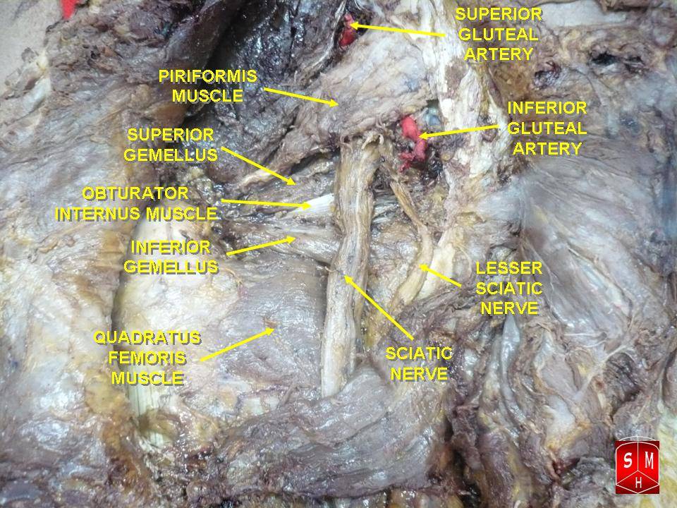
- Superior gemellus arises from the dorsal surface of the ischial spine and inserts with obturator internus and inferior gemellus as the conjoint tendon on the medial surface of the greater trochanter.
- It is supplied by the nerve to obturator internus, L5 and S1, which also supplies obturator internus.
- Superior gemellus, obturator internus and inferior gemellus together form the triceps coxae.
- The deep branch of the medial circumflex femoral artery passes anterior (deep) to the conjoint tendon and posterior to the obturator externus tendon — so the conjoint tendon may be divided safely if obturator externus is preserved.
- The nerve to obturator internus crosses the ischial spine lateral to the pudendal nerve and internal pudendal vessels.
- “The gemelli are accessory to obturator internus: they compensate for the mechanical loss caused by the tendon turning around the lesser sciatic notch.
- “Obturator internus turns roughly 90 degrees around the lesser sciatic notch, which acts as a trochlear pulley lined by a bursa.
- “Superior gemellus is the smaller and more variable twin, and is absent or hypoplastic in a proportion of specimens.
- “The nerve to obturator internus leaves the pelvis through the greater sciatic foramen and re-enters through the lesser sciatic foramen — one of only three structures that do so.
Overview
Superior gemellus is the upper of the two small, flat, triangular muscles — the gemelli, Latin for twins — that flank the tendon of obturator internus as it emerges from the lesser sciatic foramen. Together the three form the triceps coxae, and their fibres fuse into a single conjoint tendon inserting on the medial surface of the greater trochanter.
On its own, superior gemellus is a small and functionally trivial muscle. Its importance is entirely relational, and there are four relationships an examiner will test:
- It is part of the conjoint tendon, the structure divided and repaired in the posterior approach to the hip.
- Its tendon lies posterior to the deep branch of the medial circumflex femoral artery, which is what makes it safe to divide provided obturator externus is preserved.
- Its nerve — the nerve to obturator internus — crosses the ischial spine immediately lateral to the pudendal nerve, the anatomical fact that underpins pudendal nerve blocks and explains combined injuries at the spine.
- It arises from the ischial spine, the key palpable landmark in pelvic and deep gluteal surgery and the site of the sacrospinous ligament attachment.
Examiners love asking why nature bothered with two tiny accessory muscles. The answer is mechanical.
- Obturator internus arises on the inner surface of the obturator membrane and the surrounding pelvic bone, inside the pelvis. Its tendon must leave the pelvis through the lesser sciatic foramen, turning through roughly 90 degrees around the smooth, cartilage-covered groove on the posterior surface of the ischium at the lesser sciatic notch.
- The notch acts as a trochlea (pulley), complete with a bursa (the bursa of obturator internus) to reduce friction, and grooves in the bone corresponding to the tendon's slips.
- The cost of the pulley: turning a tendon through a right angle wastes force and reduces effective excursion. The gemelli, arising outside the pelvis on either side of the tendon, add muscle fibres to the tendon distal to the pulley, restoring the power lost at the turn.
- They are therefore accessory muscles — the classical description is that the gemelli are "accessory to obturator internus" — and this, not their independent action, is what they are for.
- Corollary: the bursa can become inflamed (obturator internus bursitis), producing deep buttock pain that is a recognised and often-missed cause of posterior hip pain, and the tendon can be a source of snapping at the lesser sciatic notch.
P-GO-GO-QShort External Rotators, Superior to Inferior
Hook:Each gemellus takes the nerve of the muscle it lies next to: superior gemellus shares with obturator internus above, inferior gemellus shares with quadratus femoris below.
PINStructures Leaving and Re-Entering at the Ischial Spine
Hook:All three leave the pelvis through the greater sciatic foramen, hook around the ischial spine and sacrospinous ligament, and re-enter through the lesser sciatic foramen. Medial to lateral: Pudendal nerve, Internal pudendal vessels, Nerve to obturator internus.
Attachments, Innervation and Relations
Origin
- Site: the dorsal (external) surface of the ischial spine, and the adjacent margin of the greater sciatic notch.
- Character: a small, flat, triangular muscle, typically 2-3 cm in its longest dimension, arising by short fibres.
- Landmark value: the ischial spine is the key reference point. It gives attachment to the sacrospinous ligament medially, forms the boundary between the greater and lesser sciatic foramina, and is the point around which the pudendal neurovascular bundle and the nerve to obturator internus hook.
Insertion
- Site: the fibres run laterally and fuse with the upper surface of the obturator internus tendon, and with the inferior gemellus below it, forming the conjoint tendon.
- The conjoint tendon inserts on the medial surface of the greater trochanter, just anterior to the trochanteric fossa and below and slightly anterior to the piriformis insertion on the superomedial trochanter.
- Footprint: the conjoint tendon footprint is roughly 1.5-2 cm in vertical dimension on the medial trochanter, immediately anterior to the trochanteric fossa where obturator externus inserts.
- Variability: superior gemellus is the smaller and more variable twin. It is hypoplastic or absent in a proportion of anatomical specimens, whereas the inferior gemellus is more consistently present and larger. The conjoint tendon is present regardless.
The Trochanteric Insertion Map
- Insertion site on the proximal femur
- Superomedial aspect of the greater trochanter, at its tip
- Surgical note
- The most superior; the landmark for the piriformis fossa and for antegrade nailing entry
- Insertion site on the proximal femur
- Medial surface of the greater trochanter, anterior to the trochanteric fossa
- Surgical note
- The structure tagged and divided in the posterior approach
- Insertion site on the proximal femur
- Trochanteric fossa, posterior to the conjoint tendon
- Surgical note
- Must be preserved — it shields the MFCA deep branch
- Insertion site on the proximal femur
- Quadrate tubercle on the intertrochanteric crest
- Surgical note
- Purely muscular; do not strip it flush from bone
- Insertion site on the proximal femur
- Lateral and anterior facets of the greater trochanter
- Surgical note
- Not part of the posterior release; abductor mechanism
- Dissection medial to the ischial spine enters the territory of the pudendal nerve and internal pudendal vessels. Perineal numbness, faecal or urinary incontinence and sexual dysfunction are the price of straying there.
- The order at the spine, medial to lateral, is pudendal nerve, internal pudendal vessels, nerve to obturator internus. The nerve you are entitled to be near is the most lateral of the three.
- In a Kocher-Langenbeck approach, the ischial spine is a useful medial landmark for the quadrilateral surface but should be palpated, not aggressively dissected.
- In deep gluteal space endoscopy, work lateral to the sciatic nerve and lateral to the spine, and identify the nerve to obturator internus as your medial boundary marker.
- The sacrospinous ligament attaches to the spine; a sacrospinous fixation suture placed too laterally injures the pudendal bundle, which is the gynaecological equivalent of the same error.
Action and Biomechanics
Actions by Plane
- Primary: external rotation of the extended hip, acting as one component of the triceps coxae.
- Secondary: abduction of the flexed hip, because the line of pull changes as the hip flexes.
- Stabilisation: with the other short rotators it forms the posterior dynamic sleeve that compresses the femoral head into the acetabulum and resists posterior translation — the hip's functional analogue of the rotator cuff.
The Flexion Reversal
- In extension, the short external rotators are efficient external rotators.
- Beyond about 60-70 degrees of hip flexion, the line of pull of piriformis and the obturator group swings so that they become internal rotators and abductors.
- Why this matters clinically: it explains why testing rotation in flexion gives different information from testing in extension, why the FAIR position (flexion, adduction, internal rotation) tensions piriformis and the obturator group against the sciatic nerve, and why deep gluteal syndrome is provoked in flexion.
Force Contribution
- Superior gemellus contributes a very small fraction of total external rotation torque. Gluteus maximus is by far the largest external rotator, with the posterior fibres of gluteus medius and the obturator group contributing the remainder.
- The gemelli's contribution is best understood as force restoration, not force generation: they add fibres to the obturator internus tendon distal to the trochlear pulley, recovering some of the mechanical loss the pulley imposes.
What Happens When It Is Lost
- Isolated loss is silent. No measurable deficit in strength or function follows division of superior gemellus alone, which is why it is divided routinely without hesitation.
- Loss of the whole posterior sleeve — the situation after an unrepaired posterior approach — removes a passive tether and a dynamic compressive contribution, and is one mechanism behind the increased dislocation rate.
- Denervation pattern: a lesion of the nerve to obturator internus produces selective atrophy of superior gemellus and obturator internus, sparing inferior gemellus and quadratus femoris (nerve to quadratus femoris) and piriformis (its own nerve). Recognising that pattern on MRI localises the lesion.
The conjoint tendon is not a bystander in hip arthroplasty — it is part of the structure whose repair is the best-evidenced technical step against posterior dislocation.
- What the sleeve does: the posterior capsule and the short external rotators together act as a passive checkrein against the flexion, adduction and internal rotation position that dislocates a posterior-approach hip, and as a dynamic compressor centring the head.
- The numbers: pooled series report a dislocation rate of roughly 4 per cent without posterior repair falling to under 1 per cent with a formal transosseous repair.
- How to repair it: tag the conjoint tendon and piriformis before dividing them, leave a cuff on the femur, reflect the sleeve posteriorly to shield the sciatic nerve during the case, and at closure reattach the capsule first, then the tendons, through transosseous drill holes in the posterior greater trochanter.
- Position for repair: hip in extension with neutral or slight external rotation, so the repair is not tensioned in the position that will later be avoided.
- What the repair does not fix: component malposition. A posterior repair does not rescue a retroverted cup or inadequate offset. Get the components right and repair the sleeve; they are complementary, not alternatives.
Surface Anatomy and Examination
Palpation
- Superior gemellus is not palpable — it lies deep to gluteus maximus and the sciatic nerve, on the ischial spine.
- The bony landmarks are what you palpate:
- The greater trochanter laterally, whose posterior and medial surfaces carry the rotator insertions.
- The ischial tuberosity inferomedially.
- The posterior superior iliac spine, from which a line to the greater trochanter approximates the upper border of piriformis, and a line to the ischial tuberosity crosses the field of the rotators.
- The ischial spine is palpable per rectum or per vaginam, not externally. This is how it is located for a pudendal nerve block.
Named Clinical Tests
- How to perform
- Side-lying, hip flexed 60 degrees, then adducted and internally rotated
- Positive finding
- Deep buttock pain with or without sciatic radiation
- What it means
- Deep gluteal syndrome — piriformis or obturator internus related sciatic irritation
- False positives
- Hip osteoarthritis; lumbar radiculopathy; gluteal tendinopathy
- How to perform
- Side-lying, resisted abduction and external rotation with the heel pressed into the couch
- Positive finding
- Deep buttock pain
- What it means
- Piriformis-related deep gluteal syndrome
- False positives
- Gluteal tendinopathy; sacroiliac pain
- How to perform
- Seated, knee extended, hip adducted and internally rotated while palpating lateral to the ischium
- Positive finding
- Posterior pain reproduced
- What it means
- Deep gluteal syndrome; more sensitive when combined with the active piriformis test
- False positives
- Hamstring tendinopathy
- How to perform
- Prone, knee flexed 90 degrees, resisted external rotation
- Positive finding
- Weakness or pain
- What it means
- Short external rotator group involvement — cannot isolate superior gemellus
- False positives
- Pain inhibition; gluteus maximus weakness
- How to perform
- Deep palpation lateral to the ischial tuberosity toward the lesser sciatic notch
- Positive finding
- Focal deep tenderness
- What it means
- Obturator internus bursitis
- False positives
- Ischial bursitis; proximal hamstring tendinopathy
- How to perform
- Ultrasound- or CT-guided local anaesthetic to the obturator internus bursa or the deep gluteal space
- Positive finding
- Substantial temporary relief
- What it means
- Confirms the deep gluteal structures as the pain source
- False positives
- Anaesthetic spread to the sciatic nerve causing transient weakness
Grading and Interpretation
- There is no isolated test for superior gemellus. External rotation strength is graded 0-5 for the group. Any examiner asking for an isolation test is testing whether you know that one does not exist.
- The clinically useful discrimination is between the deep external rotator group (deep, posterior, provoked in flexion and internal rotation) and the abductor mechanism (lateral, provoked by single-leg stance), and between both of those and the hip joint itself (groin pain, C-sign, restricted internal rotation in flexion).
Differential Diagnosis of Deep Buttock Pain
- Key discriminator
- Sitting-related pain with sciatic radiation; positive FAIR and active piriformis tests
- Confirmatory finding
- Sciatic nerve signal change or entrapment on MR neurography; response to diagnostic injection
- Key discriminator
- Focal tenderness deep to the ischium, worse on resisted external rotation
- Confirmatory finding
- Fluid in the obturator internus bursa on MRI
- Key discriminator
- Long-stride pain relieved by short strides
- Confirmatory finding
- Quadratus femoris oedema with narrowed ischiofemoral space
- Key discriminator
- Pain on sitting on hard surfaces, tender on the tuberosity
- Confirmatory finding
- High signal at the conjoint hamstring origin
- Key discriminator
- Perineal and genital pain in a sitting position, relieved on standing or sitting on a toilet seat
- Confirmatory finding
- Response to a pudendal nerve block at the ischial spine
- Key discriminator
- Pain below the PSIS, positive cluster of provocation tests
- Confirmatory finding
- Response to an image-guided intra-articular block
- Key discriminator
- Dermatomal pain, positive straight-leg raise, neurological signs
- Confirmatory finding
- Concordant lumbar MRI
Complications
Neurological
- Sciatic nerve injury — the commonest neurological complication of the posterior approach. Mechanisms are direct trauma during release, retractor pressure, traction from limb lengthening, and thermal injury. The peroneal division is more vulnerable because of its more lateral position, larger fascicles and less protective connective tissue.
- Pudendal nerve injury — from a perineal post, medial dissection past the ischial spine, or retractor placement. Presents as perineal numbness, and in severe cases sexual and continence dysfunction.
- Nerve to obturator internus injury — functionally silent in itself, but its proximity to the pudendal nerve makes it a warning marker.
- Nerve to quadratus femoris injury — from deep capsular dissection; produces selective quadratus femoris and inferior gemellus atrophy.
- Superior gluteal nerve injury — from extending the dissection above piriformis; produces abductor weakness and a Trendelenburg gait.
Vascular
- MFCA deep branch injury causing femoral head osteonecrosis, if obturator externus is divided or the release is taken flush against the femur.
- Inferior gluteal artery avulsion, which retracts into the pelvis and cannot be controlled through the posterior wound; requires packing and, if uncontrolled, angiographic embolisation or a pelvic approach.
- Superior gluteal artery injury at the notch, with the same problem of proximal retraction.
- Internal pudendal vessel injury at the ischial spine from medial dissection.
Related to the Repair
- Dislocation if the repair is omitted or fails — the risk the repair addresses.
- Repair failure from placing sutures in poor-quality tendon, dividing the tendon flush at the bone so there is no cuff, or tensioning the repair in internal rotation.
- Suture knot irritation over the posterior trochanter.
- Heterotopic ossification in the split gluteus maximus and the rotator bed, particularly after acetabular fracture surgery.
Preventing Each
- Mechanism
- Retraction, traction from lengthening, direct or thermal injury
- Prevention
- Hip extended and knee flexed; reflect the rotator sleeve over the nerve; limit lengthening
- Mechanism
- Perineal post traction; dissection medial to the ischial spine
- Prevention
- Large padded post, minimum traction force and time; stop at the spine
- Mechanism
- Division of obturator externus or release flush at the femur
- Prevention
- Preserve obturator externus; divide short of the insertion, leaving a cuff
- Mechanism
- Unrepaired posterior sleeve, component malposition
- Prevention
- Formal transosseous repair plus correct version, offset and head size
- Mechanism
- No cuff left on bone; poor suture purchase; tensioning in internal rotation
- Prevention
- Divide short of the insertion leaving a cuff, tag before dividing, repair in extension and neutral rotation
- Mechanism
- Avulsion of the superior or inferior gluteal artery at the notch
- Prevention
- Do not extend dissection above piriformis; identify the pedicle before splitting maximus deeply
Clinical Relevance
The Conjoint Tendon in Surgery
- In the posterior (Moore or Southern) approach to the hip, the conjoint tendon — superior gemellus, obturator internus and inferior gemellus — is the principal structure released, along with piriformis.
- Sequence:
- Split the iliotibial band and gluteus maximus fascia; split gluteus maximus bluntly along its fibres.
- Internally rotate the hip to bring the rotators into view and to tension their tendons.
- Identify the sciatic nerve — it lies superficial to the rotators and deep to gluteus maximus. Palpate rather than dissect it out.
- Tag piriformis and the conjoint tendon with heavy non-absorbable sutures before dividing.
- Divide them a short distance medial to the trochanteric insertion, leaving a cuff on bone.
- Reflect the tagged sleeve posteriorly — it now protects the sciatic nerve for the remainder of the case.
- Perform the capsulotomy, tagging its corners.
- Quadratus femoris is usually left intact. Obturator externus should be preserved.
Piriformis-Sparing Modification
- Works through the interval between piriformis above and the conjoint tendon below, dividing only the conjoint tendon (or in some descriptions, only a portion of it).
- Superior gemellus is therefore the first structure divided in a piriformis-sparing approach — it is the upper border of the released sleeve.
- Justification: soft tissue preservation, better repair quality and possibly faster early recovery. Not a vascular justification, since piriformis lies superficial to the MFCA deep branch.
The Repair
- Transosseous drill holes in the posterior greater trochanter, or suture anchors.
- Repair the capsule first, then the tendon sleeve.
- Position: hip in extension, neutral or slight external rotation.
- Check stability through flexion, adduction and internal rotation before closing, and document the safe arc.
- Evidence: pooled data show the dislocation rate falls from roughly 4 per cent without repair to under 1 per cent with it.
Kocher-Langenbeck Approach
- The same rotator release is used to expose the posterior column and posterior wall of the acetabulum.
- Additional considerations:
- Keep the hip extended and the knee flexed throughout to protect the sciatic nerve.
- The ischial spine is a medial landmark for the quadrilateral surface; palpate it, do not dissect medial to it.
- Some surgeons use a trochanteric flip osteotomy rather than a rotator release, which preserves the entire posterior sleeve including obturator externus and gives superior superior-acetabular access.
Because of where the artery runs.
- The MFCA deep branch passes anterior (deep) to the conjoint tendon and posterior to the obturator externus tendon.
- Therefore piriformis, the gemelli and obturator internus can all be divided without endangering the femoral head — provided obturator externus is preserved.
- Divide away from the femoral insertion, leaving a cuff on bone, to preserve the trochanteric anastomosis. In the Ganz surgical dislocation they are not divided at all.
Medial to lateral: pudendal nerve, internal pudendal vessels, nerve to obturator internus.
- All three leave through the greater sciatic foramen and re-enter through the lesser.
- The pudendal nerve is the most medial and the one you must not injure.
- The ischial spine is the medial limit of safe posterior hip dissection.
Surgical Relevance
Structures at Risk with Distances
- Location relative to a landmark
- Superficial to the conjoint tendon, descending below piriformis onto quadratus femoris
- How to protect it
- Reflect the tagged rotator sleeve posteriorly over it; hip extended, knee flexed
- Location relative to a landmark
- Medial to the internal pudendal vessels at the ischial spine
- How to protect it
- Do not dissect medial to the ischial spine; pad the perineal post and limit traction
- Location relative to a landmark
- Between the pudendal nerve and the nerve to obturator internus at the spine
- How to protect it
- Same medial limit; avoid blind retraction over the spine
- Location relative to a landmark
- Anterior (deep) to the conjoint tendon; posterior to the obturator externus tendon
- How to protect it
- Preserve obturator externus; divide the conjoint tendon short of the femur, leaving a cuff
- Location relative to a landmark
- Emerging below piriformis at the superomedial corner of the field
- How to protect it
- Identify before deep blunt splitting of gluteus maximus
- Location relative to a landmark
- Emerging above piriformis at the greater sciatic notch
- How to protect it
- Do not extend the dissection above piriformis; the artery retracts into the pelvis if torn
- Location relative to a landmark
- Deep to the conjoint tendon, on the posterior hip capsule
- How to protect it
- Avoid blind cautery and stripping of the capsule
Approaches Involving Superior Gemellus
- Posterior (Moore or Southern) approach: superior gemellus is the upper component of the conjoint tendon, tagged, divided and repaired.
- Piriformis-sparing posterior approach: superior gemellus is the first structure divided, defining the upper margin of the released sleeve.
- Kocher-Langenbeck approach for acetabular fractures: same release, with the ischial spine as the medial landmark and obsessive sciatic nerve protection.
- Ganz surgical hip dislocation (trochanteric flip): the entire posterior sleeve is preserved, including superior gemellus and obturator externus. Access is gained through the trochanteric osteotomy instead. This is why the technique has such a low osteonecrosis rate.
- Deep gluteal space endoscopy: the conjoint tendon forms part of the floor of the space; the sciatic nerve is followed across it, and the tendon may be released to decompress the nerve.
- Antegrade femoral nailing (piriformis entry): the piriformis fossa entry point sits just medial to the tip of the greater trochanter and superior to the conjoint tendon footprint. A malpositioned entry point damages the rotator insertions and can breach the medial trochanteric cortex.
Technique Checklist for the Rotator Release and Repair
- Position lateral decubitus, pelvis rigidly held; confirm the pelvis has not rolled before assessing component version.
- Internally rotate the hip to tension and present the rotators.
- Palpate the sciatic nerve on the posterior surface of the rotators; do not skeletonise it.
- Tag piriformis and the conjoint tendon with heavy braided non-absorbable suture before dividing.
- Divide a short distance medial to the trochanteric footprint, leaving a cuff. Preserve obturator externus and quadratus femoris.
- Reflect the sleeve posteriorly as the sciatic nerve's protective blanket.
- Tag the capsule corners at the capsulotomy.
- At closure: transosseous drill holes in the posterior trochanter; capsule repaired first, then the tendon sleeve, with the hip extended and in neutral or slight external rotation.
- Test stability through the dislocating arc and document the safe range.
- Do not rely on the repair to compensate for component malposition.
The Ischial Spine as a Surgical Landmark
- Boundary between the greater and lesser sciatic foramina.
- Attachment of the sacrospinous ligament.
- Origin of superior gemellus on its dorsal surface.
- Point around which the pudendal bundle hooks — the target for a pudendal nerve block and the site of entrapment in pudendal neuralgia.
- Radiographic landmark: the ischial spine is used to assess acetabular version on an anteroposterior pelvic radiograph — a visible ischial spine sign indicates acetabular retroversion, provided the film is well centred and not rotated.
Guidelines, Registries & Global Practice
Anatomical Variation Across Populations
- Superior gemellus is the most variable of the short external rotators. It is reported as hypoplastic or absent in a minority of cadaveric specimens across European, North American and Asian series, with wide variation in reported prevalence reflecting differing definitions of what counts as absent. The inferior gemellus is more consistently present and larger.
- Sciatic nerve variants relative to piriformis are described in all populations. A meta-analysis of 44 cadaveric studies gave a pooled prevalence of 90 per cent for the typical pattern, with variants more common in East Asian populations (31 per cent pooled prevalence of total variants) and no significant difference by sex or laterality. The surgical message is to expect a variant in roughly one in ten patients, and more often in East Asian populations.
- The extracapsular course of the deep branch of the medial circumflex femoral artery was constant in all 24 hips in the defining cadaveric study, which is what permits the Ganz principles to be taught as rules.
- Ischial spine morphology varies, and a prominent spine is associated with acetabular retroversion — the basis of the radiographic ischial spine sign.
Differences in Described Practice
- Position
- Preserve the whole posterior sleeve using a trochanteric flip osteotomy with the external rotators left undivided; where rotators are divided, leave a cuff on the femur and never divide obturator externus.
- Position
- Multiple approaches acceptable; posterior soft tissue repair is standard when the posterior approach is used.
- Position
- Posterior approach with formal capsular and rotator repair is the most widely used technique in the United Kingdom.
- Position
- Kocher-Langenbeck with rotator release preserving obturator externus and quadratus femoris; ischial spine as the medial landmark; hip extended and knee flexed throughout.
- Position
- Divide only the conjoint tendon, beginning with superior gemellus, and preserve the piriformis tendon; justified on soft tissue grounds rather than vascular ones.
- Position
- The conjoint tendon forms part of the deep gluteal space floor and may be released to decompress the sciatic nerve; patient selection by diagnostic injection.
Registry and Practice Signals
- National joint registries (NJR, AOANJRR, the Nordic registries and the AJRR) consistently list dislocation among the leading indications for early revision after primary total hip arthroplasty, which is the practice-level argument for the posterior repair.
- Registries also show declining dislocation rates over time across all approaches, attributable to larger femoral heads, dual-mobility bearings in high-risk patients, improved component positioning and the widespread adoption of posterior soft tissue repair.
- There is no registry for deep gluteal syndrome or for endoscopic sciatic nerve decompression. The evidence base is single-centre case series, and the consistent signal is that outcomes depend on selection — a positive diagnostic injection and the absence of significant lumbar pathology.
High- and Limited-Resource Practice
- Well-resourced settings: MR neurography for suspected deep gluteal syndrome, image-guided diagnostic blocks, endoscopic sciatic nerve decompression, dual-mobility bearings and navigation or robotics for component positioning.
- Limited-resource settings: the anatomical rules cost nothing and matter most. Tagging the conjoint tendon before dividing it, dividing short of the femur to leave a cuff, preserving obturator externus, and performing a transosseous repair with a drill and a suture are available in every operating theatre in the world and deliver the single largest reduction in dislocation risk. Diagnosis of deep gluteal syndrome can be made clinically with the FAIR and active piriformis tests plus a landmark-guided injection.
MCQ Practice Points
Q: Where does superior gemellus arise and insert? A: From the dorsal surface of the ischial spine, inserting via the conjoint tendon on the medial surface of the greater trochanter, anterior to the trochanteric fossa.
Q: What innervates superior gemellus? A: The nerve to obturator internus, L5 and S1, which also supplies obturator internus. The inferior gemellus, by contrast, takes the nerve to quadratus femoris.
Q: What is the triceps coxae? A: Superior gemellus, obturator internus and inferior gemellus, fusing into the conjoint tendon.
Q: Which three structures leave the pelvis through the greater sciatic foramen and re-enter through the lesser? A: The pudendal nerve, the internal pudendal vessels and the nerve to obturator internus. Medial to lateral in that order at the ischial spine.
Q: What is the functional purpose of the gemelli? A: They are accessory to obturator internus. They add muscle fibres to the tendon distal to the trochlear pulley at the lesser sciatic notch, restoring force lost when the tendon turns through 90 degrees.
Q: Where does the MFCA deep branch run relative to the conjoint tendon? A: Anterior (deep) to it, and posterior to the obturator externus tendon. This is why the conjoint tendon can be divided safely and obturator externus cannot.
Q: What happens to the short external rotators beyond 60-70 degrees of hip flexion? A: They become internal rotators and abductors. This underlies the FAIR test and the flexion-provoked nature of deep gluteal syndrome.
Q: MRI shows selective atrophy of obturator internus and superior gemellus. Where is the lesion? A: The nerve to obturator internus. If quadratus femoris and inferior gemellus were atrophic instead, the lesion would be the nerve to quadratus femoris.
Q: Which of the two gemelli is more often absent? A: The superior gemellus. It is the smaller and more variable twin; the conjoint tendon is present regardless.
Q: What does a visible ischial spine on a well-centred anteroposterior pelvic radiograph suggest? A: Acetabular retroversion — the ischial spine sign. It is only valid on a well-centred, non-rotated film.
Exam Viva Scenarios
Practise clinical reasoning and management decisions out loud
“You are teaching a registrar the posterior approach to the hip. They have split gluteus maximus and can see the short external rotators. Talk them through what happens next and why.”
“During a Kocher-Langenbeck approach for a posterior column acetabular fracture, your assistant asks how far medially they can safely retract and dissect. What is your answer and what is the anatomy behind it?”
“A 38-year-old cyclist has six months of right buttock pain radiating to the posterior thigh, much worse after 20 minutes of sitting. Lumbar MRI is normal. Straight-leg raise is negative but the FAIR test reproduces the pain. What is your working diagnosis and how do you proceed?”
Core Anatomy
- Origin: dorsal surface of the ischial spine
- Insertion: conjoint tendon on the medial greater trochanter
- Nerve: nerve to obturator internus, L5-S1
- Triceps coxae = superior gemellus + obturator internus + inferior gemellus
The Ischial Spine
- Medial to lateral: pudendal nerve, internal pudendal vessels, nerve to obturator internus
- All three exit the greater and re-enter the lesser sciatic foramen
- Sacrospinous ligament attaches here
- Medial limit of safe posterior dissection
Vascular Rules
- MFCA deep branch is anterior to the conjoint tendon
- It is posterior to the obturator externus tendon
- Conjoint tendon safe to divide; obturator externus is not
- Divide short of the femoral insertion, leaving a cuff on bone
Clinical
- Gemelli are accessory to obturator internus, compensating for the trochlear pulley
- Beyond 60-70 degrees of hip flexion the rotators become internal rotators
- Posterior repair drops dislocation from about 4 per cent to under 1 per cent
- Selective obturator internus and superior gemellus atrophy = nerve to obturator internus lesion
Evidence Base
Anatomy of the Medial Femoral Circumflex Artery and Its Surgical Implications
- 24 cadaver hips dissected after neoprene-latex injection of the femoral or internal iliac arteries
- The extracapsular course of the deep branch of the medial femoral circumflex artery was constant, with a trochanteric branch at the proximal border of quadratus femoris in every case
- The deep branch crosses posterior to the obturator externus tendon and then anterior to the conjoint tendon of inferior gemellus, obturator internus and superior gemellus
- It perforates the capsule at the level of the superior gemellus and divides into two to four subsynovial retinacular vessels on the posterosuperior neck
- Obturator externus protected the deep branch from disruption OR STRETCHING during dislocation in any direction after serial release of all other soft-tissue attachments of the proximal femur INCLUDING A COMPLETE CIRCUMFERENTIAL CAPSULOTOMY - the release was total, which is what makes the protective claim striking
- The trochanteric branch is the intra-operative landmark the paper offers: it marks the level of the obturator externus tendon, which the deep branch crosses posteriorly
Surgical Dislocation of the Adult Hip: A Technique with Full Access to the Femoral Head and Acetabulum Without the Risk of Avascular Necrosis
- Technique of anterior dislocation through a posterior approach with a trochanteric flip osteotomy, based on detailed anatomical studies of the blood supply
- The external rotator muscles are not divided, and the medial femoral circumflex artery is protected by the intact obturator externus
- Reported in 213 hips over seven years, including 19 with simultaneous intertrochanteric osteotomy
- Femoral head perfusion was verified intra-operatively and no patient had developed avascular necrosis at the time of reporting
- Morbidity was low and the exposure permitted treatment of conditions not amenable to other methods
Posterior Approach to Total Hip Replacement Using Enhanced Posterior Soft Tissue Repair
- Two senior surgeons independently adopted an identical enhanced posterior soft tissue repair
- In the first series the dislocation rate fell from 4 per cent in 395 patients to 0 per cent in a further 395
- In the second series it fell from 6.2 per cent in 160 replacements to 0.8 per cent in 124
- Both reductions were highly statistically significant
Does Surgical Approach Affect Total Hip Arthroplasty Dislocation Rates?
- Meta-analysis of five studies directly comparing the posterior approach with and without soft tissue repair
- Dislocation rates were 0.49 per cent with repair and 4.46 per cent without
- The posterior approach without repair carried an 8.21 times greater relative risk of dislocation (95 per cent confidence interval 4.05 to 16.67, fixed-effects model) - a fourfold-wide interval, which is why the direction is more trustworthy than the magnitude
- A separate review of 11 studies gave comparable dislocation rates for the anterolateral (0.70 per cent), direct lateral (0.43 per cent) and repaired posterior (1.01 per cent) approaches
The Endoscopic Treatment of Sciatic Nerve Entrapment / Deep Gluteal Syndrome
- 35 patients with sciatic nerve entrapment treated by endoscopic decompression of the subgluteal space
- Entrapment was addressed by resection of fibrovascular scar bands, piriformis tendon release, obturator internus or quadratus femoris release, or release of hamstring tendon scarring
- Mean modified Harris Hip Score improved from 54.4 to 78.0 and mean visual analogue pain score fell from 6.9 to 2.4
- 83 per cent of patients had no postoperative sciatic sitting pain
- Mean symptom duration before surgery was 3.7 years
Sciatic Nerve Variants and the Piriformis Muscle: A Systematic Review and Meta-Analysis
- Systematic review and meta-analysis of 44 cadaveric studies of sciatic nerve variants relative to piriformis
- The typical pattern, with the undivided sciatic nerve passing below piriformis, had a pooled prevalence of 90 per cent
- In greater than 10 per cent of the population the nerve coursed through or above the piriformis
- Variants were more common among East Asian populations, with a 31 per cent pooled prevalence of total variants
- No significant differences were found with respect to sex or laterality