The 'low back pain of the wrist' — a location, not a diagnosis. Discriminate by exact point of tenderness, provocative test and ulnar variance.
- Standardise the film, and control the grip: power grip raised ulnar variance by a mean 0.64 mm on CT, whereas a radiostereometric cadaver study found forearm rotation did not significantly alter it.
- The fovea sign is the most sensitive clinical test for a foveal TFCC tear and one of the least specific. Use it to rule out, never to rule in.
- Ulnar impaction syndrome is a load problem, not just an anatomical one: positive ulnar variance plus lunate ulnar-corner subchondral sclerosis and cyst formation on the PA view.
- A displaced or non-united ulnar styloid BASE fracture implies avulsion of the deep radioulnar ligaments and therefore DRUJ instability — a tip fracture does not.
- Hook of hamate fracture presents as ulnar-sided pain in a club/bat/racquet athlete with possible ulnar nerve motor symptoms; it is invisible on standard PA and needs a carpal tunnel view or CT.
- Ulnar-sided pain with a normal wrist may be referred: cubital tunnel, Guyon canal lesion, or C8 radiculopathy.
- “State the exact anatomical point of maximal tenderness before offering any diagnosis — it is the viva's discriminator.
- “Always ask for a pronated grip view: it dynamically unmasks positive ulnar variance in impaction.
- “ECU subluxation is diagnosed dynamically: supination, ulnar deviation and flexion cause a painful snap.
- “Lunotriquetral ballottement (Reagan) tests LT ligament; ulnocarpal stress test is non-specific and positive in almost everything ulnar-sided.
Recognising the Pattern
Definition. Pain localised to the ulnar third of the wrist, bounded by the ulnar head and styloid proximally, the pisiform and hamate hook volarly, and the triquetrum dorsally. It is an anatomical region containing at least eight discrete pain generators within about 2 cm of one another.
Referred pain. Ulnar-sided pain with a normal wrist may be referred from the cubital tunnel, a Guyon canal lesion or a C8 radiculopathy. Pain referred from the cubital tunnel or the C8 root is diffuse rather than point-tender, with numbness in an ulnar distribution above the wrist crease.
Next Investigation
Standardised PA and lateral radiographs, always first. Insist on the shoulder abducted 90 degrees, the elbow flexed 90 degrees, the wrist in neutral rotation and no grip, with the pisiform overlying the volar third of the scaphoid. On these films assess the ulnar variance, lunate ulnar-corner sclerosis and cysts, any ulnar styloid fracture and its level, DRUJ congruity on a true lateral, and Kienböck changes. The film changes management by identifying impaction that needs shortening.
Rotation is the weaker variable. The classical teaching that forearm rotation shifts ulnar variance comes from Epner 1982, which gives a direction (pronation reduces measured negative variance) and no magnitude. A radiostereometric cadaver study designed to test exactly this found that rotation significantly altered palmar tilt but did not significantly alter ulnar variance (slopes 0.01 and 0.02, both non-significant).
Grip has the measured effect. Power grip increased variance on CT by a mean 0.64 mm, and the mechanism is proximal migration of the radius rather than distal migration of the ulna: the radiocapitellar distance shortened by an almost identical 0.62 mm. Still take a standardised neutral-rotation PA, but control the grip, and never diagnose impaction on a film taken with the hand gripping.
Apparent positive variance. A pronated film, the direction Epner described, or a previous distal radius fracture with radial shortening can make the variance look positive.
The pronated grip view. Always ask for it when impaction is suspected. It unmasks positive ulnar variance dynamically and often converts an apparently neutral wrist to 1–2 mm positive, supporting a diagnosis of dynamic ulnar impaction and justifying ulnar shortening. The neutral PA is read without grip; this view is read as the dynamic one.
Presenting it in a viva. "This is a wrist series in neutral rotation. Ulnar variance is positive at approximately 3 mm. There is subchondral sclerosis and a small cyst in the ulnar proximal corner of the lunate and the ulnar pole of the triquetrum, with no fracture and preserved carpal alignment. The appearances are those of ulnar impaction syndrome, and I would confirm with a pronated grip view and MRI."
Carpal tunnel view or dedicated CT for the hook of hamate. Normal PA and lateral films do not exclude a hook fracture. Any tenderness over the hook needs one of these views, and tenderness with pain on resisted small finger flexion needs CT, which is definitive and also shows non-union. It changes management: a symptomatic non-union is treated by excising the hook fragment.
Bilateral CT of both DRUJs. The test of choice for suspected DRUJ instability, taken in neutral, pronation and supination and read by the epicentre or radioulnar line method with the contralateral side as control. Confirmed instability drives foveal repair or ligament reconstruction.
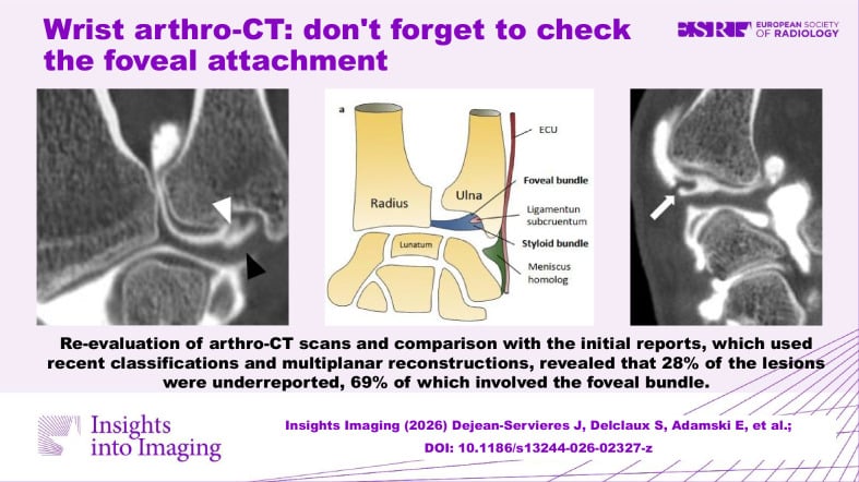
MRI or MR arthrogram. At 3 Tesla with a dedicated wrist coil, for suspected TFCC and LT tears, occult ganglia, ECU subsheath rupture and lunate oedema. It distinguishes foveal detachment, which is repairable, from degenerative central perforation, which is debrided and unloaded.
Central TFCC perforation prevalence rises steadily with age and is frequently asymptomatic beyond the fifth decade. Correlate MRI with the fovea sign and the ulnar variance, not with signal alone.
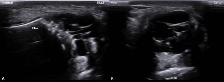
Dynamic ultrasound. The only test that shows ECU subluxation in real time, during active supination. It is also useful for ganglia and for guided injections.
Wrist arthroscopy. The reference standard when imaging is equivocal but symptoms persist. The trampoline and hook tests define foveal integrity, and it is diagnostic and therapeutic in the same sitting.
When no further imaging is needed. Clear-cut ECU tendinopathy without instability, or a first presentation of localised ulnocarpal pain after a minor strain with normal radiographs and a stable DRUJ, is treated with splintage and activity modification and reviewed at six weeks before escalating.
The Differential
How many diagnoses are really in play. In a prospective series of 110 consecutive patients presenting with ulnar-sided wrist pain, seventeen different diagnoses were made, and six accounted for 85 per cent: TFCC injury, ulnocarpal abutment, pisotriquetral arthritis, triquetral fracture or nonunion, DRUJ arthritis and ECU pathology. The long tail in the table is real but rare. Most of the diagnostic work is separating six conditions whose examination findings overlap almost completely.
- Typical age / setting
- Any age; after distal radius or Galeazzi fracture, or forced supination injury
- Discriminating feature
- Painful dorsopalmar translation of the ulnar head with the forearm in neutral, compared with the other side; ulnar head prominence
- What confirms it
- Bilateral CT of the DRUJ in neutral, full pronation and full supination — epicentre or radioulnar line method
- Typical age / setting
- Adult after distal radius fracture
- Discriminating feature
- Fracture line at the styloid base with greater than 2 mm displacement and DRUJ widening — not a tip avulsion
- What confirms it
- PA radiograph plus examination under anaesthesia or MRI showing deep radioulnar ligament detachment
- Typical age / setting
- Golfer, cricketer, baseball or racquet player; young adult
- Discriminating feature
- Point tenderness over the hook with pain on resisted ring and small finger flexion; possible ulnar motor weakness or FDP rupture
- What confirms it
- Carpal tunnel view or CT — plain PA is normal
- Typical age / setting
- 20–40 years, often negative ulnar variance
- Discriminating feature
- Lunate sclerosis, loss of lunate height and collapse — pain is more central-dorsal but often reported ulnarly
- What confirms it
- Radiographs plus MRI showing diffuse low T1 signal in the whole lunate
- Typical age / setting
- Young adult, fall on outstretched hand or power-tool torque injury
- Discriminating feature
- Positive fovea sign plus DRUJ laxity; pain worse with rotation under load
- What confirms it
- MR arthrogram or wrist arthroscopy with a positive trampoline and hook test
- Typical age / setting
- Over 40 years; associated with positive ulnar variance
- Discriminating feature
- Pain on axial load and ulnar deviation without DRUJ instability; often coexists with chondromalacia
- What confirms it
- MRI: central perforation with lunate and ulnar head chondral change
- Typical age / setting
- Adults, positive ulnar variance, or after radial shortening malunion
- Discriminating feature
- Positive variance on neutral PA, accentuated on pronated grip view; ulnar lunate corner sclerosis and cysts
- What confirms it
- Radiographs plus MRI showing lunate ulnar-corner oedema
- Typical age / setting
- Racquet and stick sports; also inflammatory arthritis
- Discriminating feature
- Tenderness in the ECU groove; painful snap on supination with ulnar deviation and flexion
- What confirms it
- Dynamic ultrasound in supination; MRI shows subsheath disruption and tendon dislocation
- Typical age / setting
- Adult after fall in extension and radial deviation; also with ulnar impaction
- Discriminating feature
- Focal LT interval tenderness with positive ballottement and shear tests; ring sign absent
- What confirms it
- Radiographs may show volar intercalated segment instability on lateral; MR arthrogram or arthroscopy
- Typical age / setting
- Over 50 years, or after pisiform fracture
- Discriminating feature
- Tenderness confined to the pisiform with a painful grind on side-to-side translation; pain on resisted FCU
- What confirms it
- 30-degree supinated oblique view showing joint space narrowing; relief with an intra-articular injection
- Typical age / setting
- Rheumatoid; middle-aged, bilateral
- Discriminating feature
- Synovitis with ulnar head erosion (caput ulnae), carpal supination and ulnar translocation
- What confirms it
- Erosive change on radiograph plus rheumatoid serology
- Typical age / setting
- Cyclist, ganglion, hook fracture
- Discriminating feature
- Sensory or motor deficit without point bony tenderness; positive Tinel over the canal
- What confirms it
- Nerve conduction studies plus MRI or ultrasound to find a ganglion
- Typical age / setting
- Young adult female
- Discriminating feature
- Painful swelling that transilluminates and varies with wrist position
- What confirms it
- Ultrasound or MRI showing a fluid-filled unilocular cyst
- Typical age / setting
- Manual worker using palm as a hammer
- Discriminating feature
- Cold intolerance, digital ischaemia, positive Allen test rather than mechanical pain
- What confirms it
- Doppler ultrasound or CT angiography of the ulnar artery and superficial arch
- Typical age / setting
- Adult, repetitive activity
- Discriminating feature
- Pain over the fifth extensor compartment reproduced by resisted small finger extension
- What confirms it
- Ultrasound in dynamic mode; MRI excluding TFCC pathology
Narrowing It Down
Seven questions, asked in order, narrow the field.
Was there a discrete injury, and what was the mechanism? Take the mechanism before touching the wrist, and ask specifically about a pronated fall, a torque kickback, hypothenar impact and a previous distal radius fracture.
- A fall on the outstretched hand with the forearm pronated, or a power tool kicking back, points to a traumatic TFCC or LT tear
- Insidious onset with heavy loading, or a previous distal radius malunion, points to ulnar impaction
- Hypothenar impact points to the hook of hamate or the ulnar artery
Palmer's 1989 classification is built on exactly this split: class 1 traumatic lesions are subclassified by location, class 2 degenerative lesions by severity. The history determines which half of the classification you are in before any imaging.
Where exactly is the point of maximal tenderness? Palpate each point separately, compare it with the other side, and reproduce the patient's exact pain at one of them.
- Fovea: the soft spot bounded by the ulnar styloid, FCU tendon, ulnar head and pisiform. A positive fovea sign suggests a foveal TFCC tear or an ulnotriquetral ligament split.
- ECU groove: dorsal-ulnar, over the distal ulna. Tenderness with a palpable snap on active supination suggests ECU subluxation or tendinopathy.
- LT interval: dorsal, just distal to the ulnar head and radial to the ECU. Pain on ballottement suggests lunotriquetral injury.
- Pisiform and pisotriquetral joint: volar, a mobile bone. Grind pain suggests pisotriquetral arthritis.
- Hook of hamate: volar, 1–2 cm distal and radial to the pisiform, in line with the ring finger axis. Tenderness here suggests a hamate fracture.
- DRUJ itself: pain on passive forearm rotation with dorsopalmar translation.
Localising the tenderness narrows the differential more than any single imaging test. Tenderness alone still discriminates poorly, because the first five points lie within about 2 cm of one another.
The fovea sign. It is the most sensitive clinical test for a foveal TFCC tear and one of the least specific, which is the central problem of this region. Tay's originating series reported 95.2 per cent sensitivity and 86.5 per cent specificity, but in independent hands specificity is 41 to 48 per cent, and in 73 patients with a positive fovea sign only 21 had a foveal tear at arthroscopy. Use it to rule out, never to rule in.
The other provocative tests. In the prospective series of 110 patients, the ulnar fovea sign and the ulnocarpal stress test were positive across several of the six common diagnoses and were not sufficiently specific to choose between them. Only the pisotriquetral shear test discriminated well.
Is the DRUJ stable? Test dorsopalmar translation of the ulnar head in neutral, pronation and supination, always against the contralateral side. A DRUJ that is stable in neutral can be grossly unstable at the extremes, so a single-position test misses the diagnosis. Instability moves a foveal TFCC tear and a styloid base avulsion to the top of the list, and mandates CT of both DRUJs in the same position.
Which ulnar styloid fracture matters. Tip avulsions are cosmetic and carry no instability implication. Only a fracture at the styloid base indicates avulsion of the deep radioulnar ligaments and DRUJ instability: a displaced or non-united base fracture implies it, especially when displaced more than 2 mm or accompanied by DRUJ widening.
What is the ulnar variance on a strictly neutral PA? Measure it on a standardised film (technique below) and add a pronated grip view if impaction is suspected. Positive variance supports ulnar impaction and a degenerative central TFCC tear, and argues for ulnar shortening osteotomy or a wafer procedure. Negative variance raises Kienböck disease, and neutral variance with a traumatic history favours a repairable peripheral tear.
Is there a mechanical clunk, and is it painful and reproducible? Reproduce the clunk yourself before believing it, and record whether it hurts. Many normal wrists produce a painless midcarpal shift, and a painless clunk is usually physiological and needs no treatment; it is the commonest reason a wrist is over-investigated. Only a painful, reproducible clunk counts.
- A painful snap on supination with ulnar deviation is ECU subluxation
- A painful clunk on ulnar deviation from radial deviation may be midcarpal instability or LT instability
Are there neurovascular features? Examine the intrinsics, test Froment sign, and ask about cold intolerance and colour change before ordering any wrist imaging. Numbness, intrinsic wasting or a positive Froment sign shifts the diagnosis to Guyon canal or cubital tunnel compression. Cold intolerance, colour change and a positive Allen test, rather than mechanical pain, shift it to ulnar artery thrombosis (hypothenar hammer syndrome). Both need entirely different imaging, and a wrist MRI ordered first will delay the diagnosis rather than make it.
Are there systemic or bilateral features? Ask about the other wrist, morning stiffness and other joints. Bilateral symmetrical symptoms with morning stiffness and synovitis suggest inflammatory arthritis: look for caput ulnae, erosions and carpal ulnar translocation on the film you already have, and request inflammatory markers and serology rather than an arthroscopy.
MCQ Practice Points
Q: Which radiographic view best demonstrates dynamic positive ulnar variance, and what actually moves?
A: The pronated grip PA view. But name the mechanism correctly: the RADIUS migrates proximally, the ulna does not descend. Paired CT of wrist and elbow showed variance increasing a mean 0.64 mm with power grip while the radiocapitellar distance shortened by an almost identical 0.62 mm, with the ulnohumeral joint unchanged. The load is transmitted through the interosseous membrane. Note also that grip, not rotation, is the variable with a demonstrated effect - a radiostereometric cadaver study found forearm rotation altered palmar tilt significantly but not ulnar variance.
Q: Which clinical sign best identifies a foveal TFCC tear, and how much can you trust a positive result?
A: The fovea sign - deep tenderness in the soft spot bounded by the ulnar styloid, FCU, ulnar head and pisiform, positive only when it reproduces the patient's own pain. Do not call it specific. Tay's originating series reported 95.2 per cent sensitivity and 86.5 per cent specificity, but for a composite target of foveal disruption or ulnotriquetral tear. Independent series report specificity of 41 to 48 per cent, and among 73 patients with a positive sign only 21 had a foveal tear - a positive predictive value near 29 per cent. It is also positive in ulnocarpal abutment (85 per cent sensitivity, 37 per cent specificity). Sensitivity is its value: a negative fovea sign is useful, a positive one is a starting point.
Q: Which ulnar styloid fracture pattern predicts DRUJ instability?
A: A fracture at the styloid base, particularly if displaced more than 2 mm, because the deep radioulnar ligaments insert at the fovea at the base. Tip avulsions carry no instability implication. The anatomical logic is the same one that makes a Palmer 1B lesion the destabilising tear - both are avulsions of the deep ligamentous attachment rather than injuries to the disc.
Q: Which imaging modality is the only one that demonstrates ECU subluxation in real time?
A: Dynamic ultrasound during active supination with ulnar deviation. MRI shows subsheath disruption but is static and usually performed in a neutral, pronated position - the position in which the tendon is reduced. The clinical corollary is the ECU synergy test: resisted radial abduction of the thumb makes ECU contract isometrically without moving the wrist, so pain provoked by it cannot arise from the ulnocarpal joint. That is how you separate extra-articular from intra-articular before imaging anything.
Q: A 30-year-old has ulnar-sided wrist pain with negative ulnar variance and lunate sclerosis. What is the diagnosis?
A: Kienbock disease. Negative ulnar variance is the classical association; MRI shows diffuse low T1 signal throughout the lunate before radiographic collapse appears. Note the trap this sets: positive variance with lunate ulnar-corner sclerosis is ulnar impaction, the opposite deformity with the opposite operation. The ulnar variance, not the examination, separates the two commonest ulnar-sided diagnoses - which is exactly why the fovea sign cannot.
Exam Viva Scenarios
Practise clinical reasoning and management decisions out loud
“You are shown this PA radiograph of the right wrist of a 46-year-old scaffolder with two years of ulnar-sided wrist pain worse on gripping and pronation. There is no history of significant trauma.”
“You are shown radiographs of a 32-year-old six months after plate fixation of a distal radius fracture. He complains of pain and weakness on twisting a door handle. The ulnar head appears prominent dorsally.”
“You are shown normal PA and lateral wrist radiographs of a 24-year-old amateur golfer with six weeks of ulnar-sided palm pain after striking the ground with a club. He has pain on gripping and mild small finger weakness.”
Localise by point tenderness
- Fovea — foveal TFCC tear or ulnotriquetral split
- ECU groove — tendinopathy or subluxation
- Dorsal LT interval — lunotriquetral injury
- Pisiform — pisotriquetral arthritis
- Hook of hamate — fracture or non-union
- DRUJ — instability or arthritis
Provocative tests
- Fovea sign — foveal TFCC
- Ballottement (Reagan) and LT shear — lunotriquetral
- Piano key and dorsopalmar translation — DRUJ instability
- Supination with ulnar deviation and flexion — ECU subluxation
- Pisiform grind — pisotriquetral joint
- Ulnocarpal stress test — sensitive but non-specific
Radiographic essentials
- Neutral-rotation PA for ulnar variance — pisiform over volar third of scaphoid
- Pronated grip view for dynamic variance
- Carpal tunnel view for hook of hamate
- 30-degree supinated oblique for pisotriquetral joint
- True lateral for DRUJ congruity and VISI deformity
Confirmatory imaging
- Bilateral three-position CT — DRUJ instability
- MR arthrogram — TFCC and LT tears, occult ganglia
- CT — hook of hamate fracture and non-union
- Dynamic ultrasound — ECU subluxation
- Arthroscopy — reference standard when imaging is equivocal
Do not miss
- Chronic DRUJ instability after distal radius malunion
- Hook of hamate non-union with impending FDP rupture
- Ulnar artery thrombosis in a manual worker
- Guyon canal or cubital tunnel compression masquerading as joint pain
- Kienböck disease with negative ulnar variance
Evidence Base
The Ulnar Fovea Sign for Defining Ulnar Wrist Pain: An Analysis of Sensitivity and Specificity
- 272 consecutive patients undergoing wrist arthroscopy by a single senior surgeon between 1998 and 2005, reviewed retrospectively
- The test: press the thumb distally into the interval between the ulnar styloid and the FCU tendon, between the volar ulnar head and the pisiform - positive only when the tenderness REPLICATES the patient's own pain, compared with the other side
- 90 foveal disruptions and 68 ulnotriquetral ligament injuries were found at arthroscopy; the sign was positive in 156 patients
- Sensitivity 95.2 per cent and specificity 86.5 per cent - BUT for a COMPOSITE target condition of foveal disruption AND/OR ulnotriquetral ligament tear
- The authors propose separating the two clinically: UT tears usually have a STABLE distal radioulnar joint, foveal disruptions an unstable one
Clinical Tests and Magnetic Resonance Imaging Have Limited Diagnostic Value for Triangular Fibrocartilaginous Complex Lesions
- 908 patients who underwent both clinical testing and wrist arthroscopy for suspected TFCC lesions - the largest series addressing this question
- POSITIVE PREDICTIVE VALUE OF EVERY CLINICAL TEST WAS 0.53 TO 0.55 - barely better than a coin toss in a population already suspected of having the lesion
- The ulnar fovea sign and MRI performed almost identically: sensitivity 0.73 to 0.76, specificity 0.41 to 0.44
- The ulna grinding test had the highest sensitivity and the lowest specificity of any test
- Diagnostic value varied by Palmer class, so a single accuracy figure conceals different performance against different lesions
Ulnar-Sided Wrist Pain: A Prospective Analysis of Diagnostic Clinical Tests
- PROSPECTIVE evaluation of 110 consecutive patients presenting with ulnar-sided wrist pain - not a pre-selected arthroscopy cohort
- SEVENTEEN different diagnoses were made; six accounted for 85 per cent - TFCC injury, ulnocarpal abutment, pisotriquetral arthritis, triquetral fracture or nonunion, DRUJ arthritis and ECU pathology
- Ulnar fovea sign: sensitivity 89 per cent and specificity 48 per cent for TFCC injury; 85 per cent and 37 per cent for ulnocarpal abutment - it is positive in BOTH
- The pisotriquetral shear test was the outlier: sensitivity 100 per cent and specificity 92 per cent for pisotriquetral arthritis
- Patients with pisotriquetral or ECU pathology localised their pain more precisely on a pain-localisation chart than the rest
- The authors conclude the ulnocarpal stress test and the ulnar fovea sign are NOT sufficiently specific, and that MRI or CT is required after examination
Is the Ulnar Fovea Sign Positive Only in Foveal Tears of the Triangular Fibrocartilage Complex?
- Retrospective review of 73 patients who had a POSITIVE ulnar fovea sign and went on to wrist arthroscopy
- ONLY 21 OF THE 73 had a foveal tear at arthroscopy - a positive predictive value of about 29 per cent
- The study asks the question the way a clinician needs it asked: given a positive sign, what is the chance of the lesion?
The ECU Synergy Test: An Aid to Diagnose ECU Tendonitis
- 55 adults with more than four months of dorsal ulnar-sided wrist pain, reviewed retrospectively against MRI, arthroscopy and response to ECU sheath injection
- The test uses SYNERGISM: with the elbow on the table, forearm supinated and wrist neutral, the patient radially abducts the thumb against resistance - ECU contracts isometrically as a stabiliser, so pain along the ECU is extra-articular
- 21 patients with a NEGATIVE synergy test but a positive ulnar examination all had intra-articular pathology on MRI or arthroscopy
- 11 patients with a positive synergy test and an otherwise normal examination all obtained more than 90 per cent relief from an ECU sheath injection
- In 22 patients with BOTH positive, injection separated the two: five had persistent intra-articular signs and all five had confirmed intra-articular pathology
Triangular Fibrocartilage Complex Lesions: A Classification
- Built from anatomical and biomechanical studies plus ten years of one surgeon's clinical experience, using examination, plain films, arthrography, arthroscopy and arthrotomy
- Class 1 TRAUMATIC lesions are subclassified by LOCATION: 1A central perforation, 1B ulnar avulsion, 1C distal ulnocarpal ligament tear, 1D radial avulsion
- Class 2 DEGENERATIVE lesions are subclassified by SEVERITY, progressing 2A to 2E through TFCC wear, chondromalacia, perforation, lunotriquetral ligament perforation and finally ulnocarpal and DRUJ arthritis
- The two axes reflect two different mechanisms - a discrete injury versus chronic ulnocarpal load
The Effect of Power Grip on Ulnar Variance: A Computed Tomography Study of the Wrist and Elbow
- Five healthy volunteers imaged by CT at the WRIST AND ELBOW simultaneously, at rest and at maximum power grip
- Ulnar variance at the wrist increased by a mean of 0.64 mm with grip (range 0.3 to 1.2 mm)
- The ulnohumeral joint did not change, but the RADIOCAPITELLAR distance shortened by a mean 0.62 mm (p equals 0.03) - almost exactly the change at the wrist
- The increase in variance is therefore PROXIMAL MIGRATION OF THE RADIUS, not distal migration of the ulna
Nonunion Rate Following Ulnar Shortening Osteotomy Using Brand-Specific Systems: A Systematic Review
- 42 studies and 1,617 ulnar shortening osteotomies, comparing conventional plates against purpose-designed systems
- Nonunion with CONVENTIONAL plates was 5 per cent (41 of 815)
- Purpose-built systems did not clearly do better, and the paper names them, which is the point of it: Stryker Trauma 0 per cent (0 of 67), Rayhack 1.9 per cent (3 of 157), Acumed 4 per cent (11 of 271), specialised AO 10.3 per cent (3 of 29) and TriMed 5 per cent (14 of 278)
- The authors conclude technique and patient factors matter more than the device, and that cost should therefore weigh in the choice