Uniform, near-complete collapse of a vertebral body with preserved adjacent discs
- Vertebra plana = collapse of the vertebral body to a thin wafer, usually less than one-third of the height of the level above, with the posterior elements and adjacent discs intact.
- Age narrows the differential but does not settle it. In the only series to ask directly, ONLY 12 OF 27 children with vertebra plana had eosinophilic granuloma and SIX had a different neoplasm - leukaemia, germ cell tumour, giant cell tumour, rhabdomyosarcoma, teratoma. LCH is the commonest single cause in a child, not the overwhelming favourite. In an adult over 40 the odds favour metastasis, myeloma or osteoporotic collapse.
- Disc space preservation with a paravertebral soft-tissue mass points to tumour; disc destruction with endplate erosion points to infection — but tuberculosis can spare the disc early and still cause collapse.
- Posterior element involvement or a soft-tissue mass in a child argues against simple LCH and should raise Ewing sarcoma, lymphoma and leukaemia. BUT DO NOT INVERT THAT: in the Baky series the degree of collapse, its symmetry, the lesion's location and the presence of pain were NONE of them associated with the final diagnosis - a reassuring morphology does not rule malignancy out.
- Vertebra plana in LCH is typically painful but neurologically intact; new neurology in any vertebra plana is a red flag demanding urgent MRI with contrast.
- Do not biopsy a solitary lesion until whole-body staging is done — the vertebral lesion may not be the safest or most representative target.
- “Say 'uniform loss of vertebral body height with preserved adjacent disc spaces and no significant kyphosis' — that is the phrase examiners want.
- “Coin-on-edge or silver-dollar appearance on the lateral is the classic descriptor.
- “LCH vertebra plana reconstitutes with remaining growth, and you can quote the numbers: 18 to 64 per cent of adjacent vertebral height before remodelling, rising to 72 to 97 per cent after skeletal maturity, in all 14 braced children Raab followed. Collapse is worst at about one year and recovery accelerates after two - warn parents it looks worse before it looks better.
- “A vertebra plana that is expanded rather than flattened is not vertebra plana — think aneurysmal bone cyst or giant cell tumour.
- “Always check the chest radiograph and skull films: LCH is often multifocal and myeloma often has skull lesions.
LCH is the commonest cause in childhood, but Ewing sarcoma, lymphoma, leukaemia and tuberculosis all produce identical flattening. A paravertebral or epidural soft-tissue mass, posterior element destruction or systemic upset moves you off LCH immediately.
Early tuberculous spondylitis spares the disc because the mycobacterium lacks proteolytic enzymes. A flat vertebra with preserved discs plus a large, thin-walled, calcified paravertebral collection is tuberculosis until proven otherwise, especially in an endemic population.
Anterior wedging with preserved posterior height is an osteoporotic or traumatic compression, not vertebra plana. Vertebra plana requires near-uniform loss of anterior AND posterior body height. Getting this wrong changes the whole differential.
A solitary flat vertebra in an adult may be one of many myeloma or metastatic deposits. Whole-spine MRI, serum and urine electrophoresis and a CT chest, abdomen and pelvis precede any needle. Untargeted biopsy of a vascular renal or thyroid metastasis can bleed catastrophically.
Recognising the Pattern
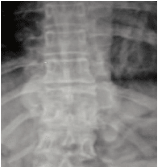
Definition. Vertebra plana is complete or near-complete collapse of a single vertebral body so that it appears as a thin, dense, wafer-like disc of bone. The residual body height is characteristically less than one-third — often less than one-quarter — of the height of the adjacent normal vertebrae. The hallmark is that the collapse is uniform: anterior and posterior body height are lost together, so segmental kyphosis is minimal and the spinal alignment is usually preserved.
Confirming it is genuinely present. Three checks on the lateral radiograph:
- Is the whole body flattened, or just the front? Anterior wedging with a preserved posterior wall is a compression fracture, not vertebra plana.
- Are the adjacent discs normal in height? In vertebra plana the discs are preserved or even appear increased in height relative to the collapsed body. Loss of disc height with endplate irregularity shifts you towards discitis-osteomyelitis.
- Are the pedicles and posterior elements intact? Look at the AP for a missing pedicle (winking owl) — that signals a destructive process extending beyond the body.
The words to say out loud. "This is a lateral radiograph of the thoracolumbar spine. There is uniform, near-complete loss of height of a single vertebral body, which appears as a dense flat wafer — the coin-on-edge or silver-dollar appearance. The adjacent disc spaces are preserved and there is no significant kyphosis. The posterior elements appear intact and I cannot see a paraspinal soft-tissue shadow on this film. This is vertebra plana. In a child of this age my leading diagnosis is Langerhans cell histiocytosis, but I must exclude Ewing sarcoma, lymphoma, leukaemia and tuberculous spondylitis."
Mimics and false positives.
- Butterfly or hemivertebra — congenital, present from birth, with a sagittal cleft and adjacent compensatory disc changes; adjacent vertebrae are often fused or malformed.
- Overlapping projection / obliquity — a poorly centred lateral can make a normal body appear thin. Always confirm on a second projection or on CT.
- Limbus vertebra and Scheuermann disease — endplate irregularity and Schmorl nodes with anterior wedging of multiple contiguous levels, never uniform pancaking of one body.
- Cupid's bow / physiological endplate concavity — an axial-plane appearance in lumbar bodies, height preserved peripherally.
- Severe osteoporotic biconcave (codfish) vertebra — the endplates cave in but the peripheral cortical ring retains height; multiple levels affected.
- Post-vertebroplasty or fused segment — always ask for the history and look for cement.
Next Investigation
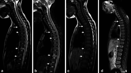
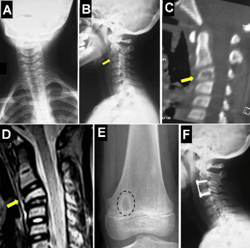
The MRI features that separate benign osteoporotic collapse from malignant collapse: benign shows a horizontal band of oedema with preserved normal fatty marrow elsewhere in the body, a fluid or vacuum cleft, a retropulsed bony fragment and normal pedicles. Malignant shows complete marrow replacement, a convex posterior cortex, pedicle involvement, a focal paravertebral mass and other lesions elsewhere.
FETISHCauses of Vertebra Plana
The Differential
- Typical age / setting
- Age 2-10, peak around 5-7; thoracic spine commonest
- Discriminating feature
- Uniform flat wafer with preserved discs, NO significant soft-tissue mass and posterior elements spared; child is well and neurologically intact
- What confirms it
- Skeletal survey or whole-body MRI for other lesions; CD1a and langerin positive on biopsy, Birbeck granules on electron microscopy
- Typical age / setting
- Adult over 50; known breast, lung, prostate, renal or thyroid primary
- Discriminating feature
- Pedicle destruction (winking owl sign) and multiple non-contiguous levels with normal intervening discs
- What confirms it
- Whole-spine MRI plus CT chest/abdomen/pelvis; bone scan or PET-CT; biopsy of the safest deposit
- Typical age / setting
- Age over 40, peak 60-70
- Discriminating feature
- Diffuse osteopenia with multiple collapses and punched-out lytic skull lesions; classically the pedicle is spared until late
- What confirms it
- Serum and urine protein electrophoresis with free light chains, whole-body low-dose CT or MRI, marrow aspirate
- Typical age / setting
- Age 5-20; sacrum and mobile spine
- Discriminating feature
- Vertebra plana WITH a large circumferential paravertebral and epidural soft-tissue mass out of proportion to the bone loss
- What confirms it
- MRI with contrast, biopsy showing small round blue cells, EWSR1 translocation on FISH
- Typical age / setting
- Any age; adults 40-70, children with systemic disease
- Discriminating feature
- Extensive marrow and epidural soft-tissue involvement with a relatively preserved bony outline — bone looks better than the patient
- What confirms it
- MRI, core biopsy with immunophenotyping, PET-CT for staging
- Typical age / setting
- Child 2-10, systemically unwell
- Discriminating feature
- Diffuse osteopenia with multiple collapses, metaphyseal lucent bands in long bones, cytopenias and organomegaly
- What confirms it
- Full blood count with film, bone marrow aspirate and trephine
- Typical age / setting
- Any age; endemic region, immunosuppression, HIV
- Discriminating feature
- Large calcified paravertebral abscess with a thin rim; early disc preservation but eventual disc destruction and subligamentous spread over multiple levels
- What confirms it
- MRI with contrast, CT-guided biopsy with culture and PCR, Mantoux or interferon-gamma release assay
- Typical age / setting
- Adults over 50, diabetes, intravenous drug use, recent bacteraemia
- Discriminating feature
- Disc space narrowing with endplate erosion involving TWO adjacent bodies — the disc is the target, unlike in tumour
- What confirms it
- MRI with contrast, blood cultures, CT-guided disc/bone biopsy before antibiotics
- Typical age / setting
- Postmenopausal women, age over 65; long-term corticosteroids
- Discriminating feature
- Multiple levels, retropulsed fragment, intravertebral vacuum cleft (Kummell) and a band of marrow oedema with preserved normal fatty marrow elsewhere
- What confirms it
- DEXA, MRI showing a fracture line rather than replaced marrow; myeloma screen to exclude the mimic
- Typical age / setting
- Any age; clear mechanism
- Discriminating feature
- Sharp fracture lines, retropulsion into the canal and posterior element fractures; disc may herniate into the body
- What confirms it
- CT for the fracture pattern, MRI for the posterior ligamentous complex
- Typical age / setting
- Child or young adult, often Ashkenazi Jewish
- Discriminating feature
- Hepatosplenomegaly with Erlenmeyer flask deformity of the distal femur and avascular necrosis of the femoral head alongside the flat vertebra
- What confirms it
- Beta-glucocerebrosidase enzyme assay and GBA1 genotyping
- Typical age / setting
- Age 10-20; posterior elements first
- Discriminating feature
- The vertebra is EXPANDED with fluid-fluid levels, not simply flattened — collapse is secondary and eccentric
- What confirms it
- MRI showing fluid-fluid levels and septations; biopsy with USP6 rearrangement
- Typical age / setting
- Age 20-40; sacrum then mobile spine body
- Discriminating feature
- Expansile lytic lesion of the vertebral BODY without matrix mineralisation, thinned but present cortical shell
- What confirms it
- MRI with low T2 signal from haemosiderin; biopsy with H3F3A mutation
- Typical age / setting
- Child or young adult on long-term steroids; blue sclerae, prior fractures
- Discriminating feature
- Multiple biconcave and flattened bodies at many levels with thin cortices throughout the skeleton
- What confirms it
- Clinical phenotype, DEXA Z-score, COL1A1/COL1A2 genetics
Narrowing It Down
- 1Step 1 — How old is the patient?
Fix the age band before looking at anything else, and state the prior out loud.
Under 10: Langerhans cell histiocytosis is the commonest single cause, with Ewing sarcoma, leukaemia and tuberculosis behind it. Age 10-30: Ewing sarcoma, aneurysmal bone cyst, giant cell tumour, tuberculosis. Over 40: metastasis, myeloma, osteoporotic collapse, pyogenic infection. BUT CALIBRATE THE PAEDIATRIC PRIOR HONESTLY - only 12 of 27 children with vertebra plana in the Baky series had eosinophilic granuloma, and six had a different neoplasm. Age narrows; it does not diagnose.
- 2Step 2 — Is it solitary or are there other lesions?
Whole-spine sagittal MRI in everyone, plus a skeletal survey or whole-body MRI in a child.
Multiple non-contiguous vertebral lesions with normal intervening discs means haematogenous disease: metastasis, myeloma, multifocal LCH or leukaemia. A single lesion with a normal remaining skeleton keeps solitary eosinophilic granuloma, plasmacytoma and primary bone tumour in play. Note how often the answer is 'not solitary' - 23 of 50 children with spinal LCH in the Moyano series had two or more lesions, and 100 vertebrae were involved across those 50 children.
- 3Step 3 — What are the discs doing?
Compare disc height and endplate integrity above and below the collapsed body, then look for a paravertebral collection.
Preserved discs point to a marrow-replacing or neoplastic process, because tumour respects the avascular disc. Loss of disc height with endplate destruction across two adjacent bodies points to pyogenic infection. Preserved discs PLUS a large paravertebral collection with a thin calcified rim and multilevel subligamentous spread is tuberculosis - which is why disc preservation alone never excludes infection.
- 4Step 4 — Are the posterior elements involved and is there a soft-tissue mass?
Examine the pedicles and laminae specifically, and look for epidural and paravertebral extension on the contrast sequences.
Classical LCH is confined to the body with minimal or no soft-tissue component. Pedicle or lamina destruction, or a circumferential epidural and paravertebral mass, argues for Ewing sarcoma, lymphoma or metastasis and escalates urgency. An expanded posterior element with fluid-fluid levels means aneurysmal bone cyst. USE THIS ONE-WAY ONLY: an adverse feature moves you off LCH, but its absence does not rule malignancy out - Baky found lesion location and collapse morphology unassociated with the final diagnosis.
- 5Step 5 — Is the vertebra flattened or expanded?
Decide whether the body is a thin dense wafer or a ballooned shell - this is a shape question, not a height question.
True vertebra plana is a thin dense wafer. An expanded, ballooned body with a thin cortical shell is a different pattern entirely: aneurysmal bone cyst, giant cell tumour or a vascular lesion. This one observation removes half the differential.
- 6Step 6 — Are there systemic features?
Examine for pallor, bruising, lymphadenopathy and organomegaly; check full blood count, film, inflammatory markers, calcium and renal function, and a myeloma screen in adults.
Fever, night sweats and weight loss suggest infection or lymphoma. Pallor, bruising and organomegaly suggest leukaemia. Anaemia, renal impairment and hypercalcaemia suggest myeloma. Hepatosplenomegaly with avascular necrosis suggests Gaucher disease. A relapsing, remitting course with multifocal lesions next to physes suggests chronic non-bacterial osteomyelitis - which collapses and, unlike LCH, does NOT reconstitute. A well child with isolated back pain and normal inflammatory markers supports LCH but does not confirm it.
- 7Step 7 — Is there neurological deficit?
Examine power, sensation, reflexes and perineal sensation in every case, and repeat it if the pain changes.
Neurology is uncommon in LCH and in osteoporotic collapse and should redirect you to a compressive mass - Ewing sarcoma, lymphoma, metastasis or an epidural abscess. Deficit converts the work-up from elective to same-day: urgent whole-spine MRI with contrast, dexamethasone where malignancy is likely, and surgical and oncological review.
MCQ Practice Points
Q: What is the commonest cause of vertebra plana in a child under 10, and how confident can you be?
A: Langerhans cell histiocytosis (solitary eosinophilic granuloma) — thoracic spine, spares the posterior elements, little soft-tissue mass, and it reconstitutes height with remaining growth. But do not say "pathognomonic". In the Baky series only 12 of 27 children with vertebra plana had eosinophilic granuloma, and six had a different neoplasm — leukaemia, germ cell tumour, giant cell tumour, rhabdomyosarcoma, teratoma. Commonest single cause, under half the total.
Q: Which radiographic feature best distinguishes neoplastic vertebral collapse from infective collapse?
A: The disc space. Tumour respects the avascular intervertebral disc, so the discs remain normal and lesions are often multiple and non-contiguous. Pyogenic infection destroys the disc and both adjacent endplates. Tuberculosis is the exception that spares the disc early — so disc preservation narrows the differential but never excludes infection, and every biopsy goes to microbiology as well as histology.
Q: On MRI, what favours a benign osteoporotic collapse over a malignant one?
A: A horizontal band of marrow oedema with preserved normal fatty marrow elsewhere in the body, a fluid or vacuum cleft, a retropulsed bony fragment and normal pedicles. Malignancy shows complete marrow replacement, a convex posterior cortex, pedicle involvement and a focal soft-tissue mass.
Q: A flat vertebra in a child is accompanied by a large circumferential epidural mass. What has changed?
A: This is atypical for LCH — Ewing sarcoma and lymphoma move to the top. Urgent contrast MRI of the whole spine, staging CT and PET, and sarcoma MDT referral with a planned biopsy tract. Note the asymmetry that makes this useful: an adverse feature moves you off LCH reliably, but a reassuring appearance does not move you onto it — collapse morphology, symmetry, location and pain were all unassociated with the final diagnosis.
Q: Which systemic disease causes vertebral collapse alongside Erlenmeyer flask femora, hepatosplenomegaly and femoral head avascular necrosis?
A: Gaucher disease. Diagnosis is by beta-glucocerebrosidase enzyme assay with GBA1 genotyping; enzyme replacement therapy is the mainstay.
Q: Which single blood test most often prevents an unnecessary spinal biopsy in an adult with vertebra plana?
A: Serum and urine protein electrophoresis with serum free light chains. A positive myeloma screen redirects you to a bone marrow aspirate and trephine rather than a spinal needle.
Q: A child has a flat vertebra that has not regained any height after three years. What should you reconsider?
A: Chronic non-bacterial osteomyelitis (CRMO), and the other non-LCH causes. LCH reconstitutes — Raab measured 18–64% of adjacent vertebral height before remodelling rising to 72–97% after maturity in every one of 14 children, with collapse worst at about one year and recovery accelerating after two. In CRMO the authors of a 170-patient cohort state they have seen no evidence of reconstitution once deformity develops; bisphosphonates prevent progression at levels not yet collapsed. Failure to remodel is a diagnostic finding, not just a poor result.
Exam Viva Scenarios
Practise clinical reasoning and management decisions out loud
“You are shown this lateral thoracic radiograph of a 6-year-old boy with three months of mid-back pain, otherwise well. A single vertebral body is reduced to a thin dense wafer with normal adjacent discs. Describe the film and give your differential.”
“You are shown this lateral lumbar radiograph of a 74-year-old woman with sudden back pain after bending. L1 is markedly flattened and there is generalised osteopenia. How do you proceed?”
“You are shown this radiograph of a 38-year-old man, recently arrived from an endemic region, with three months of thoracic back pain, night sweats and weight loss. There is a flattened vertebral body and a fusiform paraspinal shadow.”
Definition and description
- Uniform near-total collapse of one vertebral body to a thin dense wafer
- Residual height usually less than one-third of the adjacent level
- Adjacent discs preserved; minimal segmental kyphosis; posterior elements usually intact
- Descriptors: coin-on-edge, silver-dollar vertebra, pancake vertebra
Age-based first answer
- Under 10 — Langerhans cell histiocytosis
- 10 to 30 — Ewing sarcoma, aneurysmal bone cyst, giant cell tumour, tuberculosis
- Over 40 — metastasis, myeloma, osteoporotic insufficiency fracture, pyogenic infection
- Any age with systemic features — infection or lymphoma
Discriminators in one line each
- LCH — flat wafer, no mass, well child, may reconstitute height
- Metastasis — pedicle destruction, multiple non-contiguous levels, known primary
- Myeloma — diffuse osteopenia, punched-out skull lesions, positive electrophoresis
- Ewing sarcoma — huge circumferential soft-tissue mass out of proportion to bone loss
- Lymphoma — extensive marrow and epidural disease with relatively preserved bone outline
- Tuberculosis — calcified thin-walled paravertebral abscess, multilevel subligamentous spread
- Pyogenic infection — disc destruction with erosion of both adjacent endplates
- Osteoporosis — vacuum cleft, fracture line, preserved fatty marrow, normal pedicles
- Aneurysmal bone cyst / giant cell tumour — expanded, not flattened
Investigation ladder
- Whole-spine MRI with contrast — nearly always first
- Chest radiograph and plain films of the whole spine
- CT for cortical detail, matrix, stability and biopsy guidance
- Bloods: FBC and film, ESR, CRP, calcium, renal function, LDH, myeloma screen
- Child: skeletal survey or whole-body MRI for multifocal disease
- Adult: staging CT chest, abdomen and pelvis before any spinal biopsy
- CT-guided core biopsy with tissue to both histology and microbiology
Red flags demanding urgent action
- Any new neurological deficit
- Epidural soft-tissue mass or cord signal change on MRI
- Fever, night sweats and raised inflammatory markers
- Rapidly progressive collapse or instability on dynamic imaging
- Hypercalcaemia, renal impairment and anaemia suggesting myeloma
When to stop investigating
- Well child, classic isolated lesion, negative skeletal survey, no neurology — brace and observe
- Elderly patient with documented low-energy injury, known osteoporosis, benign MRI pattern and negative myeloma screen — no biopsy
- Otherwise, tissue diagnosis before any definitive treatment
Evidence Base
Vertebra Plana in Children May Result from Etiologies Other Than Eosinophilic Granuloma
- 27 children under 18 with vertebra plana at one institution 1976 to 2017, mean age 9 years, trauma excluded
- ONLY 12 OF 27 had eosinophilic granuloma - under half. It is the commonest single cause, not the default
- SIX of 27 had a DIFFERENT neoplasm: acute lymphoblastic leukaemia, primary germ cell tumour, giant cell tumour, rhabdomyosarcoma and teratoma
- 17 were biopsied - 6 confirmed eosinophilic granuloma, 6 showed another cause, and FIVE WERE NON-DIAGNOSTIC
- NO RADIOGRAPHIC FEATURE SEPARATED THEM: degree of collapse (under 50 per cent versus 50 to 100 per cent), symmetry of collapse, lesion location and pain were all unassociated with the final diagnosis
- The authors' conclusion is explicit - a child with vertebra plana and back pain needs a comprehensive work-up and potentially tissue biopsy
Vertebra Plana: A Narrative Clinical and Imaging Overview Among Possible Differential Diagnoses
- Narrative literature review analysing 602 articles reporting vertebra plana, examining demographics, presentation, imaging and final diagnosis
- States the conclusion plainly: VERTEBRA PLANA IS NOT A PATHOGNOMONIC FEATURE OF LANGERHANS CELL HISTIOCYTOSIS
- Offers the mnemonic HEIGHT OF HOMO for the differential - Histiocytosis, Ewing's sarcoma, Infection, Giant cell tumour, Haematological neoplasms, Tuberculosis, Osteogenesis imperfecta, Fracture, Haemangioma, Osteoblastoma, Metastasis, Osteomyelitis (chronic)
- Both oncological and non-oncological conditions must be considered in every case
Vertebral Remodeling in Eosinophilic Granuloma of the Spine: A Long-Term Follow-Up
- 14 children with biopsy-proven spinal eosinophilic granuloma (16 vertebral lesions), aged 1.2 to 11.3 years, all treated conservatively in a custom brace, mean follow-up 5.6 years
- THE RECONSTITUTION NUMBERS: affected vertebral height was 18.2 to 63.8 per cent of the adjacent vertebra before remodelling, and 72.2 to 97 PER CENT after skeletal maturity
- The collapsed vertebra grew at a rate of 1.5 to 6 times (average 3) against 1.1 to 2.7 (average 1.4) in unaffected vertebrae - it catches up by growing faster
- Partial to nearly complete reconstitution was seen in ALL cases
- The authors conclude bracing alone permits optimal remodelling, so curettage, bone grafting and multisegmental instrumented fusion are NOT necessary
- Two caveats they state themselves: surgical decompression and short fusion are needed if there is neurological impairment, and complete staging plus BIOPSY remain mandatory for diagnosis
Characteristic Reconstitution of the Spinal Langerhans Cell Histiocytosis in Young Children
- 13 children with biopsy-proven LCH and 16 affected vertebrae, mean age at first visit 3.6 years, and an unusually long mean follow-up of 10.2 years
- THE TIMELINE: collapse is MOST SEVERE at about one year from onset, and reconstitution accelerates from two years onward - so a vertebra that is still flattening in the first year is behaving as expected
- The ANTERIOR wall recovers faster than the central height
- During the collapse phase the ADJACENT discs thicken and the ADJACENT vertebrae also grow taller - the segment compensates, and both effects diminish once the affected vertebra has reconstituted
- Local kyphosis shifted to LORDOSIS within the first three years
Histiocytosis in the Pediatric Spine: A Clinical and Radiographic Analysis of 50 Patients
- Consecutive series of 50 children with spinal LCH treated 1984 to 2016, minimum two-year follow-up, mean age 5 years 2 months
- NEARLY HALF HAD MULTIPLE LESIONS: 27 had a single spinal lesion, 23 had two or more, and 100 vertebrae were involved in total - which is the argument for whole-body staging in every case
- Thoracic spine most affected; the lesion was in the vertebral BODY in 88 per cent
- Pain in 87 per cent, with reduced range of movement, deformity and neurological deficit also seen
- BIOPSY WAS PERFORMED IN 48 OF THE 50 - it was the routine, not the exception, at a high-volume paediatric spine unit
- 39 had medical treatment, 28 used an orthosis, SIX required surgery, and six (12 per cent) RECURRED at a mean of 3 years 5 months (range 2 to 12 years)
- Neurological symptoms, torticollis and deformity resolved in all cases after medical or surgical treatment
Spine Involvement and Vertebral Deformity in Patients Diagnosed with Chronic Recurrent Multifocal Osteomyelitis
- 170 children with chronic recurrent multifocal osteomyelitis at one institution 2003 to 2020, from a prospectively maintained database
- 48 (28.2 PER CENT) had SPINAL involvement - CRMO in the spine is not rare
- Of those 48, vertebral BODY lesions in 27 (56.3 per cent); the remainder were in the sacrum or posterior elements
- Vertebral body height loss on radiograph in 23 of those 27, ranging from minimal anterior wedging to circumferential collapse - that is, to VERTEBRA PLANA
- THE FINDING THAT SEPARATES IT FROM LCH: 'Once deformity has developed, we have seen NO evidence of reconstitution of the height of the collapsed vertebra'
- Bisphosphonates were successful in PREVENTING progression of vertebral height loss - so the treatment window is before collapse, not after
Langerhans Cell Histiocytosis of the Spine in Adults: A Rare and Controversial Disorder - A Systematic Review
- Systematic review of every reported case of adult-onset spinal LCH - 47 patients, mean age 34.7 years (range 20 to 69), 64 per cent male
- The LUMBAR spine was most often involved, the vertebral body in 53 per cent, and 42 of 47 (89 per cent) had a single vertebra affected - the other 5 (10.6 per cent) had multiple levels, so multifocal adult disease exists and warrants staging even though it is uncommon
- ONLY THREE OF 47 had vertebra plana morphology - the flat vertebra of childhood LCH is NOT how the adult disease usually looks
- Lytic destruction is the characteristic radiological feature; local pain, radiating pain and restricted movement the usual symptoms
- 29 of 47 had surgery; outcome was good to excellent in all but seven, who recurred or progressed
- THE PRACTICE POINT: non-operative treatment, which is standard in children, was associated with progression or recurrence in adults