The Only Muscle Joining Spine to Limb - and the Plexus Runs Through It
- Origin has two layers: a superficial layer from the vertebral bodies and discs of T12 to L4, and a deep layer from the transverse processes of L1 to L5. The lumbar plexus lies BETWEEN them.
- Psoas major is supplied by direct branches from the ventral rami of L1, L2 and L3 (sometimes L4); iliacus is supplied by the femoral nerve - the two halves of iliopsoas have different nerves.
- The femoral nerve emerges at the LATERAL border of psoas low down, in the groove between psoas and iliacus; the obturator nerve emerges at the MEDIAL border at the pelvic brim; the genitofemoral nerve pierces the ANTERIOR surface.
- In the lateral transpsoas approach the plexus lies dorsally in the psoas at upper levels and migrates ventrally at L4-L5, so the safe working zone moves anteriorly as you descend.
- Iliopsoas impingement after total hip arthroplasty is diagnosed by the response to an image-guided infiltration test, not by the presence of cup overhang - in the original prospective series an overhanging cup was present in only 6 of 9 cases and 7 of 9 were cured without revising the acetabulum.
- “Psoas major is the only muscle that connects the axial skeleton directly to the lower limb - which is why a lumbar spine problem can present as groin pain and a hip problem can present as back pain.
- “The psoas sign - pain on passive hip extension or resisted hip flexion - is not specific to appendicitis; it is positive with any retroperitoneal irritation of the muscle, including a psoas abscess and a leaking aneurysm.
- “Releasing the iliopsoas tendon at the level of the hip joint divides less tendon than releasing at the lesser trochanter, because only about 40 per cent of the unit is tendinous at the joint - which is why transcapsular release preserves more hip flexion power.
- “Thigh pain and hip flexor weakness after a transpsoas interbody fusion are usually from the psoas dissection itself and settle within weeks; a true femoral nerve injury with quadriceps weakness and knee extension loss is far less common and far more serious.
Overview
Psoas major is a long, fusiform muscle arising from the lumbar spine, descending along the pelvic brim, passing beneath the inguinal ligament and inserting with iliacus onto the lesser trochanter of the femur. It is the only muscle that connects the axial skeleton directly to the lower limb, and that single fact explains much of its clinical behaviour: lumbar pathology refers to the groin, hip pathology refers to the back, and an abscess in one compartment tracks freely into the other.
Its most surgically consequential feature is that it is not a solid muscle. It arises in two layers - a superficial layer from the vertebral bodies and discs, and a deep layer from the transverse processes - and the lumbar plexus is formed and runs between them. A surgeon crossing psoas to reach the lumbar disc is therefore not passing beside a plexus but through one.
At its distal end the muscle is equally consequential. The tendon crosses the iliopectineal eminence and the anterior hip capsule on its way to the lesser trochanter, which makes it the structure that snaps, the structure that is impinged by a prominent acetabular component, and the structure retracted (with the femoral nerve behind it) in every anterior approach to the hip.
Iliopsoas - psoas major plus iliacus sharing a common tendon - is the strongest and most important hip flexor, generating a greater flexion moment than rectus femoris, sartorius, tensor fasciae latae and pectineus combined in most positions.
Two mechanical features make it irreplaceable:
- Its moment arm is preserved through the whole arc. Rectus femoris has a good flexion moment arm at low flexion angles but loses effectiveness beyond about 90 degrees, and its two-joint action means it is compromised whenever the knee is extending. Iliopsoas retains a flexion moment arm throughout flexion, and it is the dominant flexor above 90 degrees. That is why a patient with an iliopsoas deficit can walk on the flat but cannot climb stairs, get out of a car, or lift the leg into bed - all tasks requiring flexion beyond mid-range against gravity.
- It acts across the spine as well as the hip. The psoas component has attachments at every lumbar level, so it functions as a segmental stabiliser of the lumbar spine as well as a hip flexor. With the femur fixed it flexes the trunk on the pelvis and, importantly, produces anterior shear and compression at the lumbar segments - a fact used in explaining the increased lumbar lordosis seen with psoas contracture and the hip-spine interactions of fixed flexion deformity.
Clinically this means an iliopsoas release is never mechanically neutral. Published series report that hip flexion power falls measurably after tenotomy, usually recovering to functionally acceptable levels by 3 to 6 months, but the loss is real and is greater when the release is at the lesser trochanter, where the unit is predominantly tendon, than at the level of the joint, where a substantial proportion of the unit is still muscle.
LAMPWhere the Lumbar Plexus Branches Leave Psoas
Hook:Femoral lateral, obturator medial, genitofemoral straight through the front.
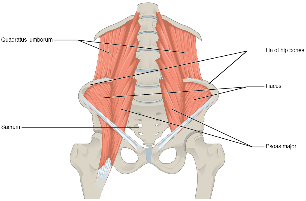
Attachments, Innervation and Relations
Origin - two layers, and the plexus between them
- Superficial (anterior) layer: the anterolateral surfaces of the vertebral bodies and the intervening intervertebral discs of T12 to L4, together with the tendinous arches that bridge the constricted parts of the bodies, beneath which the lumbar arteries and veins and the sympathetic rami communicantes pass.
- Deep (posterior) layer: the anterior surfaces and lower borders of the transverse processes of L1 to L5.
- The lumbar plexus is formed in the plane between these two layers. The nerve roots emerge from the intervertebral foramina, pass behind the superficial layer and in front of the deep layer, and unite within the muscle substance.
Course
Descends along the pelvic brim, crossing the sacroiliac joint and the superior pubic ramus at the iliopectineal eminence, where it lies in a shallow groove between the eminence and the anterior inferior iliac spine. It then passes beneath the inguinal ligament in the lacuna musculorum, lateral to the iliopectineal arch, and lies directly on the anterior hip capsule before turning to its insertion.
Insertion
- A strong tendon onto the lesser trochanter of the femur, shared with iliacus as the iliopsoas tendon.
- Iliacus fibres attach onto the lateral side of the psoas tendon and continue directly onto the femur just distal to the lesser trochanter, so the composite unit is not a single tendon but a tendon with a muscular sleeve on its lateral aspect.
- At the level of the hip joint and labrum the unit is approximately 44.5 per cent tendon and 55.5 per cent muscle belly; at the lesser trochanter it is predominantly tendon. This gradient is the whole reason release level matters.
Psoas minor
A separate, slender muscle present in roughly 40 to 60 per cent of people. Arises from the bodies of T12 and L1 and the disc between them and inserts by a long thin tendon into the pectineal line and the iliopubic eminence, blending with the iliac fascia. It has no action on the hip - it is a weak flexor of the lumbar spine - and it is a frequent source of confusion at operation because it lies on the anterior surface of psoas major.
Because the lumbar plexus is formed between the superficial and deep layers of psoas, any operation that crosses the muscle is passing through a nerve plexus. Three practical rules follow:
- Directional, triggered electromyographic neuromonitoring is mandatory for the lateral transpsoas approach. Blunt, sequential dilatation with continuous stimulation, not sharp dissection, is the technique.
- The plexus is not evenly distributed. It lies dorsally within psoas at the upper lumbar levels and migrates ventrally at L4-L5, so a docking position that is safe at L2-L3 is dangerous at L4-L5.
- Time under retraction is a risk factor in its own right. Prolonged psoas retraction produces both direct nerve compression and muscle ischaemia; limit retraction time and release the retractor intermittently in longer cases.
Action and Biomechanics
Actions
- Action
- Hip flexion - the primary flexor
- Mechanism
- Long lever from the lumbar spine to the lesser trochanter; moment arm preserved throughout the arc
- Loss Produces
- Difficulty with stairs, getting out of a car, lifting the leg into bed - tasks needing flexion beyond mid-range
- Action
- Dominant flexor as rectus femoris loses effectiveness
- Mechanism
- Retained moment arm at high flexion angles
- Loss Produces
- The deficit is greatest in deep flexion, which is why the sit-to-stand and stair tests expose it
- Action
- Flexion of the trunk on the pelvis; anterior pelvic tilt
- Mechanism
- Reverse origin-insertion action
- Loss Produces
- Weak sit-up; altered pelvic tilt
- Action
- Segmental stabilisation of the lumbar spine
- Mechanism
- Attachments at every lumbar level, close to the axis of rotation
- Loss Produces
- Loss of segmental control; contributes to the hip-spine syndrome
- Action
- Weak external rotation and adduction of the hip
- Mechanism
- Depends on hip position and the tendon's wrap around the femoral neck
- Loss Produces
- Minor in isolation
- Action
- Fixed flexion deformity with compensatory lumbar hyperlordosis
- Mechanism
- Shortened muscle-tendon unit tethering the pelvis
- Loss Produces
- Thomas test positive; anterior pelvic tilt; back pain
Length-tension and the consequences of contracture
- A psoas contracture tethers the pelvis in anterior tilt and forces a compensatory lumbar hyperlordosis to keep the trunk upright. Over time this alters lumbar segmental loading and is one of the mechanical links in the hip-spine syndrome.
- Conversely, a fused or stiff lumbar spine removes the compensation available at the pelvis, which is why patients with a fused lumbar spine and a total hip replacement have a substantially higher dislocation risk - the pelvis cannot tilt to accommodate the arc of movement.
- Psoas contracture is also the driver of the fixed flexion deformity in cerebral palsy and in longstanding hip arthritis; a Thomas test quantifies it.
Synergists and antagonists
- Synergists for hip flexion: iliacus (the other half of the unit), rectus femoris, sartorius, tensor fasciae latae, pectineus and the adductor longus in the flexed position.
- Antagonists: gluteus maximus and the hamstrings.
- Trunk synergists: rectus abdominis flexes the trunk but flattens the lumbar lordosis, whereas psoas flexes the trunk and increases it - an important distinction in rehabilitation prescription.
What happens when it is released
Iliopsoas tenotomy reduces hip flexion power measurably. Published experience is consistent:
- Strength deficit is greatest in the first three months and usually recovers to functionally acceptable levels by 3 to 6 months.
- The deficit is greater after release at the lesser trochanter than after release at the level of the hip joint, because more of the unit is tendinous distally.
- Patients whose occupation or sport demands repeated deep hip flexion against resistance (dancers, footballers, climbers) should be counselled that the deficit may be permanent and functionally noticeable.
Surface Anatomy and Examination
Palpation
- The muscle belly is not directly palpable in most people. In a thin patient it can be indented deeply lateral to the rectus abdominis and medial to the anterior superior iliac spine, with the hip flexed to relax the abdominal wall - an unpleasant examination that should be reserved for a specific question.
- The distal tendon is palpable in the femoral triangle, immediately lateral to the femoral artery, as a firm cord deep in the groin with the hip flexed and externally rotated.
- The snapping of an internal snapping hip is felt over the iliopectineal eminence and the anterior hip, not over the greater trochanter.
Examination
Quantifies fixed flexion deformity.
- Supine; the patient pulls the contralateral hip into maximal flexion to flatten the lumbar lordosis. The examiner observes the tested thigh.
- Positive: the tested thigh lifts off the couch; measure the angle.
- Means: hip flexion contracture, most often iliopsoas. The modified Thomas test with the leg hanging off the couch separates iliopsoas (thigh does not reach horizontal) from rectus femoris (knee will not flex past 90 degrees) and tensor fasciae latae (thigh abducts).
Detects retroperitoneal irritation.
- Patient lies on the contralateral side; the examiner passively extends the hip. Alternatively, supine resisted straight leg raise.
- Positive: deep pain in the flank, back or iliac fossa.
- Means: irritation of the muscle - psoas abscess, retrocaecal appendicitis, retroperitoneal haematoma, a leaking aortic aneurysm, or a spinal infection tracking into the sheath. Not specific to any one of them.
The iliopsoas pain and power test.
- Supine, the patient raises the straight leg to about 15 to 20 degrees and holds against downward pressure at the ankle; then repeats with the hip flexed past 90 degrees against resistance.
- Positive: anterior groin pain reproducing the complaint, or measurable weakness compared with the other side.
- Means: iliopsoas tendinopathy, impingement after arthroplasty, or hip flexor weakness. Pain in the seated resisted flexion position beyond 90 degrees is a good screen for iliopsoas impingement after hip replacement.
Localises the snap.
- Move the hip from a flexed, abducted, externally rotated position into extension, adduction and internal rotation (the FABER-to-extension arc), with a hand over the anterior hip and the iliopectineal eminence.
- Positive: an audible or palpable clunk anteriorly, reproducing the patient's snap, often reproducible by the patient at will.
- Means: internal snapping hip - the iliopsoas tendon catching over the iliopectineal eminence or the anterior capsule and femoral head. Confirm with dynamic ultrasound.
Pitfalls
- Painless snapping is common and needs no treatment. Only a painful, functionally limiting snap warrants intervention.
- External snapping hip (iliotibial band over the greater trochanter) is lateral, visible and often more dramatic; internal snapping is anterior and deep. Do not confuse them.
- The differential for anterior groin pain is long: femoroacetabular impingement, labral tear, hip osteoarthritis, adductor or rectus abdominis-related groin pain, inguinal or femoral hernia, stress fracture of the femoral neck or pubic ramus, and referred lumbar pain. Image the hip before diagnosing an iliopsoas problem.
- After hip arthroplasty, groin pain is iliopsoas impingement until proven otherwise, but must be distinguished from infection, loosening, and pseudotumour - so inflammatory markers, radiographs and cross-sectional imaging come first.
Complications
Femoral nerve injury
- Where: transpsoas surgery at L4-L5; the anterior acetabular retractor in anterior hip approaches; the middle window of the ilioinguinal approach; a compressive iliacus or retroperitoneal haematoma.
- Consequence: loss of knee extension, absent knee jerk, saphenous sensory loss, and an unstable knee in stance. This is a life-changing injury in a young patient.
- Avoidance: correct docking zone with neuromonitoring, retractors on bone, limited retraction time, and prompt investigation of any post-operative quadriceps weakness.
Genitofemoral and cutaneous nerve injury
- The genitofemoral nerve on the anterior surface of psoas is injured by an anteriorly placed retractor blade, producing groin, scrotal or labial numbness and loss of the cremasteric reflex.
- The lateral femoral cutaneous nerve produces meralgia paraesthetica, common and usually transient after the direct anterior hip approach and after iliac crest graft harvest.
Thigh symptoms after transpsoas surgery
- Anterior thigh pain, numbness and hip flexor weakness are common and usually reflect psoas trauma rather than a plexus lesion. They resolve over weeks to a few months in most patients. Warn about them; do not dismiss a progressive or quadriceps-predominant deficit as the same thing.
Vascular injury
- Segmental lumbar veins and the ascending lumbar vein during lateral exposure; iliac vessels anteriorly at L4-L5; the corona mortis at the pelvic brim in acetabular surgery. Venous injury is more common than arterial and is harder to control.
Visceral injury
- Ureteric injury during a retroperitoneal approach, and bowel injury if the peritoneum is breached. Sweep the peritoneum forward digitally and identify the ureter before dilating.
Complications of iliopsoas release
- Hip flexor weakness - expected, greatest in the first 3 months, greater after a distal release, and occasionally permanent in athletes.
- Recurrence of snapping or pain - often from an unrecognised second tendon slip or from a persisting bony prominence such as an unrevised prominent cup.
- Heterotopic ossification and hip instability after extensive anterior capsular release.
- Persistent groin pain after tenotomy for impingement where the true cause was infection, loosening or an adverse reaction to metal debris - which is why those must be excluded first.
Complications of psoas abscess management
- Delayed diagnosis is the dominant problem, and the reason for a low threshold for cross-sectional imaging in unexplained back or hip pain with fever.
- Recurrence where the source was not treated; fistula formation from bowel disease; sepsis in the immunocompromised.
Clinical Relevance
Primary versus secondary
- Primary (haematogenous): more common in children, in the immunosuppressed, in diabetics and in intravenous drug users, and commoner in tropical and low-resource settings. Staphylococcus aureus is the dominant organism.
- Secondary (contiguous spread): from Crohn's disease, diverticulitis, appendicitis, urinary tract or renal infection, vertebral osteomyelitis and discitis, septic arthritis of the hip or sacroiliac joint, or infected spinal instrumentation. Organisms reflect the source and are frequently polymicrobial or enteric.
- Tuberculous psoas abscess is the classic form. In Pott's disease of the thoracolumbar spine, a cold abscess tracks down within the psoas sheath and may point below the inguinal ligament in the femoral triangle, presenting as a fluctuant groin swelling with no local signs of inflammation. The sheath is a closed conduit from the spine to the thigh - the anatomical explanation for a presentation that otherwise makes no sense.
Presentation
- The classic triad of fever, back or flank pain and a limp is present in only a minority of patients; the presentation is characteristically insidious and occult, and diagnostic delay drives the mortality and morbidity.
- Hip held in flexion with pain on passive extension (the psoas sign); pain referred to the groin, hip or knee.
- Insidious onset, weight loss and night sweats in tuberculous disease.
- A high index of suspicion is required - the diagnosis is frequently delayed, and delay is the main driver of morbidity.
Investigation
- Contrast-enhanced CT of the abdomen and pelvis is the investigation of choice: it demonstrates the collection, its extent, and usually its source.
- MRI is superior for spinal involvement and for distinguishing an abscess from a tumour.
- Blood cultures, inflammatory markers, and image-guided aspiration for microbiology including mycobacterial culture before starting antibiotics wherever the patient is stable.
- Look for the source in every case - a psoas abscess with no obvious cause needs the bowel, spine, urinary tract and hip examined.
Management
- Image-guided percutaneous drainage plus targeted antimicrobial therapy is first-line for most collections.
- Open drainage for multiloculated collections, failure of percutaneous drainage, or where the source requires surgical treatment.
- Treat the source: bowel disease, spinal infection, or an infected implant.
- Antituberculous chemotherapy where tuberculosis is confirmed or strongly suspected; drainage is often still required for a large collection but the mainstay is medical.
Surgical Relevance
The lateral transpsoas approach (LLIF / XLIF / DLIF)
The problem
The lateral retroperitoneal transpsoas approach reaches the lumbar disc by splitting psoas major - and therefore by passing directly through the territory of the lumbar plexus, which lies between the muscle's two layers.
The anatomical rule
The plexus is not centred in the muscle. At the upper lumbar levels it lies dorsally within psoas, close to the posterior third of the vertebral body. As you descend, it migrates ventrally, so that at L4-L5 it lies much closer to the middle of the disc. Simultaneously the iliac vessels and the genitofemoral nerve limit the corridor anteriorly. The safe corridor therefore narrows and moves anteriorly as you go caudally.
The working zones
Cadaveric and imaging work divides the lateral surface of the disc into quarters from anterior to posterior. The consistent findings are:
- L1-L2 to L3-L4: the safest docking position is the middle to posterior part of the disc (the mid-posterior quarter), because the plexus is still dorsal and the great vessels are the more anterior constraint.
- L4-L5: the safe position moves forward to the midpoint of the vertebral body - the demarcation between the mid-anterior and mid-posterior quarters (Zone II to Zone III) - because the plexus has migrated ventrally and the femoral nerve is now close to the middle of the disc space.
- L5-S1 is not accessible by this approach - the iliac crest blocks the lateral trajectory and the iliac vessels occupy the corridor.
- The genitofemoral nerve lies on the anterior surface of psoas and is at risk from a retractor blade placed too far anteriorly at any level.
- Where the Plexus Lies
- Dorsal within psoas
- Safe Docking Position
- Middle to posterior quarter of the disc
- Principal Hazard
- Genitofemoral nerve anteriorly; the diaphragm and pleura above L1
- Where the Plexus Lies
- Dorsal within psoas
- Safe Docking Position
- Middle to posterior quarter
- Principal Hazard
- Genitofemoral nerve anteriorly
- Where the Plexus Lies
- Beginning to move ventrally
- Safe Docking Position
- Middle of the disc, slightly posterior
- Principal Hazard
- Femoral nerve becoming closer to the corridor
- Where the Plexus Lies
- Ventrally migrated; femoral nerve close to the mid-disc
- Safe Docking Position
- The MIDPOINT of the vertebral body - the Zone II to Zone III demarcation, not within Zone II
- Principal Hazard
- Femoral nerve posteriorly and the iliac vessels anteriorly - the narrowest corridor, and the level with the most thigh symptoms
- Where the Plexus Lies
- Not applicable
- Safe Docking Position
- Not accessible laterally
- Principal Hazard
- Iliac crest blocks access; iliac vessels occupy the corridor
Guidelines, Registries & Global Practice
Anatomical variation
- Psoas minor is absent in roughly half of individuals, with reported prevalence varying widely between populations and dissection series.
- Accessory psoas slips, an iliopsoas accessory tendon and a bifid distal tendon are all described; a second slip is a recognised cause of persistent snapping after an apparently complete release.
- The position of the lumbar plexus within psoas varies between individuals as well as between levels, which is precisely why intra-operative neuromonitoring rather than a fixed anatomical rule governs docking.
- Psoas morphology changes with degenerative spinal disease - a "rising psoas sign", where the muscle sits more anteriorly at L4-L5, is described in degenerative scoliosis and narrows the working corridor further.
- Bursal communication with the hip joint in a minority of people explains why an arthritic effusion can present as a groin mass.
Practice framing across bodies
- Position Relevant to Psoas Major
- Directional triggered electromyographic neuromonitoring is regarded as standard for the lateral transpsoas approach; docking position must be adjusted by level.
- Position Relevant to Psoas Major
- Persistent groin pain after hip arthroplasty requires exclusion of infection and loosening before attribution to iliopsoas impingement; cross-sectional imaging of cup position is recommended.
- Position Relevant to Psoas Major
- Iliopsoas release is reserved for a painful, functionally limiting snap after failed non-operative treatment, with release at the level of the joint preferred.
- Position Relevant to Psoas Major
- Percutaneous drainage with targeted antimicrobial therapy is first-line for psoas abscess; the source must be identified and treated.
- Position Relevant to Psoas Major
- In the ilioinguinal approach the iliopsoas and femoral nerve occupy the middle window; the iliopectineal fascia must be divided under direct vision.
Resource-dependent practice
- Well-resourced settings: intra-operative neuromonitoring for transpsoas surgery, metal-artefact-reduction CT for cup overhang, dynamic ultrasound for snapping hip, and image-guided percutaneous abscess drainage.
- Limited-resource settings: the lateral transpsoas approach should not be undertaken without neuromonitoring - this is one of the few situations where the absence of a technology is a genuine contraindication to the technique rather than an inconvenience. Psoas abscess, by contrast, is diagnosable with clinical examination and ultrasound and drainable percutaneously under ultrasound guidance, and tuberculous disease is treated medically.
Global epidemiology
- Tuberculous psoas abscess remains common in South Asia, sub-Saharan Africa and other high-burden regions, and is the presentation to think of first there; in high-income settings, secondary abscess from Crohn's disease and from spinal instrumentation infection predominates, with primary staphylococcal abscess in intravenous drug users and the immunosuppressed.
- Lateral interbody fusion has been adopted at very different rates internationally, and the reported incidence of thigh symptoms varies with how systematically they are sought - series that examine for them prospectively report far higher rates than those relying on spontaneous reporting.
- Iliopsoas impingement after arthroplasty is likely under-reported everywhere, because it is a diagnosis that is only made if it is specifically considered.
MCQ Practice Points
Q: Describe the two layers of the psoas origin and what lies between them. A: A superficial layer from the vertebral bodies and discs of T12 to L4, and a deep layer from the transverse processes of L1 to L5. The lumbar plexus lies between them.
Q: What is the nerve supply of psoas major, and how does it differ from iliacus? A: Psoas major - direct branches from the ventral rami of L1, L2 and L3 (sometimes L4). Iliacus - the femoral nerve (L2, L3). The two halves of iliopsoas have different nerves.
Q: At which border of psoas do the femoral and obturator nerves emerge? A: Femoral at the LATERAL border, obturator at the MEDIAL border. The genitofemoral nerve pierces the anterior surface.
Q: Where do you dock at L4-L5 in a transpsoas approach, and why is it different from L2-L3? A: The midpoint of the vertebral body, the Zone II to Zone III demarcation, because the lumbar plexus migrates ventrally at lower levels. At L2-L3 the safe zone is the midpoint of Zone III, the middle posterior quarter.
Q: What proportion of the iliopsoas unit is tendon at the level of the hip joint? A: Approximately 40 per cent, the remaining 60 per cent being muscle - which is why a transcapsular release preserves more flexion power than a release at the lesser trochanter.
Q: What degree of anterior acetabular overhang mandates revision rather than tenotomy for iliopsoas impingement? A: There is no verified numerical threshold. The decision rests on the response to an image-guided infiltration test and on identifying a specific implant abnormality; most patients in the original series were cured without revising the cup.
Q: What is notable about the iliopsoas bursa? A: It is the largest bursa in the body and communicates with the hip joint in a minority of people - a figure classically quoted as around 15 per cent.
Q: Where does psoas minor insert, and what does it do at the hip? A: The pectineal line and iliopubic eminence. It has NO action at the hip - it is a weak flexor of the lumbar spine, and it is absent in roughly half of people.
Q: An adult presents with an isolated avulsion of the lesser trochanter after trivial trauma. What must you exclude? A: A pathological fracture - most often a metastasis. In an adolescent the same injury is a normal apophyseal avulsion.
Q: Why can lumbar spine pathology present as groin pain and hip pathology as back pain? A: Because psoas major is the only muscle connecting the axial skeleton directly to the lower limb, and the L1 to L3 supply overlaps the sensory territory of the groin.
Exam Viva Scenarios
Practise clinical reasoning and management decisions out loud
“You are planning a lateral transpsoas interbody fusion at L3-L4 and L4-L5. Describe the anatomy that determines where you dock and what you will tell the patient about the risks.”
“A 58-year-old woman has groin pain nine months after an uncemented total hip replacement. The hip felt good for two weeks and then began to hurt whenever she got out of a car or climbed stairs. Passive movement is painless. How do you assess her?”
“A 19-year-old man presents with two weeks of increasing right hip and back pain, a limp, low-grade fever and weight loss. He holds the hip flexed and resists passive extension. What is your differential and how do you proceed?”
Anatomy
- Superficial layer: bodies and discs T12-L4; deep layer: transverse processes L1-L5
- Lumbar plexus lies BETWEEN the two layers
- Insertion: lesser trochanter with iliacus as the iliopsoas tendon
- Nerve: direct ventral rami L1-L3 (iliacus is femoral nerve)
Plexus Exits
- Femoral nerve - LATERAL border, low, between psoas and iliacus
- Obturator nerve and lumbosacral trunk - MEDIAL border at the brim
- Genitofemoral nerve - pierces the ANTERIOR surface
- Lateral femoral cutaneous, iliohypogastric, ilioinguinal - lateral border above
Transpsoas Surgery
- Plexus dorsal at upper levels, migrates ventrally caudally
- Safe zone: midpoint of Zone III (middle posterior quarter) L1-L2 to L3-L4; MIDPOINT of the vertebral body at L4-L5
- L5-S1 not accessible laterally - iliac crest and vessels
- Neuromonitoring mandatory; limit retraction time; thigh symptoms common and usually transient
Hip
- Primary hip flexor, dominant above 90 degrees
- Snapping over the iliopectineal eminence - release at joint level (40 per cent tendon)
- Impingement after THA: infiltration test diagnoses it; most cured without revising the cup
- Psoas abscess: sheath conducts a Pott's cold abscess to the femoral triangle
Evidence Base
Defining the Safe Working Zones Using the Minimally Invasive Lateral Retroperitoneal Transpsoas Approach
- Twenty lumbar segments dissected, with the area between the anterior and posterior edges of the vertebral body divided into four equal zones
- All parts of the lumbar plexus, including the nerve roots, lay within psoas DORSAL to the posterior fourth of the vertebral body (Zone IV)
- The safe anatomical zone from L1-2 to L3-4 is the middle posterior quarter (midpoint of Zone III)
- At L4-5 the safe zone is the MIDPOINT OF THE VERTEBRAL BODY (the Zone II to Zone III demarcation)
- The genitofemoral nerve emerges from the medial border of psoas at L3-4 and runs along the anterior medial fourth of L4 and L5 (Zone I), so it is at risk in Zone II at L2-3 and Zone I at L3-4 and L4-5
An Anatomic Study of the Lumbar Plexus with Respect to Retroperitoneal Endoscopic Surgery
- Thirty cadavers analysed, with axial sections cut parallel to the lumbar disc spaces and the plexus distribution mapped on computer images
- At L2-3 and above, all parts of the lumbar plexus and the nerve roots lay from the dorsal fourth of the vertebral body and dorsally
- The genitofemoral nerve descends obliquely forward through psoas major, emerging on its abdominal surface between the cranial third of L3 and the caudal third of L4
- The safety zone of the psoas major to prevent nerve injury, excluding the genitofemoral nerve, is at L4-L5 and above
Anterior Iliopsoas Impingement after Total Hip Arthroplasty - Diagnosis and Conservative Treatment
- Prospective study of 206 painful total hip arthroplasties; after excluding loosening (67 per cent) and infection (21.7 per cent), 9 cases (4.3 per cent) were attributable to iliopsoas impingement
- Mean delay to pain was 7.3 months, and the most suggestive sign was groin pain on active and resisted hip flexion with a painful arc of 30 to 70 degrees
- Diagnosis was confirmed by CT-guided extra-articular infiltration at the anterior border of the cup; an overhanging cup was present in only 6 of the 9
- Infiltration alone gave complete relief in 4 and partial relief in 1; tenotomy in 4 gave complete relief in 3 and partial in 1, with successful relief in 7 of 9 overall and NO loss of flexion force at follow-up
- The authors concluded an anterior cup overhang is NOT necessary for the diagnosis, and that cure can be achieved without changing the cup
Cross-Sectional Analysis of the Iliopsoas Tendon and Its Relationship to the Acetabular Labrum
- Eight hip joints dissected with cross-sectional measurement of the iliopsoas muscle-tendon complex
- In every specimen the tendon lay directly anterior to the anterosuperior capsulolabral complex at the 2 to 3 o'clock position
- Mean tendon length from the lesser trochanter to the acetabular labrum was 75.4 mm
- At the level of the labrum the iliopsoas is 44.5 per cent tendon and 55.5 per cent muscle belly
- The authors concluded that about 45 per cent of the tendon-muscle complex needs release at that level to divide the entire tendinous portion
An Analysis of Postoperative Thigh Symptoms after Minimally Invasive Transpsoas Lumbar Interbody Fusion
- Review of 59 patients undergoing transpsoas interbody fusion over 3.2 years, using patient-drawn pain diagrams at matched intervals
- 62.7 per cent had postoperative thigh symptoms; at first follow-up pain affected 39.0 per cent, numbness 42.4 per cent, paraesthesias 11.9 per cent and weakness 23.7 per cent
- By 3 months these had fallen to 15.5, 24.1, 5.6 and 11.3 per cent respectively
- Thigh pain, numbness and weakness were most prevalent after L4-5 transpsoas fusion, though not significantly so
- About half of patients had resolution of symptoms by approximately 3 months and more than 90 per cent by 1 year
Iliopsoas Abscesses
- Review article on the epidemiology, aetiology, clinical features and management of iliopsoas abscess
- Iliopsoas abscess is described as a relatively uncommon condition presenting with vague clinical features
- Its insidious onset and occult characteristics cause diagnostic delay
- Diagnostic delay results in high mortality and morbidity
- NOTE - the indexed abstract is brief, so only these summary statements are claimed here