A growth plate that is too thick, too irregular or too lucent — one radiographic sign, a dozen causes
- The physis is a radiolucent line; 'widening' means the lucent zone is taller than expected for age and site — always compare with the contralateral side and with the adjacent physes.
- Symmetrical, multi-site, metaphyseal-cupping widening equals a metabolic cause until proven otherwise; focal, single-site widening equals trauma, infection or tumour.
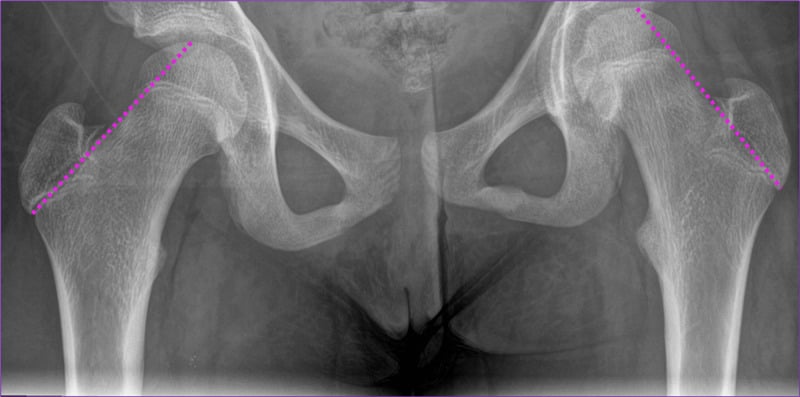
- A Salter–Harris I injury and an early slipped capital femoral epiphysis both present as physeal widening with a normal-looking epiphysis — the frog-lateral hip and the physeal irregularity are the tell.
- Metaphyseal osteomyelitis produces a blurred, irregular metaphyseal margin with adjacent soft-tissue swelling; the child is systemically unwell and unable to weight-bear.
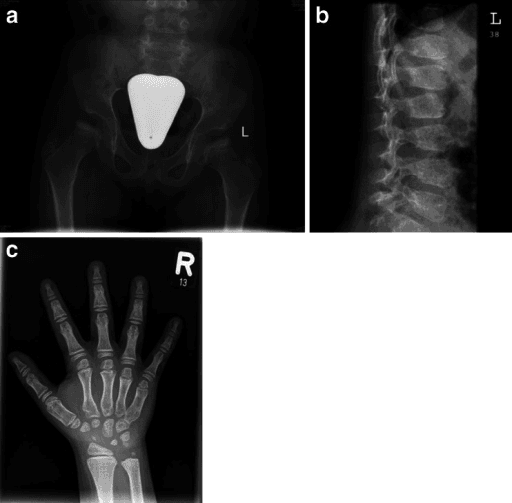
- Widening of the physis in a well child with short stature and no pain points to a skeletal dysplasia — check the spine and the pelvis before you commit.
- The one metabolic mimic in which the classic test INVERTS is hypophosphatasia: the radiograph resembles rickets but the alkaline phosphatase is LOW, not raised. A low ALP is not an assay error, bisphosphonates are contraindicated, and early loss of primary teeth is one of the three highest-agreement diagnostic features — so ask about it.
- In a young overhead athlete, physeal widening is NOT a diagnosis on imaging alone. Bilateral shoulder MRI in 23 asymptomatic Little League players aged 10 to 12 showed throwing-side abnormalities in 52 per cent, including proximal humeral physeal oedema or widening in 5 — and prior shoulder pain did NOT predict who had an abnormal scan. Correlate clinically; the exposure that predicted abnormality was year-round single-sport play.
- Conversely, do not dismiss a widened distal radial physis in a gymnast as training adaptation. Caine's authors are explicit that it is a stress fracture of the growth plate, and the consequence to warn about is growth arrest with positive ulnar variance — permanent, not a lost season. It was present in only 5 of 60 competitive gymnasts, so it is neither universal nor benign.
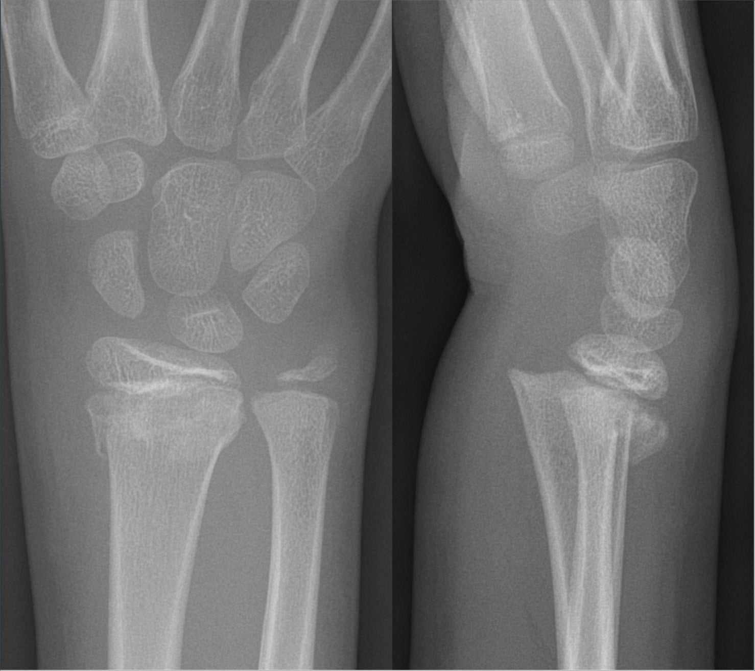
- “Say the site precisely: 'widening of the distal ulnar and distal radial physes with metaphyseal cupping and fraying' is worth more than 'abnormal wrist'.
- “The distal femur and distal radius grow fastest, so metabolic disease shows there first and most severely.
- “Rickets fray, leukaemia band, scurvy line — three metaphyseal patterns you must be able to name apart.
- “In an adolescent athlete with wrist pain, widened distal radial physis is gymnast's wrist — a chronic Salter–Harris I stress injury, not rickets.
- “Never call physeal widening in a neonate without checking that you are not looking at a normal, wide, unossified epiphysis.
Physeal lucency is genuinely thicker in infants and in early puberty (the pubertal growth spurt physis is at its most active and its most vulnerable). Age-match before you call widening. The neonatal proximal femur and proximal humerus are largely unossified — that is cartilage, not disease.
An off-axis beam projects the physis tangentially and it looks broad and blurred. Confirm on a true orthogonal view. A genuinely widened physis is widened on both projections.
Rickets, hypophosphataemia and renal osteodystrophy are systemic and therefore bilateral. A single abnormal physis is trauma, infection, tumour or focal dysplasia. Ask for the other side.
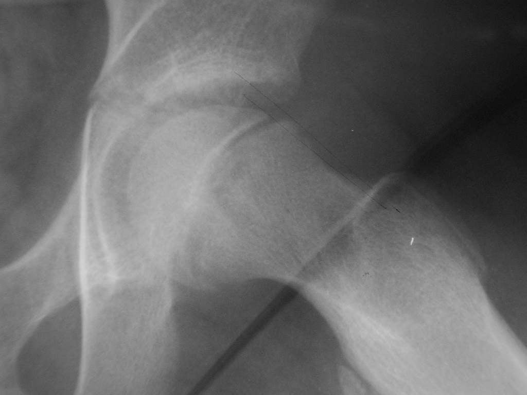
Early SCFE shows only physeal widening and irregularity before any slip is visible on the AP. Frog-lateral or cross-table lateral is mandatory in any adolescent with hip, thigh or knee pain and a wide-looking capital physis.
Recognising the Pattern
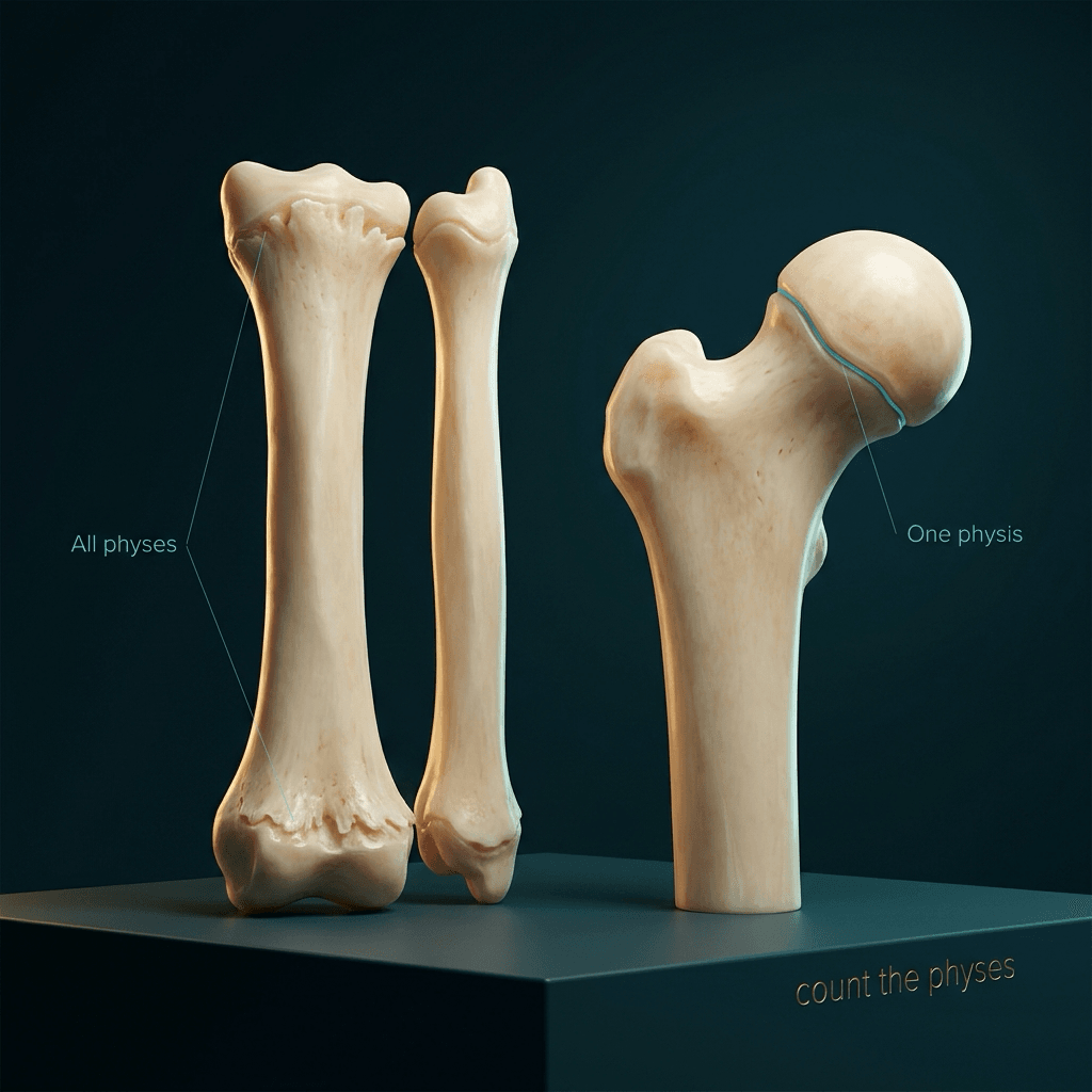
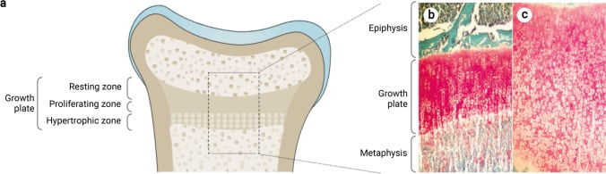
What the physis normally looks like. The growth plate is a transverse radiolucent band between the ossified epiphysis and the metaphysis. Its metaphyseal border — the zone of provisional calcification — is a crisp, dense, straight white line. The physeal lucency is thin and uniform, typically only a few millimetres, thickest at the fastest-growing sites (distal femur, proximal tibia, distal radius, proximal humerus).
What "abnormal" actually means. Four separable abnormalities, and you should name which one you see:
- Widening — the lucent zone is taller than expected. Causes: metabolic (unmineralised hypertrophic zone accumulates), traumatic (physeal separation), or chronic stress.
- Loss of the zone of provisional calcification — the crisp white metaphyseal line becomes blurred, indistinct or absent. This is the earliest sign of rickets and it precedes visible widening.
- Cupping and fraying — the metaphysis becomes concave and its margin shaggy and ill-defined. Combination of widening plus loss of the provisional zone plus mechanical splaying under load.
- Irregularity or bridging — undulating, sclerotic or bony-bar formation across the plate. Post-traumatic, post-infective, or dysplastic.
How to confirm it is genuinely present. Compare with (a) the contralateral limb, (b) the adjacent physes on the same film — in the wrist, the distal radius and distal ulna should be affected together in metabolic disease — and (c) an age-matched normal. Confirm on two orthogonal projections. Look at the metaphyseal margin rather than the lucency alone, since obliquity blurs the lucency but rarely abolishes the zone of provisional calcification.
The words to say out loud. "This is an AP radiograph of the left wrist in a skeletally immature patient. There is widening of the distal radial and distal ulnar physes with loss of the zone of provisional calcification, metaphyseal cupping and fraying. The bones are generally under-mineralised with coarsened trabeculae. There is no fracture, periosteal reaction or lytic lesion. The changes are symmetrical in distribution at the fastest-growing physes, which points to a metabolic cause — rickets being the commonest. I would examine the knees and the costochondral junctions and check calcium, phosphate, alkaline phosphatase, PTH and 25-hydroxyvitamin D."
Mimics — the false positives.
- Normal unossified epiphysis in the neonate and infant.
- Obliquity / projectional blurring on a rotated film.
- Overlying soft-tissue shadow or plaster reducing metaphyseal definition.
- Normal metaphyseal irregularity at the distal femur (the metaphyseal cortical irregularity or "cortical desmos" on the posteromedial distal femur) — a normal variant frequently mistaken for erosion.
- Physiological sclerosis of the neonatal metaphysis and the "bone-in-bone" appearance of the newborn.
- Motion artefact in an uncooperative toddler.
Next Investigation
CHARMSCHARMS — causes of dense or abnormal metaphyseal bands
Hook:CHARMS for bands; fraying and cupping is rickets territory
The Differential
- Typical age / setting
- Any age; peak under 5 years; febrile, non-weight-bearing
- Discriminating feature
- Single joint, blurred metaphyseal margin with adjacent lucency and soft-tissue swelling; joint space widening from effusion; child systemically unwell
- What confirms it
- Ultrasound effusion plus joint aspiration; CRP and ESR; MRI shows marrow oedema and subperiosteal collection
- Typical age / setting
- 10-16 years; obese or endocrinopathic adolescent; hip, thigh or knee pain
- Discriminating feature
- Widened, irregular proximal femoral physis with loss of epiphyseal height on AP; Klein's line fails to intersect the epiphysis
- What confirms it
- Frog-lateral or cross-table lateral hip; measure the slip angle; screen for hypothyroidism and renal disease in atypical ages
- Typical age / setting
- Any skeletally immature child after a discrete injury
- Discriminating feature
- Unilateral, focal widening with point tenderness over the physis; often a metaphyseal Thurston-Holland fragment (SH II)
- What confirms it
- Orthogonal views plus contralateral comparison; MRI or stress views if radiographs are equivocal
- Typical age / setting
- 2-10 years; bone pain, pallor, malaise, refusal to walk
- Discriminating feature
- Dense or lucent transverse metaphyseal bands with permeative destruction and periosteal reaction; multiple sites, often at multiple bones
- What confirms it
- Full blood count and film, bone marrow aspirate; urinary catecholamines for neuroblastoma; MRI of marrow
- Typical age / setting
- Infants under 18 months, usually non-ambulant
- Discriminating feature
- Corner or bucket-handle avulsion at the metaphyseal margin, often multiple and at different healing stages; posterior rib fractures
- What confirms it
- Full skeletal survey with repeat at 11-14 days; safeguarding referral; coagulation and metabolic screen to exclude mimics
- Typical age / setting
- 6 months to 3 years, and again in adolescence; dark skin, breastfed, limited sunlight
- Discriminating feature
- Bilateral symmetrical widening, cupping and fraying at the fastest-growing physes; generalised osteopenia; rachitic rosary and bowing
- What confirms it
- Low or low-normal calcium, low phosphate, markedly raised alkaline phosphatase, low 25-OH vitamin D, raised PTH
- Typical age / setting
- Presents at walking age; positive family history; normal growth velocity early
- Discriminating feature
- Rachitic changes with low phosphate but NORMAL calcium and NORMAL or only mildly raised PTH; marked lower-limb bowing out of proportion
- What confirms it
- Serum phosphate low with inappropriately high renal phosphate wasting (low TmP/GFR); raised FGF23; PHEX mutation
- Typical age / setting
- Child with known chronic kidney disease, growth failure
- Discriminating feature
- Rachitic physes PLUS subperiosteal resorption of the radial border of the middle phalanges, rugger-jersey spine and vascular or soft-tissue calcification
- What confirms it
- Raised urea and creatinine, high phosphate, high PTH, low 1,25-dihydroxyvitamin D
- Typical age / setting
- 9-16 year old competitive athlete; activity-related pain, no trauma
- Discriminating feature
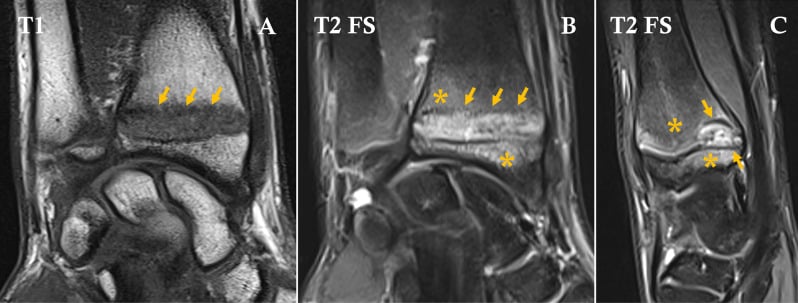
- Widened, irregular physis at the loaded site only (distal radius, proximal humerus) with metaphyseal sclerosis and cystic change; settles with rest
- What confirms it
- Focal tenderness and history of training load; MRI shows physeal oedema; radiographs normalise after 3 months of rest
- Typical age / setting
- Months to years after a physeal injury or septic arthritis
- Discriminating feature
- Focal physeal narrowing or bridging with adjacent widening, progressive angular deformity and limb-length discrepancy
- What confirms it
- MRI physeal map or CT to size and site the bar; Harris growth arrest lines converge towards the bar
- Typical age / setting
- Toddler with a very restricted diet or neurodevelopmental disorder; irritable, refuses to walk
- Discriminating feature
- Dense white metaphyseal line (Frankel), lucent band beneath it (Trümmerfeld), ring epiphyses (Wimberger) and dense subperiosteal haemorrhage
- What confirms it
- Low serum ascorbate; dramatic clinical response to vitamin C within days
- Typical age / setting
- Toddler onwards, short stature, waddling gait, NO pain and NO biochemical abnormality
- Discriminating feature
- Rickets-like metaphyseal irregularity with entirely normal calcium, phosphate and alkaline phosphatase; normal epiphyses; spine spared in Schmid
- What confirms it
- Normal bone biochemistry with a rachitic-looking skeletal survey; genetic testing (COL10A1 in Schmid)
- Typical age / setting
- Perinatal to childhood; premature loss of deciduous teeth with roots intact
- Discriminating feature
- Rachitic changes with a LOW alkaline phosphatase — the reverse of every other rickets
- What confirms it
- Low serum ALP, raised urinary phosphoethanolamine and plasma pyridoxal-5-phosphate; ALPL mutation
- Typical age / setting
- Neonate or young infant; hepatosplenomegaly, rash, pseudoparalysis
- Discriminating feature
- Symmetrical metaphyseal lucent bands with Wimberger sign (medial proximal tibial metaphyseal erosion) and diaphyseal periostitis
- What confirms it
- Maternal and infant treponemal serology; CSF examination
Narrowing It Down
- 11. Is it real, and is it one physis or many?
Confirm on two views and against the contralateral side, then count how many physes are involved before naming anything.
ONE abnormal physis means a local process — fracture, slipped capital femoral epiphysis, infection, tumour, chronic stress. MANY abnormal physes, symmetrically distributed, means a systemic metabolic or dysplastic process. This single question splits the differential in half and should be the first sentence out of your mouth.
- 22. How old is the child?
Fix the age and the ambulant status before proposing a diagnosis.
Under 18 months and non-ambulant with metaphyseal corner lesions — think non-accidental injury. Six months to 3 years with symmetrical cupping — nutritional rickets. Two to 10 years with malaise and marrow signs — leukaemia. Ten to 16 with hip or knee pain — slipped capital femoral epiphysis. Adolescent athlete — chronic physeal stress. Age alone excludes more than half the list.
- 33. Which physis, and does it follow the growth-rate rule?
Ask whether the fastest-growing physes are involved: distal femur, proximal tibia, distal radius and ulna, proximal humerus, costochondral junctions.
Metabolic disease strikes the fast growers first. If the abnormality sits at a slow-growing physis in isolation, or ignores the fast growers, metabolic disease is unlikely. Site also localises stress injury — distal radius in gymnasts, proximal humerus in throwers, proximal tibia in runners.
- 44. What is the metaphyseal margin doing?
Look specifically at the zone of provisional calcification and decide whether the pattern is FRAYING, a BAND, or a corner lesion — they are three different diseases.
Loss of the zone of provisional calcification with fraying and cupping equals rickets. A dense white band with a lucent band beneath equals scurvy or a healing insult. Lucent bands with permeative destruction and periosteal reaction equals leukaemia or neuroblastoma. Corner or bucket-handle avulsion equals inflicted injury. Blurred margin with soft-tissue swelling in a hot limb equals osteomyelitis.
- 55. Is the child systemically unwell, in pain, or well?
Take the temperature, watch the child weight-bear, and plot the height — three observations that cost nothing.
Febrile and non-weight-bearing pushes infection and malignancy to the top and mandates bloods and aspiration. Painful but well points to trauma, stress injury or slipped capital femoral epiphysis. Painless with short stature and a normal-looking child points to a skeletal dysplasia — and predicts entirely normal biochemistry.
- 66. What does the biochemistry say — and which way does the ALP move?
Send calcium, phosphate, alkaline phosphatase, parathyroid hormone, 25-OH vitamin D and renal function together, and read the ALP as a two-directional test.
Alkaline phosphatase is the pivot. HIGH with low phosphate and low vitamin D equals nutritional rickets. HIGH with low phosphate but normal calcium and vitamin D equals X-linked hypophosphataemia. HIGH with high phosphate and renal failure equals renal osteodystrophy. **LOW equals HYPOPHOSPHATASIA** — do not discard it as an assay error; ask about early loss of primary teeth, send natural substrates and ALPL sequencing, and remember bisphosphonates are contraindicated. Entirely NORMAL biochemistry with rachitic-looking metaphyses equals metaphyseal chondrodysplasia.
- 77. If still unresolved — image the marrow, but know what a positive scan is worth
Order MRI only after steps 1 to 6 have narrowed the field, and interpret it against the clinical picture rather than in place of it.
MRI shows physeal oedema in stress injury, marrow replacement in leukaemia, subperiosteal abscess in osteomyelitis, and maps a physeal bar for surgical planning. In the young athlete, calibrate it: bilateral MRI of 23 ASYMPTOMATIC Little League players found throwing-side abnormalities in 52 per cent, with proximal humeral physeal oedema or widening in 5 — and prior shoulder pain did not predict who had them. A positive scan in this group confirms a clinical diagnosis; it does not make one.
MCQ Practice Points
Q: Which single radiographic feature best separates a metabolic cause of physeal widening from a traumatic one?
A: Symmetry and multiplicity. Metabolic disease is systemic and therefore affects all fast-growing physes bilaterally and symmetrically. A traumatic, infective or neoplastic cause is focal and unilateral. Always request or examine the contralateral side before committing.
Q: A 4-year-old has rachitic-looking metaphyses. Alkaline phosphatase is LOW. Diagnosis?
A: Hypophosphatasia. Every other cause of rickets raises alkaline phosphatase; hypophosphatasia is defined by deficient tissue non-specific alkaline phosphatase activity from pathogenic ALPL variants. The international working group's three highest-agreement features are a pathogenic ALPL variant, elevation of natural substrates, and early loss of primary teeth — the last costs nothing to ask about. Diagnosis is made on two major, or one major plus two minor, criteria. Enzyme replacement with asfotase alfa is the specific treatment, and bisphosphonates are contraindicated. Caveat: those criteria are proposed by expert consensus and have not been prospectively validated, so no sensitivity or specificity attaches to them.
Q: An MRI in a 12-year-old baseball pitcher shows proximal humeral physeal widening. Does that make the diagnosis?
A: No — calibrate it against the base rate. Bilateral shoulder MRI in 23 ASYMPTOMATIC Little League players aged 10 to 12 found throwing-side abnormalities absent from the other shoulder in 52 per cent, including proximal humeral physeal oedema or widening in five, with the dominant arm 8.5 times more likely to be abnormal. Crucially, 61 per cent had experienced prior shoulder pain and that did not predict an abnormal scan. What did predict it was exposure: none, one or both of year-round play and single-sport specialisation gave a 25, 71 and 100 per cent chance of abnormality. Imaging confirms a clinical diagnosis here; it cannot make one, and it cannot clear a symptom-free child. Twenty-three players, so treat the percentages as small counts.
Q: A 14-year-old elite gymnast has wrist pain and a widened, irregular distal radial physis. Ulnar variance is positive. Mechanism, and how common is it?
A: Chronic repetitive axial loading causes a Salter–Harris I stress injury of the distal radial physis with impaired endochondral ossification. Caine's authors are explicit that this is a stress fracture and not normal adaptive change in gymnasts — the consequence to warn about is premature physeal arrest with relative ulnar overgrowth, positive ulnar variance and ulnar impaction, which is permanent. It is also less common than its fame implies: in a survey of 60 competitive gymnasts only five had stress-related change and four of those were minimal. Management is activity modification and rest, typically 3 months, with immobilisation if symptoms are severe.
Q: Which two features on a paediatric metaphyseal film should prompt an urgent full blood count and film?
A: Diffuse lucent metaphyseal bands at multiple sites, and permeative destruction with periosteal reaction — particularly in a child with bone pain, pallor, fever or refusal to walk. Acute lymphoblastic leukaemia and metastatic neuroblastoma both present this way and are commonly misattributed to juvenile idiopathic arthritis or osteomyelitis. Blood film and bone marrow aspiration are diagnostic.
Q: Which metaphyseal pattern indicates scurvy rather than rickets?
A: Scurvy produces a DENSE white metaphyseal line (the Frankel line, from failed matrix resorption) with a LUCENT band immediately beneath it (the Trümmerfeld zone), sclerotic ring epiphyses (Wimberger ring) and dense subperiosteal haemorrhage. Rickets does the opposite: it obliterates the dense metaphyseal line and produces cupping and fraying with generalised osteopenia.
Q: Rachitic-looking metaphyses, short stature, waddling gait, and completely normal calcium, phosphate and alkaline phosphatase. Diagnosis?
A: Metaphyseal chondrodysplasia, Schmid type — the commonest. An autosomal dominant COL10A1 disorder of type X collagen with normal biochemistry, normal epiphyses and a spared spine, often mistakenly treated as rickets for years. It presents at walking age with coxa vara and genu varum, and does not respond to vitamin D.
Q: You diagnose nutritional rickets in a toddler. What else does that oblige you to do?
A: Look beyond the patient. The global consensus frames nutritional rickets as a preventable public health problem rather than an isolated diagnosis, and ranks maternal and infant risk factors specifically — so check the siblings, ask about maternal vitamin D status during pregnancy and lactation, and consider the wider community and its access to supplementation or fortified food. Note also that the diagnosis is defined by clinical, biochemical and radiographic features together: a cupped, frayed physis is one component, not the diagnosis on its own.
Exam Viva Scenarios
Practise clinical reasoning and management decisions out loud
“You are shown an AP radiograph of both knees in a 20-month-old child. The distal femoral and proximal tibial physes are widened and the metaphyses are cupped and frayed with loss of the crisp metaphyseal white line. There is genu varum. Describe the film and give your differential.”
“You are shown an AP pelvis of a 13-year-old boy with a BMI above the 98th centile who has had four weeks of right knee pain and a limp. The right proximal femoral physis looks wider and more irregular than the left, and the epiphysis appears slightly shorter. What is your concern and what do you do next?”
“You are shown an AP radiograph of the left hip and proximal femur of a 3-year-old with fever and refusal to weight-bear. The proximal femoral metaphysis has an ill-defined lucent area with blurring of the physeal margin, and the soft-tissue planes are displaced laterally. Work through this.”
First three questions
- Is it real? Two orthogonal views plus the contralateral side
- One physis or many? Focal equals trauma/infection/tumour; multiple and symmetrical equals metabolic
- How old and how well is the child?
The four abnormal appearances
- Widening — unmineralised hypertrophic zone or physeal separation
- Loss of the zone of provisional calcification — earliest rachitic sign
- Cupping and fraying — established rickets under load
- Irregularity or bridging — post-traumatic or post-infective bar
Biochemistry decision tree
- High ALP, low phosphate, low vitamin D, high PTH — nutritional rickets
- High ALP, low phosphate, normal calcium and vitamin D — X-linked hypophosphataemia
- High ALP, high phosphate, uraemia, very high PTH — renal osteodystrophy
- LOW ALP — hypophosphatasia
- Entirely normal — metaphyseal chondrodysplasia
Must-not-miss list
- Septic arthritis or metaphyseal osteomyelitis — febrile, non-weight-bearing, ultrasound then aspirate
- SCFE — adolescent with knee pain, frog-lateral hip, non-weight-bearing on suspicion
- Leukaemia or neuroblastoma — lucent bands plus permeative change, do a blood film
- Non-accidental injury — corner and bucket-handle lesions in the non-ambulant infant
Sites that show metabolic disease first
- Distal femur and proximal tibia (knee)
- Distal radius and ulna (wrist)
- Proximal humerus
- Costochondral junctions — the rachitic rosary
- These are the fastest-growing physes; screen with an AP knee and AP wrist
Mimics to exclude before calling it abnormal
- Normal unossified epiphysis in the neonate
- Obliquity and projectional blurring
- Distal femoral metaphyseal cortical irregularity (normal variant)
- Physiological wide physis of the pubertal growth spurt
When no further imaging is needed
- Classical symmetrical rickets with confirmatory biochemistry — treat and repeat the wrist film at 6-8 weeks
- Physeal stress injury that is pain-free after rest — clinical follow-up alone
Evidence Base
Shoulder MRI Abnormalities in Asymptomatic Little League Baseball Players
- 23 ASYMPTOMATIC Little League baseball players aged 10 to 12 had BILATERAL shoulder MRI, read blinded by two radiologists, with each player acting as his own control
- 12 of 23 (52 PER CENT) had abnormalities in the throwing shoulder that were absent from the non-dominant side; the dominant arm was 8.5 TIMES more likely to be abnormal
- Among 17 positive findings, FIVE were oedema or WIDENING OF THE PROXIMAL HUMERAL PHYSIS - the very sign this page is about - alongside 4 labral tears and 4 partial-thickness cuff tears
- Risk stacked steeply: players with none, one or both of year-round play and single-sport specialisation had a 25, 71 and 100 PER CENT chance of an abnormal MRI
- 61 per cent had previously experienced shoulder pain, but PRIOR PAIN WAS NOT ASSOCIATED WITH AN ABNORMAL MRI
Trends in the Presentation, Management, and Outcomes of Little League Shoulder
- 95 patients with proximal humeral epiphysiolysis identified from one high-volume children's hospital, 1999 to 2013; 93 male, mean age 13.1 years (range 8 to 16), with diagnosed cases rising annually
- 97 per cent were baseball players (86 per cent pitchers, 8 per cent catchers) but 3 PER CENT WERE TENNIS PLAYERS - and concomitant elbow pain occurred in 13 per cent
- GLENOHUMERAL INTERNAL ROTATION DEFICIT WAS PRESENT IN 30 PER CENT and every such patient was given physiotherapy
- Mean time to full symptom resolution was 2.6 months and to return to competition 4.2 months, with rest advised in 99 per cent
- Recurrence was 7 per cent overall at a mean 7.6 months. The GIRD group recurred at 14 per cent against 5 per cent, an odds ratio of 3.6 - but the confidence interval was 0.7 to 17.1 and the difference was NOT statistically significant (p equals 0.11)
Stress Changes of the Distal Radial Growth Plate: A Radiographic Survey and Review of the Literature
- Radiographic survey of 60 young competitive gymnasts (39 female, 21 male) examining skeletal age and distal radial physeal stress change
- ONLY FIVE GYMNASTS had stress-related change of the wrist, and FOUR OF THOSE FIVE WERE JUDGED MINIMAL - the sign was much less common than the condition's prominence suggests
- Skeletal maturation was significantly delayed in the girls (p less than 0.001) compared with chronological age
- The authors argue the widening and irregularity represent a genuine STRESS FRACTURE of the physis and are NOT normal adaptive change in gymnasts
- Long-term consequences they describe include symmetrical or asymmetrical growth retardation or arrest, POSITIVE ULNAR VARIANCE and its sequelae; they state the true incidence 'remains unclear' and call for a large prospective study
Global Consensus Recommendations on Prevention and Management of Nutritional Rickets
- Evidence-based global consensus developed by 33 nominated experts across paediatric endocrinology, nutrition, epidemiology, public health and health economics, representing 11 international scientific organisations
- Built on a systematic literature search with recommendations graded by the GRADE system, so strength of recommendation and quality of evidence are separately stated throughout
- Defines nutritional rickets and its DIAGNOSTIC CRITERIA, distinguishing it from osteomalacia and from the non-nutritional rickets that mimic it radiographically
- Ranks risk factors, with particular emphasis on maternal and infant risk - the group in whom the disease is both commonest and most preventable
- Frames rickets as a PREVENTABLE global public health problem and calls for supplementation and food fortification programmes rather than case-by-case treatment alone
Proposed Diagnostic Criteria for Hypophosphatasia in Children and Adolescents: The HPP International Working Group
- International multidisciplinary working group reviewing 93 papers published 2005 to 2020, with two reviewers independently assessing each full text
- Hypophosphatasia is caused by pathogenic ALPL variants producing LOW activity of tissue non-specific alkaline phosphatase - the one metabolic bone disease on this differential in which ALP is DEPRESSED rather than raised
- The three features with highest agreement and prevalence were a pathogenic or likely pathogenic ALPL variant, ELEVATION OF NATURAL SUBSTRATES, and EARLY LOSS OF PRIMARY TEETH, together with reduced bone mineral density
- These were organised into THREE MAJOR and SIX MINOR criteria
- Diagnosis is made with TWO MAJOR, or ONE MAJOR PLUS TWO MINOR criteria. No formal diagnostic guideline for children existed before this