More than one bone lesion — age drives the differential before morphology does
- Over 40 with multiple lytic lesions: metastasis or myeloma until proven otherwise — never biopsy a bone lesion before staging for a primary.
- Under 30, multiple lesions are far more likely benign: fibrous dysplasia, Langerhans cell histiocytosis, enchondromatosis, osteochondromatosis, non-ossifying fibromas.
- Multiplicity does not equal benignity. Multifocal osteosarcoma, multifocal Ewing sarcoma and metastatic neuroblastoma all present as polyostotic disease in children.
- A solitary aggressive-looking lesion plus a whole-body test that reveals more lesions changes the diagnosis, not just the stage.
- Myeloma is classically cold on technetium bone scan — a normal bone scan with multiple lytic lesions on radiograph is myeloma until disproven.
- Skeletal survey (or whole-body MRI / low-dose whole-body CT) is the myeloma imaging pathway; bone scan is the metastasis pathway.
- “Ground-glass matrix in multiple bones with bowing = polyostotic fibrous dysplasia; add café-au-lait and precocious puberty for McCune-Albright.
- “Multiple lytic skull lesions with bevelled edges and a 'geographic skull' = Langerhans cell histiocytosis.
- “Brown tumours are the great mimic: check calcium, phosphate and PTH before biopsying multiple lytic lesions.
- “Unilateral hand and foot enchondromas with expansile deformity = Ollier disease; add soft-tissue haemangiomas = Maffucci.
A biopsy of one of many lesions in a 65-year-old, before a CT chest-abdomen-pelvis and myeloma screen, wastes time and may seed a tract in the wrong compartment. Stage first, biopsy the most accessible lesion afterwards.
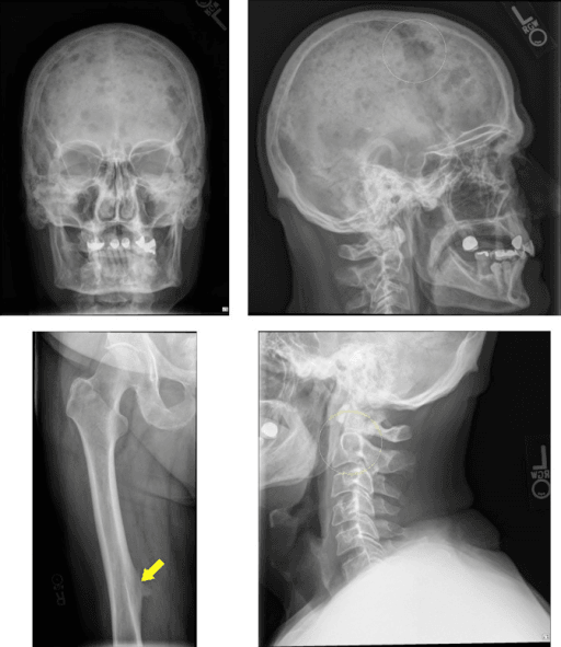
Multiple lytic lesions with subperiosteal resorption of the radial border of the middle phalanges, a salt-and-pepper skull and loss of the lamina dura is brown tumour disease. A blood test, not a biopsy, makes the diagnosis.
Myeloma, renal cell metastases, thyroid metastases and highly lytic disease may show minimal osteoblastic response. Never exclude polyostotic malignancy on a negative bone scan alone.
Metastatic neuroblastoma in a child under 5, and multifocal osteosarcoma in an adolescent, both produce multiple aggressive lesions. Permeative destruction and periosteal reaction override the reassurance of number.
Recognising the Pattern
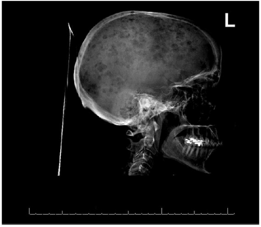
Definition. Polyostotic disease means two or more separate bones carry lesions, or a single bone carries widely separated non-contiguous lesions. This is distinct from multifocal within one bone (skip lesions) and from diffuse marrow replacement, where the marrow signal is abnormal throughout without discrete lesions.
Confirming the pattern is genuine. Before committing, satisfy yourself that:
- The lesions are in bone, not overlying soft tissue, bowel gas or a skin marker. Repeat the film with a marker if the lesion moves.
- The lesions are real, not composite shadows — sacral foramina, the trochanteric fossa, the herald patch of a ribbon rib, and vascular grooves in the skull all mimic lucency on a single projection. Orthogonal views resolve most.
- The normal variants are excluded: pseudocysts of the humeral head and calcaneus (the neutral triangle), Ward's triangle in the femoral neck, and the pelvic digit are all trabecular rarefaction, not lesion.
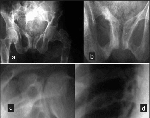
How to say it out loud. "This is a radiograph of the pelvis and both femora in a skeletally mature patient. There are multiple well-defined lytic lesions involving the iliac wings and both proximal femora. They are geographic with a narrow zone of transition, there is no matrix mineralisation, no periosteal reaction and no soft tissue mass. The distribution is axial and proximal appendicular, which corresponds to red marrow. In a patient over 40 my primary concerns are metastatic disease and myeloma, and I would want to exclude hyperparathyroidism."
Then commit to a lead diagnosis. Never end a description without one.
Mimics of the pattern (false positives).
- Multiple non-ossifying fibromas in an adolescent — eccentric, cortically based, sclerotic rim, asymptomatic; if numerous plus café-au-lait think Jaffe-Campanacci.
- Bone islands (enostoses) — dense, brush-bordered, cold on bone scan; the osteopoikilosis pattern gives symmetric periarticular dense foci and is a normal-life-expectancy sclerosing dysplasia.
- Fat-containing marrow (haemangioma, focal red marrow rests) — vertebral corduroy or polka-dot pattern; red marrow rests follow marrow signal on all sequences and lack restricted diffusion.
- Insufficiency fractures in the sacrum and pubic rami of an osteoporotic patient can look like multiple lucencies or, on bone scan, the Honda sign — a fracture pattern, not lesions.
- Degenerative subchondral cysts and gouty erosions cluster at joints and communicate with articular surfaces.
Next Investigation
- Best for
- Osteoblastic metastases (prostate, breast), stress lesions, Paget mapping
- Blind spot
- Purely lytic disease — myeloma, renal cell, aggressive lytic thyroid
- Practical rule
- Use when the suspected primary is prostate or breast
- Best for
- Lytic myeloma lesions, cortical integrity, fracture risk
- Blind spot
- Early marrow infiltration before bone destruction
- Practical rule
- Standard myeloma staging in most units
- Best for
- Marrow infiltration, myeloma before lysis, spinal cord compromise
- Blind spot
- Availability, motion, cost
- Practical rule
- Best sensitivity overall; use in myeloma and paediatric multifocal disease
- Best for
- Lymphoma, response assessment, occult primary
- Blind spot
- Low-grade chondroid lesions, some myeloma
- Practical rule
- Answers the 'is it active?' question
FEEMHIFEEMHI — Multiple Lytic Bone Lesions
Hook:A long-established radiology mnemonic for multiple lucent bone lesions. Add bone infarcts and multiple non-ossifying fibromas as the non-aggressive extras, and remember the paediatric aggressive causes — neuroblastoma metastases, leukaemia and multifocal osteosarcoma — sit outside the mnemonic.
The Differential
- Typical age / setting
- Over 40; breast, prostate, lung, renal, thyroid
- Discriminating feature
- Axial and proximal appendicular distribution (red marrow); lesions distal to elbow and knee are rare and suggest lung or renal primary
- What confirms it
- CT chest-abdomen-pelvis plus bone scan; biopsy of most accessible lesion with immunohistochemistry
- Typical age / setting
- Over 50, peak 60s-70s
- Discriminating feature
- Uniform punched-out lytic lesions with no reactive sclerosis; classically cold on technetium bone scan; endosteal scalloping of long bones
- What confirms it
- Serum and urine electrophoresis with free light chains; bone marrow aspirate; whole-body low-dose CT or whole-body MRI
- Typical age / setting
- Child under 5
- Discriminating feature
- Permeative metaphyseal lucency with periosteal reaction, plus skull sutural widening and orbital involvement (raccoon eyes)
- What confirms it
- Urinary catecholamines (VMA/HVA), abdominal imaging for adrenal mass, MIBG scan
- Typical age / setting
- Adolescent, second decade
- Discriminating feature
- Multiple synchronous sclerotic metaphyseal lesions with aggressive periosteal reaction and cloud-like osteoid
- What confirms it
- Biopsy plus staging CT chest; high alkaline phosphatase and LDH
- Typical age / setting
- Any age; CRMO in children 4-14
- Discriminating feature
- CRMO waxes and wanes, is often symmetrical and is culture-negative. The medial clavicle is CHARACTERISTIC but not COMMON - in Kong's 80 cases the leading sites were tibia 70%, femur 65%, fibula 42.5%, and the clavicle did not reach the top five. Fever in 57.5% does not make it pyogenic
- What confirms it
- MRI whole body showing multifocal marrow oedema; CRMO is a diagnosis of exclusion after biopsy and culture
- Typical age / setting
- Presents first two decades; monomelic tendency
- Discriminating feature
- Ground-glass matrix, long lesion in a long bone, no periosteal reaction, shepherd's crook deformity of proximal femur
- What confirms it
- Radiographic appearance is usually sufficient; GNAS mutation; look for café-au-lait and endocrinopathy (McCune-Albright)
- Typical age / setting
- Adults; renal failure or parathyroid adenoma
- Discriminating feature
- Lytic lesions plus subperiosteal resorption of radial border of middle phalanges, distal clavicle erosion, salt-and-pepper skull
- What confirms it
- Serum calcium, phosphate, PTH, alkaline phosphatase; parathyroid imaging
- Typical age / setting
- Peak 5-15 years
- Discriminating feature
- Skull lesions with bevelled edge from unequal table involvement; vertebra plana; lesion appearance changes rapidly over weeks
- What confirms it
- Skeletal survey; biopsy with CD1a and langerin (CD207) positivity, Birbeck granules
- Typical age / setting
- Childhood presentation
- Discriminating feature
- Asymmetric, often unilateral, hand and foot involvement with expansile columnar lucencies extending from the physis; Maffucci adds soft-tissue haemangiomas with phleboliths
- What confirms it
- Clinical and radiographic; monitor for chondrosarcomatous transformation (pain, cortical destruction, growth)
- Typical age / setting
- Childhood; autosomal dominant
- Discriminating feature
- Lesions are surface osteochondromas with cortex and medulla continuous with host bone, metaphyseal, pointing away from the joint; short forearm with ulnar shortening
- What confirms it
- Family history; EXT1/EXT2 mutation; MRI for cartilage cap greater than 1.5 cm suggests transformation
- Typical age / setting
- Over 55
- Discriminating feature
- Cortical thickening, trabecular coarsening and bone expansion — the bone gets bigger, which no malignancy does; flame sign in long bone, osteoporosis circumscripta in skull
- What confirms it
- Radiographs plus raised alkaline phosphatase with normal calcium and phosphate; bone scan shows intense uptake
- Typical age / setting
- Ages 5-20; incidental
- Discriminating feature
- Eccentric cortically based metaphyseal lesions with thin sclerotic scalloped rim; asymptomatic; regress with age
- What confirms it
- Radiograph alone; no biopsy needed; Jaffe-Campanacci if with café-au-lait
- Typical age / setting
- Sickle cell, steroids, alcohol, SLE, caisson
- Discriminating feature
- Serpiginous sclerotic rim around central lucency, medullary and metadiaphyseal, sparing cortex
- What confirms it
- MRI double line sign; identify risk factor; haemoglobin electrophoresis if sickle suspected
- Typical age / setting
- Any age; leukaemic lines in children
- Discriminating feature
- Permeative moth-eaten destruction with disproportionately small radiographic change relative to large soft tissue mass; metaphyseal lucent bands in leukaemia
- What confirms it
- Marrow biopsy, flow cytometry, PET-CT
- Typical age / setting
- Adults
- Discriminating feature
- Sarcoid gives lace-like lytic phalangeal lesions with tunnelled trabeculae; mastocytosis gives diffuse mixed sclerosis with flushing and urticaria pigmentosa
- What confirms it
- Chest radiograph and ACE for sarcoid; serum tryptase and marrow biopsy for mastocytosis
Narrowing It Down
- 1Step 1 — How old is the patient?
Establish the age before reading the film in any detail, and let it set the prior.
Over 40: metastasis, myeloma, Paget, brown tumours dominate. Ages 5-20: LCH, fibrous dysplasia, multiple NOFs, enchondromatosis, osteochondromatosis. Under 5: neuroblastoma metastases, LCH, osteomyelitis. Answering this alone reduces a 15-item list to 4.
- 2Step 2 — Is the pattern aggressive or non-aggressive?
Assess zone of transition, cortical integrity, periosteal reaction and any soft tissue mass — on every lesion, not just the largest.
Geographic with a narrow zone and no periosteal reaction pushes towards fibrous dysplasia, NOFs, enchondromas, infarcts. Permeative or moth-eaten with periosteal reaction pushes towards metastasis, myeloma, round cell tumour, infection.
- 3Step 3 — Where in the skeleton, and where within the bone?
Map the distribution against red marrow, then locate each lesion within its bone (epiphysis, metaphysis, diaphysis).
Axial plus proximal appendicular follows red marrow and favours haematogenous seeding - metastasis, myeloma, lymphoma. Hands and feet favour enchondromatosis, sarcoid, and uncommonly lung or renal metastases. Epiphyseal favours chondroblastoma or infection; metaphyseal favours NOF, LCH, osteosarcoma; diaphyseal favours fibrous dysplasia, Ewing, LCH, infarct.
- 4Step 4 — What is the matrix?
Look inside the lesion for mineralisation and name its character; use CT if the plain film is equivocal.
Ground glass equals fibrous dysplasia. Rings-and-arcs or popcorn equals chondroid - enchondromatosis. Cloud-like or ivory equals osteoid - osteosarcoma or blastic metastases. No matrix at all with sharply punched-out margins equals myeloma or LCH.
- 5Step 5 — Is the bone expanded or thickened?
Compare the overall calibre and cortical thickness of the affected bone with the contralateral side.
Bone that is LARGER than normal with coarse trabeculae and cortical thickening is Paget disease; malignancy destroys but does not enlarge in this manner. Expansile bubbly lesions in the hands suggest enchondromas or brown tumours.
- 6Step 6 — What do the bloods say?
Order calcium, phosphate, PTH, alkaline phosphatase, full blood count, ESR/CRP, protein electrophoresis with free light chains, and PSA in men.
This panel separates myeloma, hyperparathyroidism, Paget and infection without any further imaging, and it is cheap. Send it before the biopsy request, not after.
- 7Step 7 — Are there systemic or syndromic features?
Examine the skin, ask about endocrine symptoms and thirst, and look for palmoplantar pustulosis.
Cafe-au-lait with ragged borders plus precocious puberty equals McCune-Albright. Diabetes insipidus plus skull lesions equals LCH. Soft-tissue haemangiomas equals Maffucci. Urticaria pigmentosa equals mastocytosis. Clavicular and metaphyseal lesions with palmoplantar pustulosis equals CRMO/SAPHO - though note the clavicle is characteristic rather than common, and long bones dominate.
MCQ Practice Points
Q: A 62-year-old woman has multiple lytic lesions on plain radiographs but a normal technetium bone scan. What is the most likely diagnosis?
A: Multiple myeloma. Myeloma lesions are produced by osteoclast activation with profound osteoblast suppression, so there is little or no reactive bone formation and therefore minimal tracer uptake. Low-dose whole-body CT or whole-body MRI is the appropriate staging modality — and note that the IMWG criteria define a myeloma-defining bone lesion by imaging thresholds, so which scan you order changes whether the patient meets the definition.
Q: Which polyostotic condition is most strongly associated with a shepherd's crook deformity?
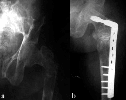
A: Polyostotic fibrous dysplasia. Repeated microfracture through mechanically weak fibro-osseous tissue in the proximal femur produces progressive varus of the femoral neck. Load-sharing intramedullary fixation is preferred to plate fixation.
Q: Multiple lytic lesions with subperiosteal resorption of the radial aspect of the middle phalanges — what is the diagnosis and the confirming test?
A: Brown tumours of hyperparathyroidism. Subperiosteal resorption is the most specific radiographic sign. Confirm with serum calcium, phosphate, alkaline phosphatase and PTH before any biopsy is considered — this is the polyostotic pattern most often biopsied unnecessarily.
Q: An 11-year-old has multiple unilateral expansile lucent lesions in the metacarpals and phalanges of one hand with associated shortening. What is the diagnosis and the chief long-term concern?
A: Ollier disease (enchondromatosis). The concern is malignant transformation to chondrosarcoma, signalled by new rest pain, rapid growth, cortical destruction or a soft tissue mass, and risk is higher in Maffucci syndrome where soft-tissue haemangiomas with phleboliths coexist. Add the surgical caution from Colello's survey: about a THIRD of the chondrosarcomas reported arose at a PREVIOUS SURGICAL SITE, and two of the four patients who metastasised had had an intramedullary nail — so treat new pain at an old operative site as a sarcoma question, and think carefully before instrumenting the whole length of an affected bone. Those figures come from a self-selected support-group survey and overstate absolute risk, but the site pattern is the lesson.
Q: Which feature best distinguishes polyostotic Paget disease from widespread blastic metastases?
A: Bone enlargement. Paget disease expands the bone with cortical thickening and coarse trabeculae, and the alkaline phosphatase is raised with a normal calcium and phosphate. Blastic metastases increase density without enlarging the bone contour.
Exam Viva Scenarios
Practise clinical reasoning and management decisions out loud
“You are shown this radiograph of the skull and pelvis of a 68-year-old man with three months of thoracolumbar back pain and fatigue. There are numerous small, uniform, round lucencies in the calvarium and multiple lytic lesions in both iliac wings without surrounding sclerosis. Describe and manage.”
“You are shown this radiograph of the pelvis and both femora of a 12-year-old girl with a limp and a leg length discrepancy. There are expansile lesions with a hazy internal matrix in the right proximal femur and right ilium, with varus angulation of the femoral neck. What is your diagnosis?”
“You are shown this lateral skull radiograph of a 6-year-old boy with a tender scalp swelling. There are several lytic lesions with sharply defined, asymmetric, bevelled margins. A lateral thoracic spine film shows a uniformly flattened T7 vertebral body with preserved adjacent discs. Discuss.”
Age-driven first line
- Over 40: metastasis, myeloma, Paget, brown tumours
- Ages 5-20: LCH, fibrous dysplasia, multiple NOFs, enchondromatosis, osteochondromatosis
- Under 5: metastatic neuroblastoma, LCH, multifocal osteomyelitis, leukaemia
- Any age with fever and raised CRP: multifocal infection until excluded
One-line discriminators
- Ground glass, bowed long bone — fibrous dysplasia
- Punched-out, no sclerosis, cold bone scan — myeloma
- Bevelled-edge skull lesion, vertebra plana — Langerhans cell histiocytosis
- Subperiosteal phalangeal resorption, salt-and-pepper skull — brown tumours
- Bone enlarged with thick cortex, high ALP — Paget disease
- Unilateral hand enchondromas — Ollier; with haemangiomas — Maffucci
- Serpiginous sclerotic rim, medullary — bone infarct
- Continuous cortex and medulla, pointing away from joint — osteochondromas
Blood panel to order every time
- Calcium, phosphate, PTH, alkaline phosphatase
- Full blood count, ESR and CRP
- Serum and urine protein electrophoresis with free light chains
- PSA in men; thyroid function if thyroid primary suspected
- Urinary catecholamines (VMA/HVA) in a child under 5
Imaging pathway
- Suspected metastasis: CT chest-abdomen-pelvis plus technetium bone scan
- Suspected myeloma: whole-body low-dose CT or whole-body MRI, not bone scan
- Paediatric multifocal disease: whole-body MRI
- Index lesion for surgery: local MRI with contrast before biopsy
- No imaging needed: classic multiple NOFs, osteopoikilosis, bone islands, familial HME
Safety rules
- Stage before you biopsy — never the reverse
- Biopsy tract must be resectable within the future incision; plan with the tumour surgeon
- Multiplicity never excludes malignancy in a child
- Any neurology with spinal lesions equals urgent whole-spine MRI
- Check calcium — hypercalcaemia of malignancy is the emergency you can treat today
Evidence Base
International Myeloma Working Group Updated Criteria for the Diagnosis of Multiple Myeloma
- Consensus of 34 international authors that redefined multiple myeloma to include validated BIOMARKERS alongside the existing CRAB features (hypercalcaemia, renal failure, anaemia, bone lesions)
- The change was driven by biomarkers identifying patients with near-inevitable progression to CRAB who would previously have been labelled smouldering myeloma and left untreated
- It also CLARIFIED AND UPDATED the radiographic variables that count as a myeloma-defining bone lesion, not only the laboratory ones
- Sets out the histological and monoclonal protein requirements for diagnosis
- Specifies the metrics any future biomarker must meet before it can enter the disease definition
Rate and Type of Surgery and Prevalence of Malignancy in Patients With Ollier's Disease and Maffucci Syndrome
- 123 evaluable survey responses from five continents - 109 Ollier disease and 14 Maffucci syndrome
- 89 per cent had at least one operation, first at a mean age of 9.1 years, with an average of 7.6 OPERATIONS EACH
- Commonest indications were lower-limb length discrepancy, deformity correction and resection of a symptomatic enchondroma
- 19 PER CENT DEVELOPED A CHONDROSARCOMA - and 35 per cent of those arose AT A PREVIOUS SURGICAL SITE
- Four patients developed metastatic disease; TWO of them had had an intramedullary nail placed at a prior procedure
Chronic Recurrent Multifocal Osteomyelitis: A Review of 80 Cases in Chinese Children
- Five new cases combined with 75 previously reported Chinese paediatric cases, 80 in total; no sex difference in incidence
- DIAGNOSTIC DELAY OF UP TO SEVEN YEARS was observed - the nonspecific presentation is the problem, not the imaging
- Bone pain in 92.5 per cent, fever in 57.5 per cent, arthritis in 23.8 per cent, rash in 13.8 per cent; CRP or ESR raised
- Most frequently affected sites were TIBIA 70.0 per cent, FEMUR 65.0 per cent, fibula 42.5 per cent, foot 36.3 per cent and pelvis 32.5 per cent
- The authors emphasise long-term follow-up, because remission rate cannot be judged early