Reduced bone density confined to one limb, one joint or one bone segment — a pattern, not a diagnosis
- Regional osteopenia means density loss limited to a region — compare directly with the contralateral side or the adjacent normal bone on the SAME film.
- The single most useful analysis is the pattern of resorption: uniform thinning, subchondral/periarticular banding, spotty (patchy) or permeative.
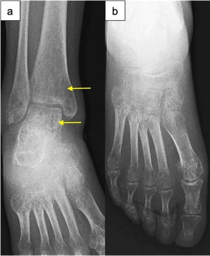
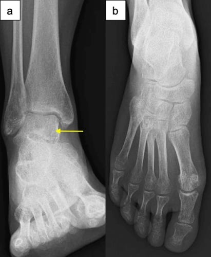
- Spotty (mottled) periarticular osteopenia after immobilisation or minor trauma with pain out of proportion equals complex regional pain syndrome until proven otherwise.
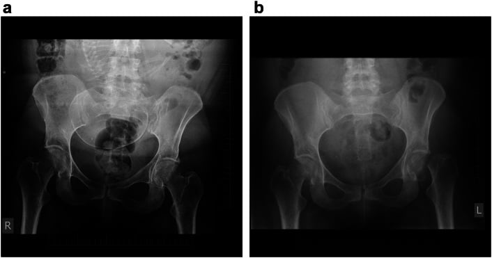
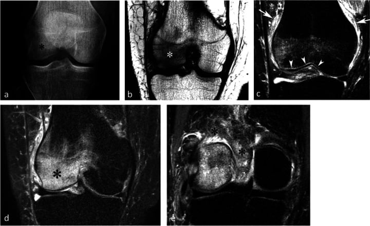
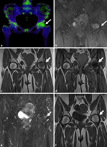
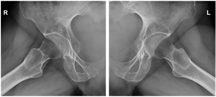
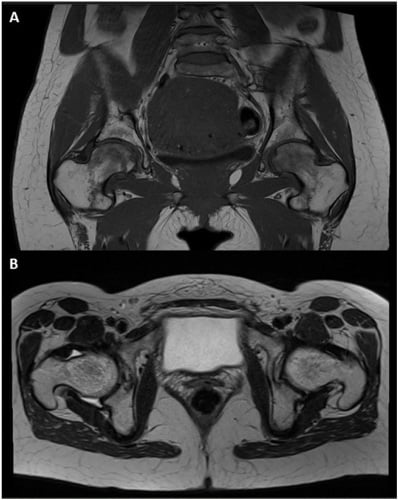
- Osteopenia limited to one hip in a third-trimester pregnant woman or middle-aged man with acute groin pain equals transient osteoporosis of the hip — MRI shows femoral head and neck oedema with preserved subchondral line.
- A permeative or moth-eaten pattern is NOT osteopenia — it is bone destruction and mandates urgent MRI, myeloma screen and consideration of biopsy.
- Radiographs need approximately 30 to 40 percent mineral loss before density change is visible; a normal film never excludes early regional bone loss.
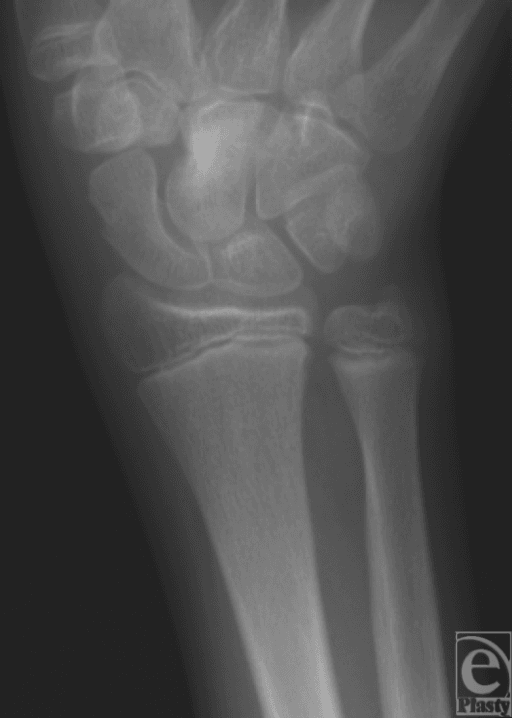
- “Say it as: 'There is regional osteopenia of the distal radius and carpus, with a patchy subchondral and periarticular distribution, intact cortices and no periosteal reaction.'
- “Always ask for the contralateral film — density is a comparative judgement and exposure factors deceive.
- “Preserved joint space with periarticular osteopenia points to disuse or CRPS; joint space loss with erosions points to inflammatory arthritis.
- “Focal osteopenia crossing the physis or subchondral plate with a joint effusion in a child equals septic arthritis or osteomyelitis until excluded.
Overexposed or digitally over-processed films, obliquity and soft tissue swelling all mimic focal density loss. Check the cortices and trabeculae of the ADJACENT bone on the same film — if they are equally faint, it is technique.
Multiple tiny lucencies in cortex and medulla read as "osteopenia" at a glance. If the cortex has endosteal scalloping or intracortical tunnelling, this is aggressive bone destruction — myeloma, lymphoma, Ewing or osteomyelitis — not metabolic bone loss.
Disuse osteopenia requires a plausible period of immobilisation, usually at least 6 to 8 weeks. If the patient has been mobilising and the bone is osteopenic, look for CRPS, infection, inflammatory arthritis or tumour.
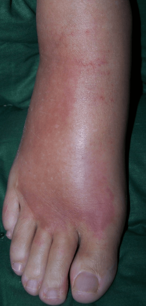
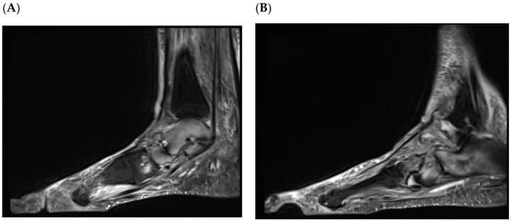
The clinical stem matters. Burning pain, allodynia, swelling, temperature and sweating change with patchy periarticular osteopenia is CRPS — a clinical diagnosis (Budapest criteria) that radiographs only support.
Recognising the Pattern
Definition. Regional osteopenia is a radiographically reduced bone mineral density confined to a single anatomical region — one limb, one joint and its periarticular bone, or one bone segment — with normal density elsewhere on the same or comparable films. It is distinct from generalised osteopenia (whole skeleton; osteoporosis, osteomalacia, hyperparathyroidism, myeloma) and from focal lucency (a discrete lesion with a definable margin).
Confirming it is genuinely present
- Compare within the film. The distal radius should not be markedly less dense than the mid-shaft or the adjacent ulna on the same exposure. Isolated faintness of one segment is real; global faintness is technique.
- Compare with the contralateral side. Ideally on a single film or matched exposure. This is the single most reliable manoeuvre.
- Look at the cortex separately from the medulla. Regional osteopenia thins the cortex from the endosteal surface and blurs the fine trabeculae, but the cortical outline remains continuous and sharp.
- Characterise the distribution:
- Uniform / diffuse thinning of the whole segment — disuse, immobilisation, paralysis.
- Periarticular / juxta-articular band — inflammatory arthritis, early septic arthritis, disuse.
- Spotty or patchy (mottled) subchondral and metaphyseal lucencies with subcortical banding — classical CRPS; also acute disuse.
- Subchondral, one joint, acute — transient osteoporosis / bone marrow oedema syndrome.
- Permeative or moth-eaten — NOT osteopenia. Aggressive marrow-replacing or infective process.
Saying it out loud in a viva
"This is a plain radiograph of the [region]. There is regional reduction in bone density involving the [segment], with a [patchy periarticular / uniform diffuse] pattern. The cortices are thinned but intact and there is no periosteal reaction, endosteal scalloping or cortical breach. The joint space is [preserved / reduced], there are [no] erosions and the soft tissues show [swelling / normal planes]. The remainder of the skeleton on this film is normally mineralised. My differential for regional osteopenia is disuse, complex regional pain syndrome, transient osteoporosis, inflammatory or septic arthritis, and I must exclude an aggressive permeative process such as myeloma, lymphoma or osteomyelitis."
Mimics — the false positives
- Why it deceives
- Whole region looks lucent
- How to exclude
- Adjacent bones equally lucent; soft tissues washed out
- Why it deceives
- Windowing exaggerates contrast
- How to exclude
- Re-window; check raw exposure index
- Why it deceives
- Less attenuation makes bone look darker
- How to exclude
- Bone trabecular pattern is preserved
- Why it deceives
- Cortical ring projected off-axis appears thin
- How to exclude
- Repeat orthogonal view
- Why it deceives
- Regional lucency of skull or long bone
- How to exclude
- Sharp advancing "blade of grass" front, bone expansion
Next Investigation
DXA is a poor tool for regional osteopenia. It quantifies but does not characterise, and regional measurements are unreliable in the presence of oedema, deformity or metalwork. Use it for fracture-risk stratification in neurological disuse osteopenia, not for diagnosis.
The Differential
- Typical age / setting
- Any age; children 3 to 12, diabetic foot, prosthetic joint
- Discriminating feature
- Osteopenia PLUS cortical destruction, periosteal reaction and a soft tissue collection; effusion appears before bone change
- What confirms it
- MRI marrow oedema with rim-enhancing collection; aspirate and cultures; CRP trend
- Typical age / setting
- Older than 55, back or rib pain, anaemia, renal impairment
- Discriminating feature
- Permeative or punched-out lucencies without sclerotic rim; endosteal scalloping; osteopenia disproportionate to age
- What confirms it
- Serum and urine electrophoresis, free light chains, whole-body MRI or low-dose CT; marrow biopsy
- Typical age / setting
- Children (leukaemic lines) and adults 40 to 70
- Discriminating feature
- Permeative marrow lesion with a large soft tissue mass but relatively preserved cortex; metaphyseal lucent bands in childhood leukaemia
- What confirms it
- MRI marrow replacement; bone marrow aspirate; biopsy of soft tissue component
- Typical age / setting
- Age 5 to 25, diaphysis of femur, pelvis, fever and raised inflammatory markers
- Discriminating feature
- Permeative diaphyseal destruction with lamellated or 'onion-skin' periosteum and a disproportionate soft tissue mass
- What confirms it
- MRI whole bone plus staging CT chest; biopsy through planned resection tract
- Typical age / setting
- Older than 50, known primary (breast, lung, renal, thyroid, prostate)
- Discriminating feature
- Focal cortical destruction or moth-eaten pattern rather than uniform density loss; pain at rest and at night
- What confirms it
- Bone scan or FDG-PET, CT chest/abdomen/pelvis, biopsy if solitary
- Typical age / setting
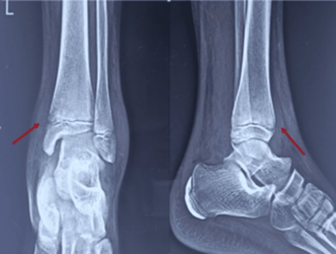
- Any age; after cast, plaster, paralysis, prolonged non-weightbearing
- Discriminating feature
- Uniform loss of the whole immobilised segment, exposure-matched to the opposite limb; painless; timeline of at least 6 to 8 weeks of immobilisation
- What confirms it
- History alone; radiographs reverse over months with remobilisation — no further imaging needed
- Typical age / setting
- Age 40 to 60, female predominance; weeks to months after distal radius fracture or minor injury
- Discriminating feature
- Spotty subchondral, subperiosteal and intracortical lucency in a patchy distribution; burning pain, allodynia, vasomotor and sudomotor change out of proportion
- What confirms it
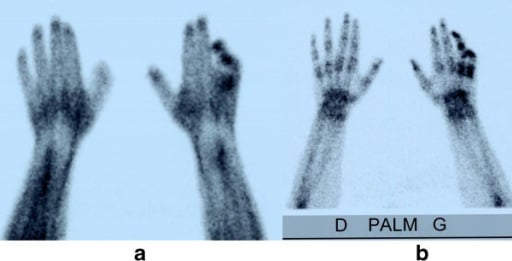
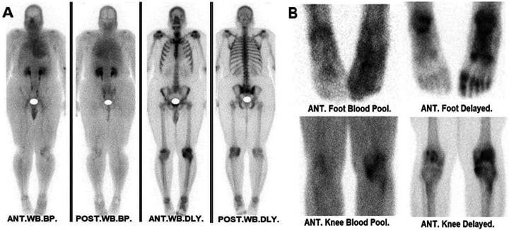
- Clinical Budapest criteria; three-phase bone scan shows diffuse periarticular uptake in the delayed phase
- Typical age / setting
- Third-trimester pregnancy; men 40 to 60
- Discriminating feature
- Osteopenia of femoral head AND neck with loss of the subchondral white line but NO subchondral collapse or crescent; self-limiting over 6 to 12 months
- What confirms it
- MRI: diffuse head and neck oedema, effusion, no focal necrotic segment or double-line sign
- Typical age / setting
- Age 30 to 60 (RA); JIA in children
- Discriminating feature
- Periarticular osteopenia WITH symmetrical joint space narrowing, marginal erosions and soft tissue swelling; MCP and PIP predilection
- What confirms it
- Anti-CCP, rheumatoid factor, ultrasound or MRI synovitis and erosions
- Typical age / setting
- Men 30 to 60, lower limb
- Discriminating feature
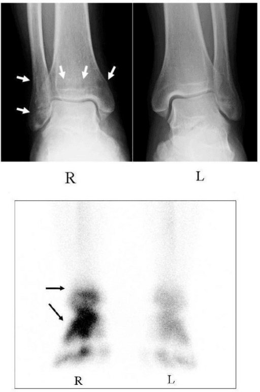
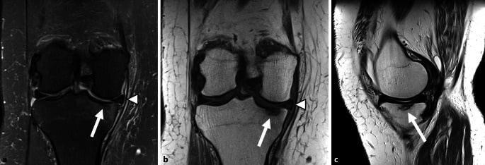
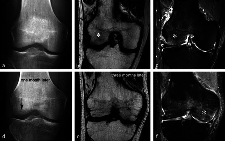
- Successive episodes of self-limiting joint pain migrating between knee, ankle and hip over months to years
- What confirms it
- Serial MRI showing oedema in a new joint as the previous resolves
- Typical age / setting
- Any age; hemiplegia, tetraplegia
- Discriminating feature
- Rapid, profound osteopenia below the level of the lesion, most marked in the distal femur and proximal tibia; fragility fracture with trivial force
- What confirms it
- History plus DXA of the affected limb; no biopsy required
- Typical age / setting
- Months to years after therapeutic radiotherapy
- Discriminating feature
- Mixed osteopenia and coarse sclerosis sharply confined to the radiation portal, with a straight geographic border ignoring anatomical boundaries
- What confirms it
- Radiotherapy field records; MRI to exclude recurrent tumour or radiation-induced sarcoma
- Typical age / setting
- Diabetic neuropathy, age 50 to 70, midfoot
- Discriminating feature
- Osteopenia with fragmentation, subluxation and debris in an insensate but relatively painless joint; skin usually intact
- What confirms it
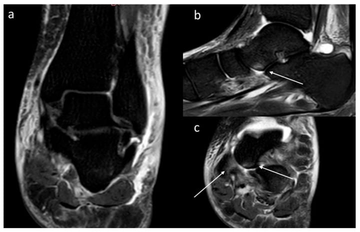
- MRI: subchondral oedema in multiple bones about a deformed joint; distinguishing from infection may need labelled white cell scan
- Typical age / setting
- Haemophilia from childhood; PVNS age 20 to 50, single joint
- Discriminating feature
- Periarticular osteopenia with epiphyseal overgrowth and widened intercondylar notch (haemophilia) or cystic subchondral erosions with preserved space (PVNS)
- What confirms it
- MRI blooming haemosiderin on gradient echo sequences
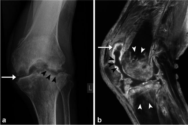
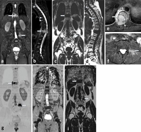
Narrowing It Down
- 1Step 1 — Is it real, or is it technique?
Compare the suspect segment with adjacent bone on the SAME film, and with the contralateral limb, before accepting that anything is abnormal.
If the neighbouring bone is equally faint the film is over-penetrated and there is nothing to explain. This step costs seconds and avoids an entire fabricated differential.
- 2Step 2 — Is it osteopenia or bone destruction?
Inspect the cortex under magnification and decide whether it is thin-but-continuous or actually breached.
Thin but continuous cortex with blurred trabeculae is true osteopenia. Endosteal scalloping, intracortical tunnelling, moth-eaten or permeative lucency, periosteal reaction or a soft tissue mass is aggressive destruction - which separates the benign metabolic group from the myeloma, lymphoma, Ewing and osteomyelitis group needing urgent MRI and bloods the same day.
- 3Step 3 — What is the distribution within the region?
Name the pattern out loud - uniform, spotty periarticular, single-joint subchondral, or straight-edged.
Uniform whole-segment loss favours disuse or neurological injury. Spotty periarticular and subcortical lucency favours CRPS. A single joint with subchondral loss and effusion favours transient osteoporosis or sepsis. A straight-edged field crossing anatomy favours radiation. This usually narrows the list to two.
- 4Step 4 — What is the joint doing?
Assess joint space, erosions, subchondral contour and any effusion separately from the bone density.
Space preserved with no erosions: disuse, CRPS, transient osteoporosis. Narrowed with marginal erosions and a symmetrical distribution: inflammatory arthritis. Narrowed rapidly with subchondral irregularity and effusion: septic arthritis. Fragmentation, subluxation and debris: neuropathic joint. This separates articular from purely osseous causes.
- 5Step 5 — What does the history say about pain and function?
Ask specifically whether the pain is proportionate to the injury, and look for vasomotor, sudomotor and trophic change.
Painless in an immobilised limb is disuse. Burning pain, allodynia, swelling, colour and sweating change is CRPS - and note the Budapest CLINICAL criteria have a specificity of 0.68 even against neuropathic pain, so meeting them does not release you from excluding infection and tumour. Acute severe groin pain with an antalgic gait in pregnancy or a middle-aged man is transient osteoporosis. Fever, rest pain, night pain and weight loss are infection or malignancy. This is the discriminator no plain film can supply.
- 6Step 6 — Solitary region or migratory / multifocal?
Ask about previous episodes in other joints, and look at whatever other films exist.
Single region and static: disuse, CRPS, radiation. Migrating between joints over months: regional migratory osteoporosis. Multiple regions with generalised osteopenia in an older patient: reconsider myeloma, hyperparathyroidism or osteomalacia, because the pattern may not be regional at all.
- 7Step 7 — Commit and investigate
State one most likely cause, name the must-not-miss you are excluding, and request a specific test rather than a panel.
MRI of the region in nearly all cases; add full blood count, ESR, CRP, calcium, phosphate, alkaline phosphatase, renal function and electrophoresis when malignancy or metabolic disease is in play. In transient osteoporosis or bone marrow oedema syndrome, add AXIAL DXA - the spine in these patients averages a Z score near minus 1.8 even when there is no back pain, so treating the problem as purely local underestimates the skeleton at risk.
MCQ Practice Points
Q: What percentage of bone mineral must be lost before osteopenia becomes visible on a plain radiograph?
A: Approximately 30 to 40 per cent. A normal radiograph therefore never excludes early or clinically significant regional bone loss — this is why MRI, which detects marrow oedema within days, is the sensitive test. Put the two facts together in a viva: the film is insensitive AND the pattern it shows is non-specific, which is exactly why the differential on this page is resolved by history and MRI rather than by radiographic appearance.
Q: Which single radiographic feature best separates transient osteoporosis of the hip from avascular necrosis?
A: The distribution of change. Transient osteoporosis involves the femoral head and neck diffusely with loss of the subchondral white line but NO subchondral fracture line; avascular necrosis shows a demarcated subchondral segment, the crescent sign, and on MRI a serpiginous rim with a double-line sign. Joint space is preserved in both early on.
Q: A 6-year-old has periarticular osteopenia of the knee, a joint effusion and metaphyseal lucent bands. What must be excluded?
A: Acute lymphoblastic leukaemia. Metaphyseal lucent bands adjacent to the physis in a child with bone pain, effusion and osteopenia require a full blood count with film and a bone marrow aspirate. Septic arthritis is the other urgent consideration, and the two can look identical on a radiograph.
Q: What distinguishes disuse osteopenia from CRPS radiographically?
A: Disuse produces uniform, diffuse thinning of the whole immobilised segment; CRPS produces a patchy, spotty subchondral, subperiosteal and intracortical lucency with an irregular mottled texture. The clinical picture — painless versus disproportionate burning pain with vasomotor change — is the separator. Be precise about how strong that separator is: the Budapest CLINICAL criteria have a sensitivity of 0.99 and a specificity of 0.68, and the RESEARCH criteria reach 0.79 by demanding more findings. Those figures were measured against non-CRPS neuropathic pain, not against disuse, infection or tumour, so a positive set of criteria narrows the field without closing it.
Q: Regional osteopenia with a straight-line border that ignores anatomical boundaries — diagnosis?
A: Radiation osteitis. The change conforms to the treatment portal, mixes osteopenia with coarse sclerosis, and carries risks of insufficiency fracture and, rarely, radiation-induced sarcoma — so new pain or a mass in an irradiated field mandates MRI.
Exam Viva Scenarios
Practise clinical reasoning and management decisions out loud
“You are shown this radiograph of the wrist in a 54-year-old woman, ten weeks after cast treatment of a distal radius fracture. The fracture has united in acceptable alignment but she has persistent burning pain, a swollen shiny hand and cannot tolerate the sleeve of her coat.”
“You are shown an anteroposterior pelvic radiograph of a 32-year-old woman at 34 weeks gestation with two weeks of severe left groin pain and an antalgic gait. There is loss of density of the left femoral head and neck.”
“You are shown a femoral radiograph in a 62-year-old man with three months of thigh pain and back pain. The bone appears diffusely lucent. He has been told he has osteoporosis.”
Define it in one line
- Reduced density confined to one limb, joint or segment, with normal density elsewhere on the same film
- Requires 30 to 40 percent mineral loss to be visible
- Always confirm against the contralateral side
Four patterns, four groups
- Uniform whole segment — disuse, paralysis, neurological injury
- Spotty periarticular and subcortical — complex regional pain syndrome
- Single joint, subchondral, acute — transient osteoporosis / bone marrow oedema syndrome
- Permeative or moth-eaten — NOT osteopenia: myeloma, lymphoma, Ewing, metastasis, osteomyelitis
Must-not-miss list
- Osteomyelitis and septic arthritis — cortical destruction, periosteal reaction, effusion, raised CRP
- Multiple myeloma — age over 55, back pain, anaemia, renal impairment, electrophoresis
- Lymphoma and leukaemia — marrow replacement with disproportionate soft tissue mass
- Ewing sarcoma — age 5 to 25, diaphyseal, lamellated periosteum, fever
- Metastasis — known primary, night pain, focal cortical destruction
Discriminators to say out loud
- Cortex intact and continuous equals benign; scalloped or tunnelled equals aggressive
- Joint space preserved equals disuse / CRPS / transient osteoporosis
- Joint space lost with marginal erosions equals inflammatory arthritis
- Fragmentation and subluxation in an insensate joint equals neuropathic (Charcot)
- Straight geographic border ignoring anatomy equals radiation osteitis
- Migration between joints over months equals regional migratory osteoporosis
Investigation plan
- Contralateral radiograph first — exclude technique
- MRI is the default next test in nearly all cases
- Bloods: FBC, ESR, CRP, bone profile, renal function, electrophoresis and free light chains if age over 55 or permeative
- Three-phase bone scan supports CRPS; CT defines cortical integrity and impending fracture
- Aspirate or biopsy only after staging, with sarcoma unit involvement
When to stop investigating
- Painless uniform osteopenia in a recently immobilised limb with intact cortices — reassure and remobilise
- Established Budapest-positive CRPS — treat, do not rescan
- Resolving transient osteoporosis on serial clinical review with protected weightbearing
Evidence Base
BMD in Transient Osteoporosis and Bone Marrow Edema Syndrome: A Scoping Review
- 561 publications screened, yielding 188 patients with quantifiable bone mineral density measured during ACTIVE disease and before any treatment
- Symptomatic hips showed a marginal mean Z or T score of MINUS 2.12 (95 per cent CI minus 2.4 to minus 1.88, n equals 48)
- In within-patient pairwise comparison the symptomatic hip was lower than the asymptomatic hip by 1.2 Z/T units (n equals 16) and by 0.173 g per cm squared - 21 PER CENT LOWER - in raw terms (n equals 19)
- THE SPINE WAS ALSO DEMINERALISED - mean Z/T score minus 1.83 (n equals 65) - despite few patients reporting back pain, and minus 2.2 in perinatal women (n equals 22)
- No BMD difference emerged between studies using OSTEOPOROSIS terminology and those using OEDEMA terminology, which the authors read as supporting a single shared disease
Validation of Proposed Diagnostic Criteria (the 'Budapest Criteria') for Complex Regional Pain Syndrome
- Structured evaluation of CRPS signs and symptoms in 113 patients with CRPS-I against 47 patients with NON-CRPS neuropathic pain - a hard comparator, not healthy controls
- The old IASP criteria had a sensitivity of 1.00 and a SPECIFICITY OF ONLY 0.41, which the authors state leads to overdiagnosis
- The Budapest CLINICAL criteria kept the sensitivity (0.99) and improved specificity to 0.68
- The Budapest RESEARCH criteria, which demand more findings, reached a specificity of 0.79 at the cost of sensitivity
- Requiring all four symptom and sign components - sensory, vasomotor, sudomotor/oedema, motor/trophic - is what produced the gain in specificity
A Rose by Any Other Name? Terminology and Ambiguity in Pregnancy- and Lactation-Associated Osteoporosis, Transient Osteoporosis of the Hip, and Bone Marrow Edema Syndrome
- Indexed by PubMed as a LETTER, not an original study - it argues a case rather than reporting data
- Its subject is that transient osteoporosis of the hip, regional migratory osteoporosis, bone marrow oedema syndrome and pregnancy- and lactation-associated osteoporosis are described in overlapping and inconsistent terms
- The names carry implicit mechanisms - 'osteoporosis' implies mineral loss, 'oedema' implies a marrow process - and the literature applies them to what may be the same patients
- Written by the authors of the BMD scoping review above, whose data found no densitometric difference between the terminology groups